
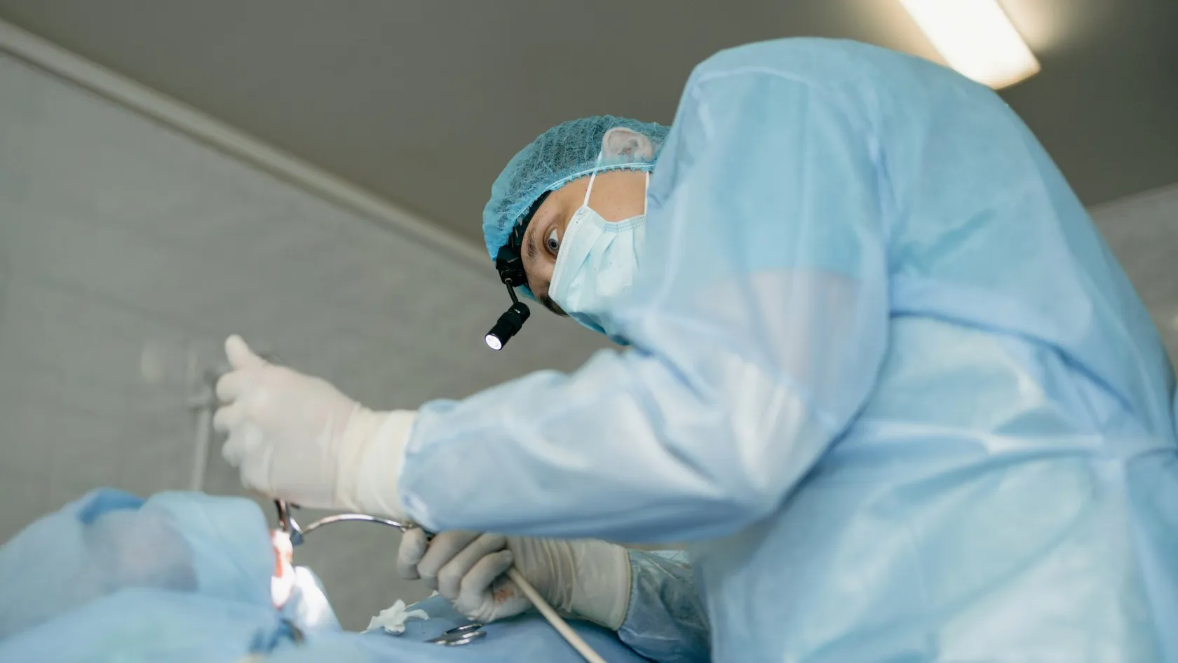
PEG Tube Insertion - Percutaneous Endoscopic Gastrostomy
A PEG is a feeding tube placed through the skin into the stomach during a gastroscopy, in twenty to thirty minutes under sedation, for patients who cannot swallow safely or cannot eat enough. This page explains who needs one and when a nasal tube serves better, what the stroke trial showed, how the tube is placed, when feeding starts, the risks, the daily care at home, the situations where guidelines advise against a PEG, and how families from abroad arrange it in Istanbul.
About This Department
A feeding tube placed through the skin into the stomach, in about twenty minutes, without an operation. For when swallowing has stopped being safe.
Percutaneous endoscopic gastrostomy, or PEG, was described in 1980 as a way to establish a feeding gastrostomy without opening the abdomen, and the first 31 patients came through it with no death and minimal morbidity (Gauderer and colleagues, Journal of Pediatric Surgery, 1980). The technique has changed little since. A gastroscope lights the stomach from within, a needle passes through the abdominal wall at the point of light, and a soft tube is drawn into place and held by a bumper on each side. European guidelines advise a percutaneous tube once feeding by a nasal tube is expected to last more than four weeks (Arvanitakis and colleagues, Endoscopy, 2021). This page explains who needs a PEG, how it compares with a nasogastric tube, how it is placed, the risks, the care at home, the situations where a PEG is not the right answer, and how families from abroad arrange it at Biruni Hospital in Istanbul.
Who needs a PEG
Some patients cannot swallow. Others can, but not enough. Both groups lose weight, strength and the ability to fight infection, and a tube into the stomach lets them be fed while the reason is treated or, when it cannot be treated, for as long as feeding serves them. The tube passes through the skin of the upper abdomen, sits inside the stomach, and takes liquid feed, water and medicines by syringe or pump. Talking, sitting, washing and, where swallowing allows, eating for pleasure all carry on around it.
The usual reasons
PEG or nasogastric tube
A nasal tube goes in at the bedside in minutes and comes out as easily. It blocks, it dislodges, it is pulled out by confused patients, and it can be passed into the airway by mistake, which the European guideline treats as a serious and avoidable event that every hospital needs a protocol to prevent. The PEG needs an endoscopy and a small procedure. Once in, it stays for months or years, is changed without another endoscopy, and is hidden under clothing.
What the stroke trial showed
Other routes
A gastrostomy can also be placed by a radiologist under X-ray guidance, when a gastroscope cannot pass a tumor in the throat, or by a surgeon at laparoscopy, when the stomach cannot be reached safely from inside. A jejunal extension through the PEG, or a direct jejunostomy, feeds beyond the stomach in patients who vomit gastric feed or inhale it. Few need that. The stomach takes bolus feeds and a normal schedule of meals, which the small bowel does not, and it remains the first choice for nearly everyone.
How it is placed
Twenty to thirty minutes, under sedation.
The pull technique, which the European guideline recommends as the standard, begins with an overnight fast, a single dose of an intravenous antibiotic and the patient lying on the back.
Is it done under general anesthesia?
What if the scope cannot pass a tumor?
When feeding starts
Feeding may begin within three to four hours after an uncomplicated placement. This is the one recommendation in the guideline supported by high quality evidence, and it replaced an older habit of waiting a day (Gkolfakis and colleagues, Endoscopy, 2021).
| When | What happens | Who does it |
|---|---|---|
| First four hours | Observation, pain relief, checking the dressing | Nursing staff |
| Four hours on | Water through the tube, then feed at a low rate | Nurse and dietitian |
| Day one | Full feeding schedule, first dressing change, training for the family | Dietitian, stoma nurse |
| Day two | Discharge for most patients with a written feeding plan | Gastroenterologist |
| Day seven to ten | Wound check and adjustment of the external bumper | Clinic or home nurse |
Risks
Low, but not zero, and the patients who need a PEG are often the ones least able to withstand a complication.
- Wound infection around the tube site, the commonest problem, largely prevented by the single dose of antibiotic and treated with local care when it occurs.
- Bleeding from the abdominal wall or the stomach lining, usually minor and stopping on its own.
- Leakage of feed around the tube, treated by adjusting the bumper, protecting the skin and sometimes resting the tube.
- Injury to the colon or liver by the needle, rare when the light and the finger sign are clear, and the procedure is abandoned when they are not.
- Peritonitis if the stomach separates from the abdominal wall before the tract has matured, which is why the tube is not changed in the first month.
- Buried bumper syndrome, in which the internal bumper is pulled into the stomach wall by a tight external fixation and grows over, months later. Prevented by keeping the outer bumper loose and pushing the tube inward a centimeter each day.
Can a PEG stop someone inhaling food?
When a PEG is not the answer
Two recommendations in the European guideline carry the word strong and deserve to be read by every family considering a tube. No PEG in advanced dementia. No PEG when life expectancy falls short of thirty days. Both rest on the same finding, gathered over thirty years from nursing homes, stroke units and cancer wards in Europe and North America. In these patients the tube does not lengthen life, does not prevent pneumonia, does not heal pressure sores and does not improve comfort, and it adds a procedure, a wound and, for a confused patient, a thing to pull at that then has to be restrained. Careful feeding by hand, with food the patient likes, at the pace the patient sets, with the family at the bedside rather than a pump, serves them better, and the guideline authors say so in the strongest language they use anywhere in the document. A hard conversation. The gastroenterology team will have it plainly with a family rather than place a tube that the evidence says will not help.
A request for a PEG is therefore reviewed before it is accepted. The team asks what the tube is for, how long it is expected to be needed, whether the patient can take part in the decision, and what the patient would have wanted. A patient with a stroke, a cancer under treatment or a neurological illness that will run for years gains from the tube. A patient in the last weeks of life, or one who no longer recognizes food, does not.
Care at home
Simple, once learned.
The family, or the patient, learns the routine before discharge, practices it on the ward under a nurse, and takes home the instructions in writing and in a video, along with the phone number of the stoma nurse for the questions that arrive in the first week, which concern leaking, redness and whether the tube has moved.
| Task | How often | Detail |
|---|---|---|
| Flush with water | Before and after every feed and medicine | 30 to 50 milliliters, to keep the tube clear |
| Clean the skin | Daily | Soap and water, dried well, no dressing once healed |
| Rotate and push in | Daily | A full turn and a centimeter inward, to prevent the bumper burying |
| Check the bumper | Weekly | One to two centimeters of play against the skin |
| Weigh | Weekly | The dietitian adjusts the feed to the trend |
- Call the same day for fever, redness spreading from the site, pain on feeding, feed leaking around the tube, or a tube that will not flush.
- Go to an emergency department if the tube falls out, because the tract closes within hours. A replacement tube or a urinary catheter placed in the tract keeps it open until a proper tube is fitted.
Coming from abroad
Two reasons bring families here for a PEG. Either the patient comes for another treatment, chiefly radiotherapy for a head and neck cancer, and the tube goes in before it starts, or the patient arrives for rehabilitation after a stroke or brain injury and needs secure feeding for the months that will take.
Cost
An estimate follows the file review and covers the endoscopy, sedation, the tube and fixation kit, the antibiotic, two nights in hospital, dietitian training and the wound check. Hospitals in this market quote PEG as a package, with feed supplies, a low-profile button and later tube changes priced separately, and a family that will be staying in Istanbul for radiotherapy or rehabilitation should ask for the tube to be quoted inside that larger package rather than on its own, since the hospital stay, the dietitian and the nursing are then already covered.
Confirm what the figure includes and whether the feed for the first weeks is part of it.
References
- Gauderer MW, Ponsky JL, Izant RJ. Gastrostomy without laparotomy. A percutaneous endoscopic technique. J Pediatr Surg. 1980;15(6):872-875.
- Dennis MS, Lewis SC, Warlow C, FOOD Trial Collaboration. Effect of timing and method of enteral tube feeding for dysphagic stroke patients (FOOD). A multicentre randomised controlled trial. Lancet. 2005;365(9461):764-772.
- Arvanitakis M, Gkolfakis P, Despott EJ, et al. Endoscopic management of enteral tubes in adult patients. Part 1. Definitions and indications. European Society of Gastrointestinal Endoscopy (ESGE) Guideline. Endoscopy. 2021;53(1):81-92.
- Gkolfakis P, Arvanitakis M, Despott EJ, et al. Endoscopic management of enteral tubes in adult patients. Part 2. Peri- and post-procedural management. European Society of Gastrointestinal Endoscopy (ESGE) Guideline. Endoscopy. 2021;53(2):178-195.
Editor's note
Written by the Biruni Hospital medical editorial team. Reviewed by Assistant Professor Koray KOÇHAN, Gastroenterology.
Medically reviewed by
Assistant Professor Koray KOÇHAN
Gastroenterology
Related Treatments
View All
Barrett's Esophagus Treatment
Most people told they have Barrett's esophagus have been told only half the story. The yearly cancer risk is 0.12 percent, and the grade of dysplasia in the biopsies decides whether the right plan is a daily tablet with endoscopy every few years, or radiofrequency ablation to remove the lining.
Capsule Endoscopy - Wireless Capsule Endoscopy
When a gastroscopy and a colonoscopy are both normal and the blood count keeps falling, the search moves to the small bowel, which neither instrument reaches. A swallowed capsule camera films that stretch in eight hours without sedation and finds a cause in six of ten patients examined for bleeding.
Colonoscopy
Ask one question before choosing where to have a colonoscopy, which is how often the doctor finds polyps. In a study of 314,872 examinations that figure ranged from 7 to 53 percent between doctors, and it predicted who later developed bowel cancer. This guide covers preparation in a hotel room, sedation, findings and the flight home.
Colorectal Polyp Removal
A large polyp in the colon does not automatically mean bowel surgery. Nearly all polyps, including flat ones wider than 20 millimeters, come out through the colonoscope, and the technique is chosen by size and shape.
Endoscopic Mucosal Resection - EMR
Endoscopic mucosal resection lifts a precancerous growth or an early cancer off the muscle wall with an injection and removes it with a snare through an ordinary endoscope, in the colon, esophagus, stomach or duodenum, without an operation. In a study of 479 patients with large colon polyps, 83.7 percent avoided surgery and none died of the procedure. This page explains what EMR removes, which lesions qualify, how it compares with ESD and surgery, evidence, risks, follow-up and care from abroad.