
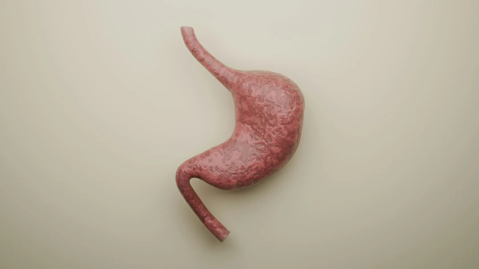
Barrett's Esophagus Treatment
Most people told they have Barrett's esophagus have been told only half the story. The yearly cancer risk is 0.12 percent, and the grade of dysplasia in the biopsies decides whether the right plan is a daily tablet with endoscopy every few years, or radiofrequency ablation to remove the lining.
About This Department
You were told Barrett's esophagus can turn into cancer. The yearly risk is 0.12 percent, and the biopsy result decides who needs treating.
Barrett's esophagus is a change in the lining of the lower food pipe caused by years of acid reflux. A Danish study of 11,028 patients in the New England Journal of Medicine put the risk of esophageal cancer at 0.12 percent a year, far below what doctors once assumed. For the smaller group whose biopsies show abnormal cells, called dysplasia, the picture differs and so does the evidence, since in a European trial radiofrequency ablation cut progression to high-grade dysplasia or cancer over three years from 26.5 percent to 1.5 percent.
People searching for Barrett's Esophagus Treatment have usually just read a pathology report. This page explains what that report means, which of the four possible results calls for an active procedure, and how a patient from another country can have one in Istanbul.
What Barrett's esophagus is
Barrett's esophagus develops when stomach acid and bile wash back into the food pipe for years. The flat, pale lining at the lower end gives way to a salmon-colored lining that resembles the inside of the intestine. Doctors call the change intestinal metaplasia. The new lining resists acid better, which explains why heartburn sometimes eases as the condition sets in. It also carries a raised risk of a cancer called esophageal adenocarcinoma. Diagnosis needs an endoscopy and biopsies. The endoscopist records the length of the segment with the Prague classification, written as C for the part that runs all the way round and M for the longest tongue, and takes four biopsies at every two centimeters. The American College of Gastroenterology puts the condition at 10 to 15 percent of people with chronic reflux, and men past 50 with a large waist, a smoking history or a close relative with the disease carry the highest risk, although many of them have never had severe heartburn.
1 in 830 each yearThe risk of esophageal adenocarcinoma among 11,028 Danish patients with Barrett's esophagus followed for a median of 5.2 years. That equals 0.12 percent a year, against the 0.5 percent that older surveillance guidelines assumed. Hvid-Jensen and colleagues, N Engl J Med, 2011.
Barrett's esophagus treatment by biopsy result
Four pathology results are possible, and each leads to a different plan. The recommendations below come from the 2023 guideline of the European Society of Gastrointestinal Endoscopy by Weusten and colleagues and the 2022 guideline of the American College of Gastroenterology by Shaheen and colleagues, which agree on the main points.
This table scrolls sideways on a narrow screen. Swipe or drag to see every column.
| Biopsy result | What it means | Recommended plan |
|---|---|---|
| No dysplasia | Barrett's lining with orderly cells | Acid-suppressing medication and a repeat endoscopy every 5 years for a segment of 1 to 3 cm, every 3 years for 3 to 10 cm. No ablation |
| Low-grade dysplasia | Mildly abnormal cells, confirmed twice | Offer radiofrequency ablation |
| High-grade dysplasia | Markedly abnormal cells, no invasion | Remove any visible lesion by endoscopic resection, then ablate the rest |
| Early cancer, stage T1a | Cancer confined to the inner lining | Endoscopic resection as curative treatment, followed by ablation of the remaining Barrett's lining |
The large majority of people with Barrett's esophagus sit in the first row at diagnosis. Their correct treatment is a tablet and a calendar.
The treatments, one by one
Acid control and surveillance
Every patient with Barrett's esophagus takes a proton pump inhibitor such as omeprazole or esomeprazole, whatever else is done, because it heals the inflammation that confuses pathologists, controls symptoms, and lets the lining regrow as normal tissue after ablation. It does not make Barrett's esophagus go away. Surveillance means a careful endoscopy at fixed intervals with high-definition equipment and a minimum of one minute of inspection for every centimeter of Barrett's lining.
Radiofrequency ablation
Radiofrequency ablation, known as RFA, burns away the Barrett's lining to a depth of half a millimeter with an electrode mounted on a balloon or on the tip of the endoscope. The wound heals under acid suppression with normal flat lining. The session takes 30 minutes under sedation. The patient goes home the same day.
Endoscopic resection
Any lump, dip or irregular patch within the Barrett's segment is cut out before anything is burned, because a visible lesion can hide a cancer and the pathologist needs the whole piece to measure its depth. Endoscopic mucosal resection lifts the area with a band or a cap and snares it. Endoscopic submucosal dissection carves out larger lesions in one piece.
Surgery and other methods
Removal of the esophagus was the standard for high-grade dysplasia until endoscopic treatment replaced it, and it remains the treatment for cancers that have grown beyond the reach of the endoscope. Freezing with a cryoballoon and burning with argon plasma are alternatives to RFA that some centers use for small residual islands. Antireflux surgery controls reflux in patients who cannot take tablets. It has not been shown to prevent cancer.
An ablation session, step by step
The day runs to a fixed order.
- Fasting. Nothing to eat for eight hours and nothing to drink for two. Blood thinners are paused on the schedule agreed with the doctor who prescribed them.
- Sedation. An anesthesiologist gives deep sedation through a vein. You sleep and breathe by yourself.
- Mapping. The endoscopist washes the lining, inspects it under high definition and marks the upper and lower limits of the Barrett's segment.
- Ablation. A balloon electrode treats a segment that runs all the way round. A smaller paddle on the endoscope treats tongues and islands. Each area receives two applications.
- Recovery. One to two hours in the recovery area, then back to the hotel with a companion.
- The first week. Liquids for 24 hours and soft food for several days. Chest discomfort and painful swallowing are expected and ease within a week, helped by doubled acid suppression and a liquid pain reliever.
Healing takes two to three months, and the next endoscopy is timed to it.
Complications and their rates
Ablation counts as safe by the standards of cancer prevention. Two trials counted its complications in detail.
This table scrolls sideways on a narrow screen. Swipe or drag to see every column.
| Complication | How often | What is done |
|---|---|---|
| Narrowing of the esophagus (stricture) | 6.0 percent in the American trial, 11.8 percent in the European trial | Stretching with a balloon during an endoscopy. Every stricture in the European trial resolved, after a median of one session |
| Chest pain and painful swallowing | Common in the first days | Liquid pain relief, soft diet, doubled acid suppression |
| Bleeding | 1 of 84 treated patients in the American trial | Treated during an endoscopy |
| Any treatment-related event | 19.1 percent in the European trial | Most were mild and managed without admission |
| Barrett's lining or dysplasia coming back | Occurs in a minority over the years | Picked up by surveillance and treated again with ablation or resection |
Resection carries higher rates of bleeding and stricture than ablation, and the larger the area removed, the higher they go. Perforation is rare with both. Food that sticks weeks after treatment signals a stricture. Report it.
How many trips it takes
One trip is not enough.
Clearing the lining takes several stages. The American College of Gastroenterology tells patients to expect one to three RFA sessions spaced two to three months apart, the European trial allowed up to five, and long segments need more than short ones. After the last session a first check endoscopy follows, and the European guideline recommends that this check happens at an expert center. For a traveler the arithmetic is plain. A patient with low-grade dysplasia in a three-centimeter segment should plan two or three visits of three to four days each over six to nine months, and a patient who needs a resection first should add one more. Anyone who cannot commit to coming back is better treated near home, even at a higher price, since a half-finished ablation leaves dysplasia behind under a false sense of security, with nobody watching it. Count the visits before you book the first one.
Each visit follows the same pattern. Day one holds the consultation and the anesthesia check, day two the procedure, and day three or four a review before the flight. Flying poses no problem once soft food goes down and the pain is controlled.
If the plan changes
The second reading of the slides often changes the diagnosis. It can go either way. A downgrade to no dysplasia cancels the ablation, which is good news even when the flight is already booked, while an upgrade, or a visible lesion found at the mapping endoscopy, means resection comes first and the specimen decides the rest. If that specimen shows cancer deeper than the inner lining, the case goes to a tumor board. Radiotherapy and chemotherapy are both given at Biruni Hospital on the same site, so that discussion and any treatment that follows can happen without sending records elsewhere. Each new step has its own consent and its own quote. You remain free to take the pathology report home.
Cost and the trip
What you pay depends on what the biopsies call for. Surveillance endoscopy with biopsies comes cheapest. Ablation costs more per session because the electrode is a single-use device, the balloon version for a full circumference costs more than the small paddle, and resection adds its own devices, a longer anesthetic and a larger pathology bill. The number of sessions multiplies all of it. A long segment that needs four sessions costs far more than a short one cleared in two. A second pathology opinion on outside slides is charged separately by most laboratories. Your own health enters through the anesthesia plan and the handling of blood thinners, diabetes and heart or lung disease. Packages published for endoscopic treatment in Istanbul cover the consultation, anesthesia, the procedure and its device, pathology, an interpreter and transfers, and leave out flights, the hotel, later sessions and the treatment of a complication.
Ask for the whole course to be quoted, session by session.
A coordinator in the international patients office manages each visit, in English, Arabic, French, Russian, Serbian, Romanian or Spanish, with interpreters for other languages. The office sends the appointment confirmation and an invitation letter naming the hospital and the treating doctor ten days before each trip, which matters more than usual here, because repeat visits may call for a multiple-entry visa. It books the hotel and the transfers. Come with a companion, since you will be sedated and should not be alone that night. Two family members are welcome in the consultation. Soft halal, vegetarian or diabetic meals are arranged on request for the days after treatment, a prayer room is on site, and a wish for a female doctor belongs in the first message.
After you fly home
Treatment ends. Surveillance does not, because Barrett's lining and dysplasia can return, and the European guideline sets out the schedule once the lining has been cleared.
- After high-grade dysplasia or early cancer, an endoscopy at 1, 2, 3, 4, 5, 7 and 10 years, after which surveillance may stop
- After low-grade dysplasia, an endoscopy at 1, 3 and 5 years
- Acid-suppressing medication every day, for life
- Weight loss where needed and no smoking, since both drive reflux and cancer risk
- A doctor the same week for food sticking, and an emergency department for vomited blood, black stools or severe chest pain
A gastroenterologist at home can do most of those endoscopies. The discharge file is written for that doctor and contains the Prague measurements before and after treatment, every pathology report, the ablation settings, photographs and the date the next endoscopy is due. Once you are back home the coordinator stays reachable on the same WhatsApp number.
Barrett's Esophagus Treatment FAQ
Can Barrett's esophagus be cured?
Do I need ablation if I have no dysplasia?
How many times do I have to come to Istanbul?
When can I fly after an ablation session?
Can I have the follow-up endoscopies in my own country?
Will someone speak my language?
References
- Weusten BLAM, Bisschops R, Dinis-Ribeiro M, et al. Diagnosis and management of Barrett esophagus: European Society of Gastrointestinal Endoscopy (ESGE) Guideline. Endoscopy. 2023;55(12):1124-1146.
- Shaheen NJ, Falk GW, Iyer PG, Souza RF, Yadlapati RH, Sauer BG, Wani S. Diagnosis and Management of Barrett's Esophagus: An Updated ACG Guideline. Am J Gastroenterol. 2022;117(4):559-587.
- Phoa KN, van Vilsteren FG, Weusten BL, et al. Radiofrequency ablation vs endoscopic surveillance for patients with Barrett esophagus and low-grade dysplasia: a randomized clinical trial. JAMA. 2014;311(12):1209-17.
- Shaheen NJ, Sharma P, Overholt BF, et al. Radiofrequency ablation in Barrett's esophagus with dysplasia. N Engl J Med. 2009;360(22):2277-88.
- Hvid-Jensen F, Pedersen L, Drewes AM, Sorensen HT, Funch-Jensen P. Incidence of adenocarcinoma among patients with Barrett's esophagus. N Engl J Med. 2011;365(15):1375-83.
Editor's note
Written by the Biruni Hospital medical editorial team. Reviewed by Assistant Professor Koray KOÇHAN, Gastroenterology.
Medically reviewed by
Assistant Professor Koray KOÇHAN
Gastroenterology
Related Treatments
View All
Capsule Endoscopy - Wireless Capsule Endoscopy
When a gastroscopy and a colonoscopy are both normal and the blood count keeps falling, the search moves to the small bowel, which neither instrument reaches. A swallowed capsule camera films that stretch in eight hours without sedation and finds a cause in six of ten patients examined for bleeding.
Colonoscopy
Ask one question before choosing where to have a colonoscopy, which is how often the doctor finds polyps. In a study of 314,872 examinations that figure ranged from 7 to 53 percent between doctors, and it predicted who later developed bowel cancer. This guide covers preparation in a hotel room, sedation, findings and the flight home.
Colorectal Polyp Removal
A large polyp in the colon does not automatically mean bowel surgery. Nearly all polyps, including flat ones wider than 20 millimeters, come out through the colonoscope, and the technique is chosen by size and shape.
Endoscopic Mucosal Resection - EMR
Endoscopic mucosal resection lifts a precancerous growth or an early cancer off the muscle wall with an injection and removes it with a snare through an ordinary endoscope, in the colon, esophagus, stomach or duodenum, without an operation. In a study of 479 patients with large colon polyps, 83.7 percent avoided surgery and none died of the procedure. This page explains what EMR removes, which lesions qualify, how it compares with ESD and surgery, evidence, risks, follow-up and care from abroad.
Endoscopic Submucosal Dissection - ESD
Removed in one piece, an early tumor can be measured at its edges and at its deepest point, and that measurement decides whether you are cured or need surgery. ESD is the endoscopic technique that makes it possible in the stomach, esophagus and bowel. See which lesions qualify, what recovery involves and how to judge an operator.