
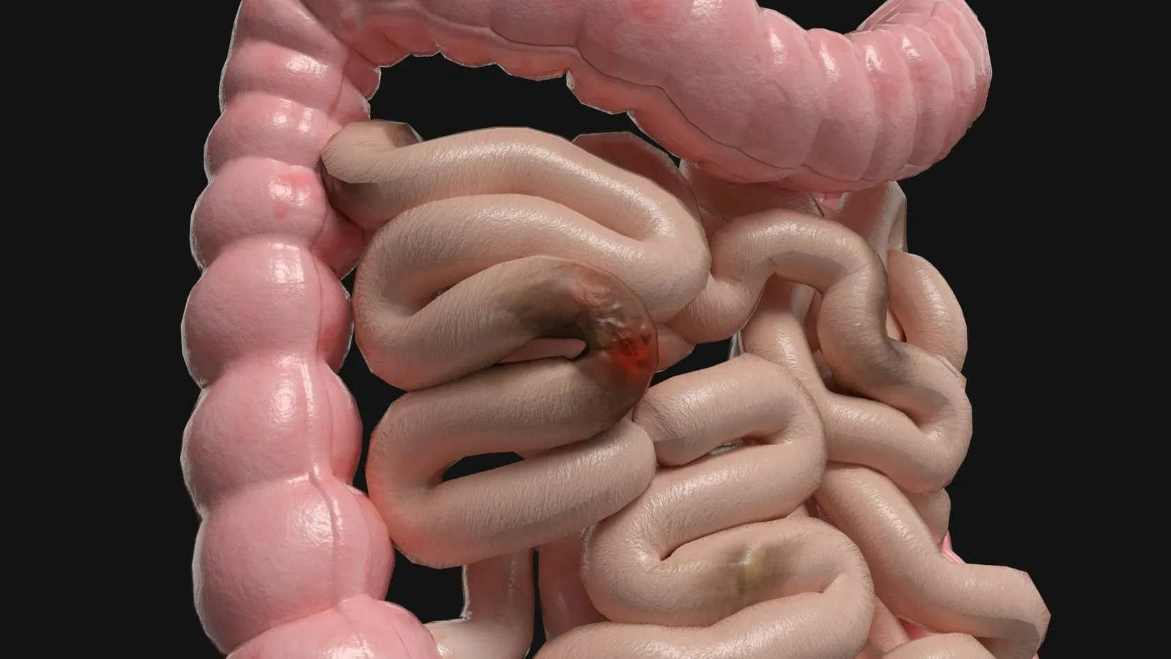
Endoscopic Mucosal Resection - EMR
Endoscopic mucosal resection lifts a precancerous growth or an early cancer off the muscle wall with an injection and removes it with a snare through an ordinary endoscope, in the colon, esophagus, stomach or duodenum, without an operation. In a study of 479 patients with large colon polyps, 83.7 percent avoided surgery and none died of the procedure. This page explains what EMR removes, which lesions qualify, how it compares with ESD and surgery, evidence, risks, follow-up and care from abroad.
About This Department
Of 479 patients with large colon polyps that would once have meant an operation, 83.7 percent avoided surgery after endoscopic mucosal resection. Nobody died of the procedure.
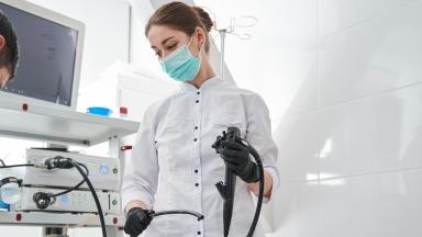
The Australian Colonic Endoscopic resection study followed every patient referred to its centers for removal of a flat or sessile polyp of 20 millimeters or more, with a mean size of 35.6 millimeters, and found that a single session cleared the lesion in 89.2 percent (Moss and colleagues, Gastroenterology, 2011). Endoscopic mucosal resection, or EMR, lifts a lesion off the muscle wall with an injection of fluid and removes it with a wire snare through an ordinary endoscope, in the esophagus, the stomach, the duodenum or the colon. It treats precancerous growths and the earliest cancers, those confined to the surface layer, without an incision, an anesthetic for surgery or a night in the hospital. This page explains what EMR removes, which lesions qualify, how it compares with the alternatives, what the research shows, the risks, the follow-up that comes with it, and how patients from abroad can arrange it at Biruni Hospital in Istanbul.
What EMR removes
The lining of the digestive tract, the mucosa, sits on a loose layer of connective tissue and, beneath that, the muscle of the gut wall. Growths that begin in the lining and have not crossed into the deeper layers can be removed from the inside. That is the whole principle. Simple to state. Harder to do well.
- Marking and lifting. The edge of the lesion is defined with enhanced imaging, and a solution containing dye and sometimes adrenaline is injected beneath it. A lesion that lifts cleanly is suitable. One that will not lift has probably invaded deeper and is left for another approach.
- Snaring. A wire loop is closed around the raised lesion and an electrical current cuts through the base. Lesions under two centimeters come away in one piece. Larger ones are removed in several pieces.
- Checking the bed. The wound is examined for bleeding, for muscle injury and for any remaining tissue, and the margin is treated with a heat probe.
- Retrieval. Every piece is retrieved and sent to the pathologist, oriented and labeled, because the report decides whether the treatment is complete.
Which lesions qualify
The commonest type of large polyp, flat and granular, carried a 1.4 percent risk of hidden invasive cancer. Lesions with a depressed area, a smooth non-granular surface or a disrupted surface pattern carried a much higher risk and were the ones that needed a different plan.
Reading the surface
Before anything is injected, the endoscopist studies the lesion under magnification and blue light. The pattern of the pits and vessels on its surface predicts, with good reliability, whether the growth is benign, whether it has become cancer, and whether that cancer has begun to invade. A lesion whose surface pattern suggests deep invasion is biopsied and staged instead of resected, because a piecemeal removal of an invasive cancer helps nobody and can make the later operation harder. In the esophagus and stomach, EUS is sometimes added to judge depth. The decision to proceed is made at the scope, by a doctor who does this often, and a patient referred with a report from elsewhere should expect the lesion to be reassessed before it is removed.
EMR, ESD or surgery
EMR removes the lining. Endoscopic submucosal dissection, ESD, cuts beneath it with a fine knife and lifts the whole lesion off the muscle in one piece, whatever its size. Surgery removes the segment of gut.
When is ESD chosen over EMR?
When surgery remains right
Four situations. A cancer that has invaded the deeper layers, a lesion that will not lift, a growth that wraps around most of the circumference of the bowel, and a recurrence that has scarred to the muscle are the usual reasons. Surgery also becomes the answer when the pathology after EMR shows invasion with high-risk features, since the lymph nodes then need removing. That outcome was uncommon in the Australian series, and most of the 16 percent who went to surgery did so because the lesion could not be completed rather than because cancer was found.
What research shows
The problem of recurrence, and its solution
Removing a large lesion in pieces leaves microscopic remnants at the edge in a proportion of cases, and 15 to 30 percent of patients had regrowth at the first check colonoscopy in earlier series.
Is recurrence dangerous?
Risks and the days after
- Bleeding. During the procedure in a minority, controlled at the time. Delayed bleeding in the days afterward in a few percent, higher for large lesions in the right colon and in patients on blood thinners, and treated by a repeat endoscopy if it does not stop.
- Perforation. A hole in the wall, in roughly one to two percent of large colon EMRs, usually recognized at once and closed with clips. Surgery is needed in a small fraction.
- Post-procedure pain. A burn to the muscle layer causes localized pain and fever for a day or two and settles with rest, fluids and antibiotics.
- Stricture. Narrowing after removal of a large area of the esophagus, treated by stretching.
- Incomplete removal. Recognized from the pathology or at surveillance, and dealt with by a further endoscopic session.
What is recovery like?
Sedation and anesthesia
EMR takes longer than a diagnostic endoscopy. Twenty minutes for a modest lesion. Well over an hour for a large one. The patient needs to stay still throughout. Deep sedation with propofol under an anesthesiologist is the usual arrangement, with the same fasting and monitoring rules as any endoscopy under sedation.
Follow-up and recurrence
The schedule
- Benign lesion, complete removal. Surveillance endoscopy at six months to inspect and biopsy the scar, then at intervals set by guideline, usually one and three years for the colon.
- Early cancer, complete removal, low-risk features. The same schedule, with closer attention, and for the esophagus and stomach a further endoscopy at three months.
- Cancer with high-risk features. Referral for surgery or, where surgery is unwise, for a discussion of the alternatives with the multidisciplinary team.
| Finding | Meaning | Next step |
|---|---|---|
| Adenoma or serrated lesion, no cancer | Precancerous growth removed before it turned | Surveillance |
| Cancer confined to the mucosa | Cured by removal in most cases | Surveillance, with attention to the scar |
| Cancer into the upper submucosa, no bad features, clear margin | Low risk of nodal spread | Surveillance, or surgery in selected patients after discussion |
| Deep submucosal invasion, vessel invasion, poor differentiation or an involved margin | Meaningful risk of spread | Surgical resection with lymph nodes |
Coming from abroad
What to send
The visit
One coordinator from the international patients office handles arrangements from the first message through discharge and answers the same WhatsApp number once you are back home. The office works in English, Arabic, French, Russian, Serbian, Romanian and Spanish and books interpreters for other languages. It arranges the visa invitation letter, airport transfers, daily transport and accommodation near the hospital. The kitchen prepares halal, vegetarian and diabetic meals, a prayer room is on site, and a request for a female physician is met wherever the rota allows.
Why the first attempt matters most
A polyp that has been biopsied repeatedly, partly removed, or burned with a heat probe at an earlier endoscopy becomes scarred to the muscle beneath it. It no longer lifts. The plane that EMR depends on has gone, and what remains is a harder, riskier procedure with a lower chance of success, or an operation that the patient might never have needed. The Australian series found that a previous attempt elsewhere nearly quadrupled the odds of failure, and the authors put the point plainly. The first EMR is the important one.
Two lessons follow for patients. When a large flat polyp is found at a colonoscopy, it should be photographed, measured and left alone until someone can remove it completely, someone who removes such lesions routinely, and a single biopsy from the edge, if any, is all that should be taken. And a patient whose polyp has already been partly removed should say so, send the earlier report, and expect a candid answer on what is still possible.
Scar is the enemy. Every attempt makes more of it.
Barrett's esophagus and the esophagus more generally
Two jobs at once.
Inside the esophagus, EMR removes the visible abnormality and at the same time provides the pathologist with a specimen deep enough to say how far any cancer has gone, which a biopsy cannot. That staging function is the reason guidelines call for EMR of any visible lesion in Barrett's esophagus before a decision on further treatment. Once the lesion is out and the depth is known, the rest of the Barrett's lining is treated with radiofrequency ablation over two or three sessions, so that the tissue that produced one cancer cannot produce another. Squamous cancers of the esophagus, common in parts of Asia, Africa and the Middle East, follow the same principle and go to ESD in most centers, because a one-piece specimen is what the pathologist needs to say whether the cancer has reached the vessels that carry it to the nodes. Removal of more than three quarters of the circumference carries a high risk of a stricture. A course of steroid injection or tablets prevents most of them.
Swallowing returns to normal within days. The follow-up endoscopies last for years.
Cost
The estimate follows the file review and depends on the size and site of the lesion, whether EMR or ESD is planned, anesthesia, pathology and any overnight stay. Hospitals in this market quote endoscopic resection as a procedure fee with anesthesia and pathology listed separately.
Confirm what the figure includes and whether a surveillance endoscopy is priced with it.
References
- Moss A, Bourke MJ, Williams SJ, et al. Endoscopic mucosal resection outcomes and prediction of submucosal cancer from advanced colonic mucosal neoplasia. Gastroenterology. 2011;140(7):1909-1918.
- Klein A, Tate DJ, Jayasekeran V, et al. Thermal ablation of mucosal defect margins reduces adenoma recurrence after colonic endoscopic mucosal resection. Gastroenterology. 2019;156(3):604-613.
- Cao Y, Liao C, Tan A, Gao Y, Mo Z, Gao F. Meta-analysis of endoscopic submucosal dissection versus endoscopic mucosal resection for tumors of the gastrointestinal tract. Endoscopy. 2009;41(9):751-757.
Editor's note
Written by the Biruni Hospital medical editorial team. Reviewed by İbrahim Yılmaz, Gastroenterology.
Medically reviewed by
İbrahim Yılmaz
Gastroenterology
Related Treatments
View All
Barrett's Esophagus Treatment
Most people told they have Barrett's esophagus have been told only half the story. The yearly cancer risk is 0.12 percent, and the grade of dysplasia in the biopsies decides whether the right plan is a daily tablet with endoscopy every few years, or radiofrequency ablation to remove the lining.
Capsule Endoscopy - Wireless Capsule Endoscopy
When a gastroscopy and a colonoscopy are both normal and the blood count keeps falling, the search moves to the small bowel, which neither instrument reaches. A swallowed capsule camera films that stretch in eight hours without sedation and finds a cause in six of ten patients examined for bleeding.
Colonoscopy
Ask one question before choosing where to have a colonoscopy, which is how often the doctor finds polyps. In a study of 314,872 examinations that figure ranged from 7 to 53 percent between doctors, and it predicted who later developed bowel cancer. This guide covers preparation in a hotel room, sedation, findings and the flight home.
Colorectal Polyp Removal
A large polyp in the colon does not automatically mean bowel surgery. Nearly all polyps, including flat ones wider than 20 millimeters, come out through the colonoscope, and the technique is chosen by size and shape.
Endoscopic Submucosal Dissection - ESD
Removed in one piece, an early tumor can be measured at its edges and at its deepest point, and that measurement decides whether you are cured or need surgery. ESD is the endoscopic technique that makes it possible in the stomach, esophagus and bowel. See which lesions qualify, what recovery involves and how to judge an operator.