When open heart surgery is too risky, or heart failure medicines no longer control a leaking mitral valve, a clip placed through a vein in the groin can close part of the leak. This guide explains the two kinds of mitral leak, who the clip suits, what the COAPT, MITRA-FR, RESHAPE-HF2 and EVEREST II trials found, how the procedure is done, the risks, the recovery and how long to stay in Istanbul.
About This Department
Cardiology
A leaking valve, closed from inside. No opening of the chest.
In the COAPT trial, deaths at two years fell from 46.1 to 29.1 percent when the clip was added to full heart failure medicines.
29.1%
Two-year death rate with the clip in COAPT, against 46.1%
1.4%
Device safety events over five years in COAPT
No incision
The clip reaches the heart through a groin vein
Free
Review of your echocardiograms before you book
Free consultation
The review costs nothing and carries no obligation. Send your echocardiogram reports and images, any transesophageal study, your list of heart medicines and recent kidney tests, and our cardiology team will tell you whether the clip suits your valve, what else to consider, and how long to plan for. A coordinator replies in your own language.
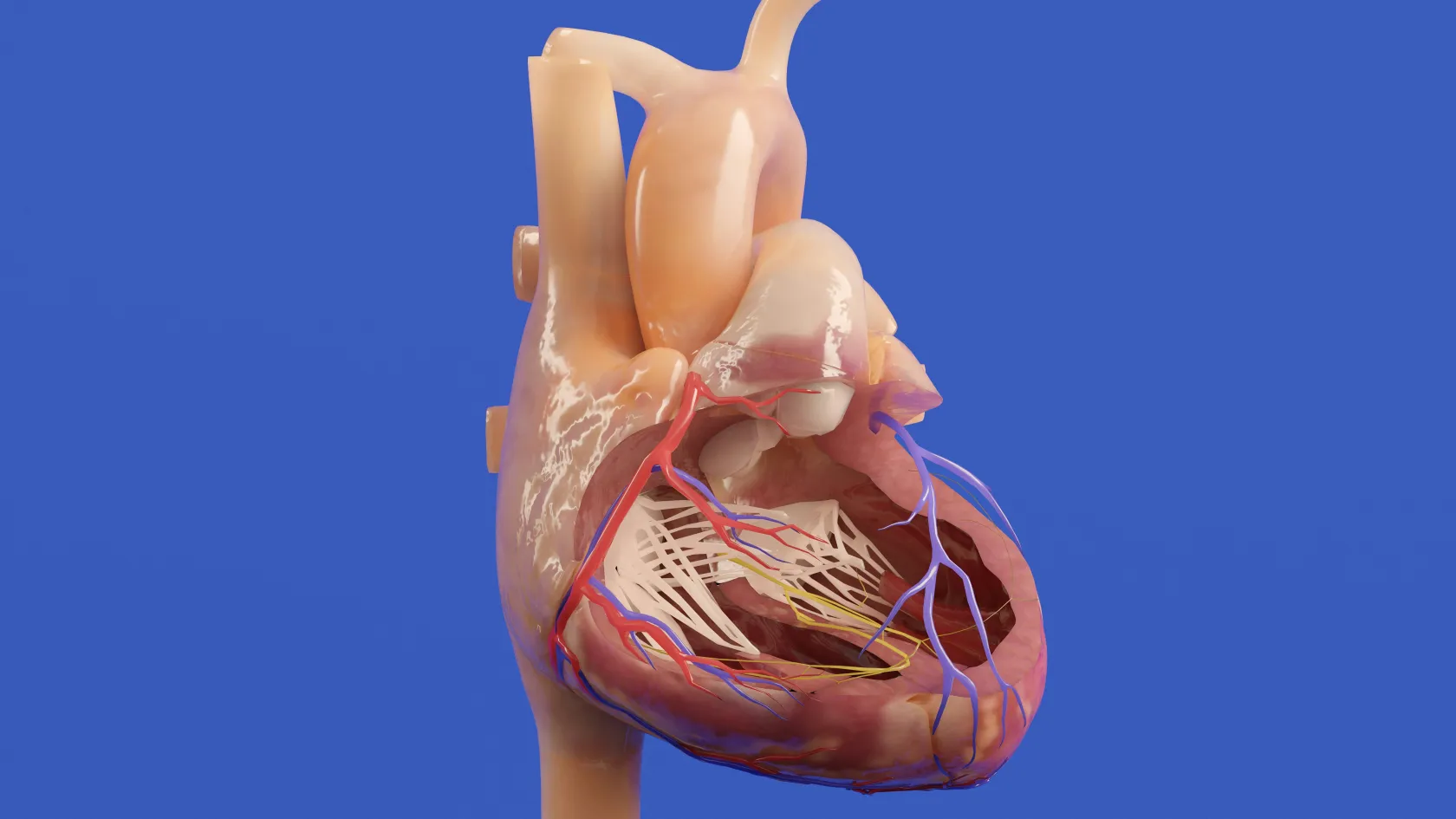
An echocardiogram has shown that your mitral valve leaks, breathlessness and tiredness have crept into ordinary days, and a cardiologist has said that open heart surgery carries too much risk or that medicines alone are no longer enough. A mitral valve clip, known to cardiologists as transcatheter edge-to-edge repair or TEER, closes part of that leak from inside the heart through a vein in the groin, with no opening of the chest and no heart-lung machine. Below you will find the two kinds of mitral leak and why the difference matters, who the clip suits, what the large randomized trials found, how it compares with surgery, how the procedure is done, the risks, the recovery, how long to stay in Istanbul, and the follow-up at home.
Two kinds of mitral leak
The mitral valve sits between the left atrium and the left ventricle, and its two leaflets should close tightly each time the heart pumps. When they do not meet, blood is pushed backward into the lungs with every beat, the heart works harder to keep up, and over months or years breathlessness, swelling of the ankles, an irregular heartbeat and repeated hospital stays for heart failure follow. Cardiologists split the problem into two kinds. In primary, or degenerative, regurgitation the valve itself is faulty, most often a leaflet that bulges back or a torn supporting cord. In secondary, or functional, regurgitation the leaflets are normal but the ventricle around them has enlarged after a heart attack or from cardiomyopathy, pulling them apart. Treatment follows the kind. For a faulty valve in a patient fit for an operation, surgical repair remains the standard, while for a leak caused by a weak ventricle, the first step is medicines for heart failure at full doses, sometimes with a pacemaker that resynchronizes the heartbeat, and the clip comes in when the leak and the symptoms persist despite them.
Signs that a leak is taking its toll include
breathlessness that comes on with less effort than before,
an enlarging heart on successive echocardiograms,
new atrial fibrillation,
and admissions to hospital for fluid on the lungs.
This table scrolls sideways on a narrow screen. Swipe or drag to see every column.
Primary and secondary mitral regurgitation compared
Feature
Primary regurgitation
Secondary regurgitation
Where the fault lies
The leaflets or their cords
An enlarged or weakened ventricle
Common causes
Prolapse, a torn cord, wear with age
Earlier heart attack, cardiomyopathy
First treatment
Surgical repair for patients fit for it
Heart failure medicines at full doses
Where the clip fits
Patients at high risk from surgery
Symptoms that persist despite those medicines
Why the kind of leak matters
The trials below tested different patients. Evidence for a faulty valve comes mostly from EVEREST II, and evidence for a weak ventricle from COAPT, MITRA-FR and RESHAPE-HF2.
How a mitral leak feels
Breathlessness on stairs and then on flat ground, tiredness, waking at night short of breath, needing more pillows, swollen ankles and a racing or irregular heartbeat. Some people feel well for years while the heart enlarges silently, which is why regular echocardiograms matter.
Who the clip suits
A heart team decides.
Interventional cardiologists, heart failure specialists, imaging cardiologists and a cardiac surgeon review each case together, weighing age, frailty, kidney and lung function, earlier heart surgery and the shape of the valve on a transesophageal echocardiogram, which shows whether the leaflets can be grasped. Valves with very thick, calcified or short leaflets, or a small opening to begin with, suit other treatments better.
Signs it may be time
Severe primary regurgitation with high surgical risk, or secondary regurgitation that persists despite full heart failure medicines, in leaflets that can be grasped.
Talk to us first
A patient fit for surgical repair of a faulty valve, a heavily calcified or narrowed valve, a ventricle so enlarged and weak that fixing the leak is unlikely to help, an active infection, or a clot inside the heart.
What to send for the first review
For the first review, send
your latest echocardiogram report and, where possible, the images,
any transesophageal echocardiogram,
a list of your heart medicines and doses,
and recent kidney tests and any coronary angiogram.
What a transesophageal echocardiogram involves
A thin ultrasound probe is passed down the throat under sedation, close behind the heart, and gives a far sharper view of the mitral leaflets than a scan through the chest wall. It takes twenty minutes. You go home the same day.
What the trials found
Leaks from a weak ventricle
Three randomized trials have tested the clip against medicines alone in heart failure with secondary regurgitation, and their results pointed in different directions. COAPT, published in 2018, enrolled 614 patients in the United States and Canada whose medicines had already been pushed to maximal doses, and over two years hospital admissions for heart failure ran at 35.8 percent per patient-year with the clip and 67.9 percent with medicines alone, and death from any cause fell from 46.1 percent to 29.1 percent. The benefit lasted. At five years, 57.3 percent of patients in the clip group had died against 67.2 percent in the medicines group, and device-specific safety events had occurred in 1.4 percent of treated patients, all within the first month.
MITRA-FR, a French trial of 304 patients published the same year, found no difference at one year, with death or unplanned admission for heart failure in 54.6 percent of the clip group and 51.3 percent of the medicines group. One widely cited explanation lies in who was enrolled, since COAPT patients had leaks that were large for the size of their ventricles and medicines adjusted to full doses before randomization, while many MITRA-FR patients had very enlarged ventricles in which the leak was a smaller part of the problem.
RESHAPE-HF2, published in 2024, added 505 patients from nine countries with moderate to severe regurgitation. Over two years, heart failure admissions or cardiovascular death occurred at 37.0 events per 100 patient-years with the clip and 58.9 with medicines alone, and quality-of-life scores rose further with the clip.
Scroll sideways on small screens to see every column.
Randomized trials of the clip for secondary mitral regurgitation
Trial
Patients
Main finding
COAPT, 2018
614
Fewer heart failure admissions and lower death rate at two years with the clip
COAPT, five years
614
Death 57.3 percent with the clip against 67.2 percent with medicines alone
MITRA-FR, 2018
304
No difference in death or admission at one year
RESHAPE-HF2, 2024
505
Fewer admissions or cardiovascular deaths and better quality of life at two years
Before recommending the clip, the heart team checks whether heart failure medicines have reached full doses, how large the leak is for the size of the ventricle, and whether a pacemaker that resynchronizes the heartbeat should come first.
The right patient matters more than the device.
Clip or open surgery
Surgery repairs more completely.
EVEREST II randomized 258 patients with grade 3 or 4 regurgitation, most of them with a faulty valve, to the clip or to surgery. At five years, freedom from death, surgery or a moderate to severe leak was 44.2 percent after the clip and 64.3 percent after surgery, a gap driven by more remaining leaks, 12.3 against 1.8 percent, and more operations to fix them, 27.9 against 8.9 percent. Most of those operations came early. After the clip, 78 percent of later surgeries happened within six months, and beyond that point the rates of surgery and of significant leaks were similar in both groups. Survival did not differ, with five-year death rates of 20.8 percent after the clip and 26.8 percent after surgery.
What this means in practice
For a faulty valve, surgery stays first. The clip is the choice when that operation carries too much risk, because of age, frailty, earlier heart surgery or other illnesses.
How the clip is placed
No cut is made in the chest. The procedure takes place in a hybrid operating room or catheter laboratory, usually under general anesthesia, so that a probe in the esophagus can show the valve in detail throughout, and it lasts one to three hours. A thin tube is passed from a vein in the groin up to the heart. It crosses the thin wall between the two upper chambers, and the clip, a small device covered in fabric, is steered to the valve. Its arms open, catch the edges of both leaflets at the point of the leak and close, joining them so the valve opens through two smaller openings instead of one, much as a surgeon's stitch would. The team measures the leak with every adjustment before releasing the clip, and adds a second or third clip when one is not enough. Each clip can be opened, repositioned and closed again many times before it is released. Once released, it stays for good. Over the following weeks a thin layer of tissue grows over the fabric, anchoring it within the heart.
Step by step
1
Anesthesia and imaging
You sleep, and a transesophageal probe gives the team a live view of the valve.
2
Access through the groin
A tube reaches the right side of the heart through the femoral vein.
3
Crossing to the left side
A small opening is made in the wall between the atria, which usually closes on its own.
4
Grasping the leaflets
The clip catches both leaflets, the leak is measured, and the clip is released once the result is right.
5
Closing the groin
A stitch or pressure closes the vein, and you wake in the recovery area.
Risks and how they are managed
Serious complications are uncommon.
Before the procedure, the team checks
kidney function and blood count,
how to pause and restart blood thinners,
the groin veins on ultrasound,
and the heart for any clot on the transesophageal scan.
Bleeding at the groin
The most common problem, and in most patients nothing more than a bruise that fades over two weeks. Pressure and a few hours of bed rest settle it.
A clip that loosens from one leaflet
Uncommon. It can be treated with a further clip or, in some patients, surgery.
Remaining leak or a narrowed valve
The team balances the two during the procedure, measuring the leak and the pressure across the valve before releasing each clip.
Stroke
Rare. Blood thinners during the procedure reduce it.
The opening between the atria
Usually small, and it closes on its own over the following months as the wall heals. A large one is closed with a device.
The recovery
Most patients walk within a day.
After a night of monitoring, an echocardiogram checks the clip and the leak before discharge, and heart failure medicines are adjusted in light of the new result, since a valve that leaks less often allows lower doses of water tablets and better tolerance of the medicines that protect the heart muscle. Short walks begin at once. Showers are fine within a day or two, baths and swimming wait until the groin has healed, and heavy lifting waits a week. A small bruise in the groin is normal and fades over two weeks. Many patients notice easier breathing within weeks, climbing stairs that had become a struggle and sleeping flat again, though those with a weak ventricle improve more gradually over three to six months as the heart remodels. Aspirin or another blood thinner is taken for some months while tissue grows over the clip. Driving returns after a week in uncomplicated cases. The medicines that protect the heart continue for life, and a cardiac rehabilitation program near home, where one exists, rebuilds stamina safely.
How long to stay in Istanbul
Plan for a week.
A patient guide published by one Istanbul hospital group says an overnight stay after the clip is common. Some patients stay longer. A transesophageal echocardiogram and tests before the procedure, one to three nights in hospital and an echocardiogram and wound check afterward fill five to seven days. Fitness to fly is confirmed at the final visit, and long flights call for an aisle seat, regular walks and plenty of water.
What decides the cost
The number of clips, the imaging before and during the procedure, anesthesia, nights in hospital and any extra tests such as a coronary angiogram set most of the figure.
A quote you can compare names the device, the tests and the stay. That figure comes only after a cardiologist reviews your echocardiograms and medicines, and the review costs nothing.
Arranging the trip
Echocardiogram reports and a list of medicines sent on WhatsApp start the review, and the coordinator who replies stays with you through the procedure and keeps in touch once you are home. The team speaks English, Arabic, French, Russian, Serbian, Romanian and Spanish, with interpreters for other languages. A family member can sleep in your room on the companion bed. The international office books the hotel, sends the visa invitation letter ten days before travel and arranges transfers, and the hospital kitchen prepares halal, vegetarian and diabetic meals, with a prayer room in the building and a female physician on request.
Once you are back home
Follow-up with your own cardiologist
Echocardiograms at one month and one year check the clip, and your own cardiologist continues the heart failure medicines, with the Istanbul team available through the coordinator to read new scans.
Sudden breathlessness, chest pain, fainting, weakness of one side of the body or difficulty speaking, fever, or a swollen, painful groin. Each needs a doctor the same day.
Mitral valve clip FAQ
Is a mitral valve clip open heart surgery?
No. The clip reaches the heart through a vein in the groin, without opening the chest or stopping the heart.
How long does a mitral valve clip last?
In EVEREST II, leaks that were controlled at six months stayed controlled as well as after surgery through five years.
Does the clip help people with heart failure live longer?
In COAPT, deaths at two years fell from 46.1 percent with medicines alone to 29.1 percent with the clip, though MITRA-FR found no difference at one year.
Will I be awake?
Most centers use general anesthesia so the valve can be imaged from the esophagus throughout.
Can I still have surgery later?
Yes, though a clip can make a later surgical repair harder, and a replacement is sometimes needed instead.
Can a family member stay with me?
Yes. One relative sleeps in your room on the companion bed, and the international office books a hotel for anyone else.
References
Stone GW, Lindenfeld J, Abraham WT, et al. Transcatheter mitral-valve repair in patients with heart failure. N Engl J Med. 2018;379(24):2307-2318.
Stone GW, Abraham WT, Lindenfeld J, et al. Five-year follow-up after transcatheter repair of secondary mitral regurgitation. N Engl J Med. 2023;388(22):2037-2048.
Obadia JF, Messika-Zeitoun D, Leurent G, et al. Percutaneous repair or medical treatment for secondary mitral regurgitation. N Engl J Med. 2018;379(24):2297-2306.
Anker SD, Friede T, von Bardeleben RS, et al. Transcatheter valve repair in heart failure with moderate to severe mitral regurgitation. N Engl J Med. 2024;391(19):1799-1809.
Feldman T, Kar S, Elmariah S, et al. Randomized comparison of percutaneous repair and surgery for mitral regurgitation. 5-year results of EVEREST II. J Am Coll Cardiol. 2015;66(25):2844-2854.
Editor's note
Written by the Biruni Hospital medical editorial team. Reviewed by Op. Dr. Mohammed N.I. SKAIK, Cardiovascular Surgery.