
IGRT Radiation Therapy - Image Guided Radiation Therapy
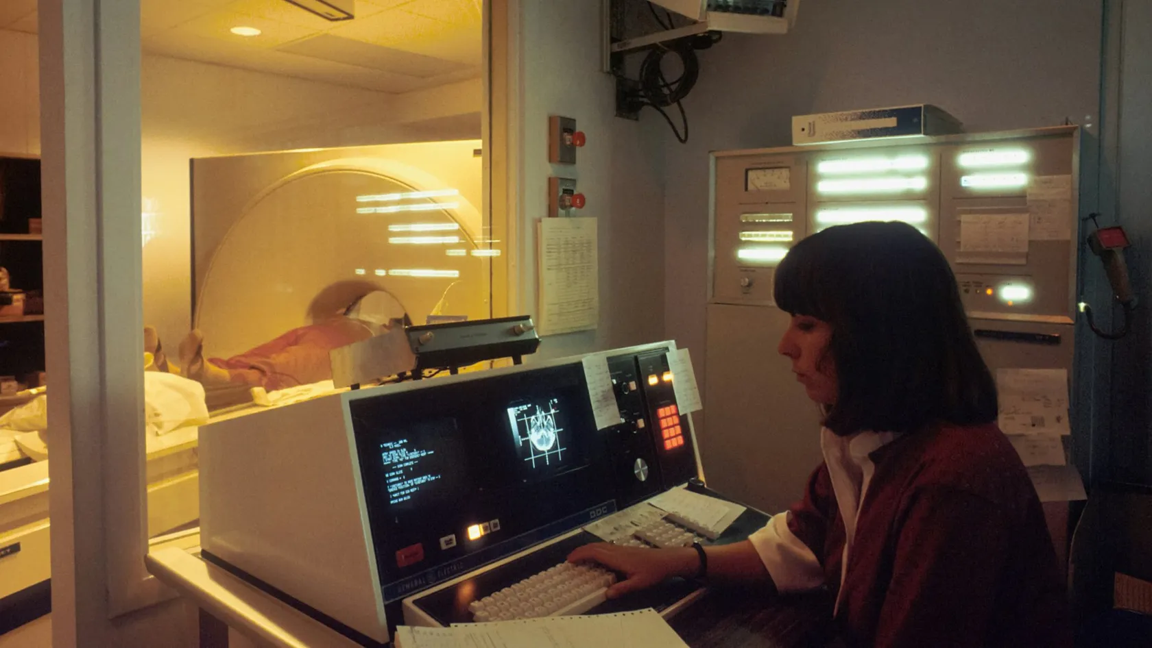
IGRT is how each radiotherapy session is aimed. A scan taken on the treatment machine, with you already in position, is matched to the planning CT, and the table shifts by millimeters before the beam turns on.
About This Department
A tumor is never in exactly the same place twice. IGRT finds it again before every session.
You need radiotherapy. The letters IGRT appear on the treatment plan or the quote. They describe how each session is aimed. Before the beam turns on, a scan taken on the treatment machine, with you already lying in position, is matched against the planning CT, and the table moves by a few millimeters until the two agree. That small step has measurable results. At Memorial Sloan Kettering, men treated for prostate cancer with daily image guidance had half the rate of lasting urinary side effects of men given the same dose without it, 10.4 percent against 20.0 percent at three years. This page explains what the machine sees, which cancers gain the most, what the imaging itself costs you in radiation, and how to plan a stay of several weeks in Istanbul around a course that runs five days a week, from the first planning scan to the flight home.
The aiming step of radiotherapy
IGRT Radiation Therapy - Image Guided Radiation Therapy sounds like a kind of radiation. It describes a way of aiming one. The beam, the dose and the biology stay the same, and what changes is that the machine checks where the target sits before every session and corrects for the difference.
The plan is made once. The body changes daily. IGRT closes the gap between the two by imaging on the treatment machine and moving the table before the beam turns on.
Every course begins with a planning CT. It is taken in the exact position you will be treated in, the radiation oncologist draws the tumor and the organs to protect on it slice by slice, and physicists then build and verify a plan, work that can fill a week. Treatment then runs for anything from one session to thirty-nine. Skin marks and room lasers line up the outside of the body each day. For decades nothing more existed. Everyone assumed the inside followed.
It does not.
Because nobody could see the target, planners used to draw a safety margin of a centimeter or more around it, which meant healthy bowel, bladder or lung received the full dose every day. Seeing the target daily lets that margin shrink to a few millimeters.
Why a tumor is never in the same place twice
Tumors move for ordinary reasons. The prostate sits between the bladder and the rectum and shifts by several millimeters, sometimes over a centimeter, depending on how full each one is. A tumor in the lower lung travels two to three centimeters with every breath. The liver and pancreas ride up and down under the diaphragm. A bladder changes shape from one hour to the next.
Slower changes matter too. Over six weeks of head and neck treatment people lose weight, swelling goes down and the tumor itself shrinks, so the anatomy on the last day no longer matches the scan from the first, and a mask that fitted tightly in week one hangs loose by week five.
Ignore this, and one of two things happens. Part of the tumor is missed on some days, or a healthy organ is overdosed on others.
How the machine sees the target
Several tools share the name image guidance, and the right one depends on what has to be seen.
| Method | What it shows | Used for |
|---|---|---|
| Cone-beam CT | A three-dimensional CT made by an X-ray tube and detector mounted on the treatment machine, showing soft tissue as well as bone | The daily standard for prostate, bladder, lung, head and neck and most body sites |
| Planar X-ray images | Two flat images at right angles, showing bone and metal markers | Quick bone matching for brain, spine and limbs, and marker matching |
| Fiducial markers | Three gold seeds the size of a grain of rice, placed in or near the tumor a week before planning | Prostate and liver, where the tumor blends into the tissue around it on X-ray |
| 4D CT and breath hold | A planning scan that records the full breathing cycle, or treatment delivered only while you hold a deep breath | Lung, liver and pancreas, and left breast to move the heart out of the field |
| Surface guidance | Cameras that track the skin in real time with no radiation | Breast, and watching for movement during any long session |
One further method exists on dedicated machines that combine an MRI scanner with the accelerator. MRI guidance shows soft tissue better than any X-ray method and is mainly used for pancreas and other upper abdominal tumors. Few centers anywhere have one.
IGRT, IMRT, VMAT and SBRT are parts of one treatment
Radiotherapy quotes list four abbreviations as if they were rival products. In practice they work as layers of a single treatment, and a modern course uses several of them at once.
IMRT and VMAT shape the dose
Intensity modulated radiotherapy bends the high-dose region around the tumor and away from the organs beside it, and VMAT does the same while the machine rotates in one continuous arc, which makes the session shorter.
IGRT aims it
A dose shaped to within millimeters is only useful if it lands within millimeters. Image guidance does that job. IMRT without it makes little sense for a moving target.
SBRT and SRS deliver it in very few sessions
Stereotactic treatment gives a very high dose in one to five sessions to a small target in the lung, liver, spine, prostate or brain. So few sessions leave no room for a miss. Every one of them is image guided, and the imaging is often repeated halfway through.
What the trials show
Prostate cancer has provided the most thorough test of image guidance, because the target moves daily and the organs at risk lie millimeters away.
470 men, 21 centers, daily imaging against weekly. Acute rectal bleeding 6 percent against 11 percent. Late rectal toxicity lower by 29 percent. Biochemical progression lower by 55 percent. De Crevoisier and colleagues, 2018.
Two studies carry most of the weight. Zelefsky and colleagues at Memorial Sloan Kettering compared 186 men treated to 86.4 Gy with daily correction on implanted markers against 190 men given the same dose two years earlier without markers, and reported the results in the International Journal of Radiation Oncology, Biology, Physics in 2012. Late urinary toxicity of grade 2 or higher at three years was 10.4 percent with image guidance and 20.0 percent without. Both groups had little rectal toxicity, 1.0 and 1.6 percent. Among high-risk patients, PSA control at three years was significantly better with guidance. The second study is a phase 3 randomized trial from 21 French centers, published in the same journal in 2018, which assigned 470 men to imaging at every session or imaging once a week. Daily imaging lowered acute rectal bleeding from 11 percent to 6 percent, lowered late rectal toxicity with a hazard ratio of 0.71, and lowered biochemical progression with a hazard ratio of 0.45.
That trial also reported something nobody expected. The daily group had worse overall survival, with a hazard ratio of 2.12, and the difference came from other cancers and not from prostate cancer. The authors could not explain it. The trial was not designed to test it. It remains a reason to keep imaging dose as low as the task allows.
Outside the prostate the evidence is indirect but consistent. A 2012 review in Seminars in Radiation Oncology by Bujold and colleagues concluded that the better agreement between planned and delivered dose leads to higher control rates, less toxicity, and treatments such as lung and liver SBRT that were not possible at all before the target could be seen.
The extra radiation from the scans
Every X-ray image adds a little radiation. A kilovoltage cone-beam CT gives the scanned region somewhere between a fraction of a centigray and a few centigray, while a single treatment session delivers around 200 centigray to the target, and far more in stereotactic treatment.
Task Group 180 of the American Association of Physicists in Medicine reviewed these doses in Medical Physics in 2018 and recommended including imaging dose in the treatment plan once it passes 5 percent of the prescribed dose, while noting that current kilovoltage procedures generally stay below that line. Physicists lower it further by scanning a shorter length of the body, using low-dose presets for children and slim patients, and choosing surface cameras or planar images on days when a full scan adds nothing.
The trade favors the patient. A few centigray of imaging buys a margin of millimeters, and that margin spares the bladder, the bowel or the lung many times the amount the scans add over the whole course.
Cancers where image guidance matters most
Large benefit
Prostate cancer, for the reasons above. Lung tumors, which move with breathing and sit beside the heart, the esophagus and the spinal cord. Liver and pancreatic tumors, invisible on plain X-ray and surrounded by bowel that tolerates little radiation, so that a miss of a few millimeters in either direction carries a cost. Head and neck cancers, where the spinal cord and the salivary glands lie within millimeters of the target and the anatomy changes over six or seven weeks. Bladder and cervical cancers, because those organs change shape daily. Tumors of the spine treated stereotactically, where the dose falls off steeply a few millimeters from the spinal cord. And any second course of radiation to an area treated before, since the organs nearby have already used up part of their tolerance. In each of these, the margin around the target decides how much healthy tissue is treated, and that margin can only shrink when the target is seen on the treatment machine every day.
Small benefit
A single palliative session to a painful bone. Skin cancers treated under direct vision. Whole-brain treatment. These still get image guidance in a simple form, and a quote that charges for daily cone-beam CT in such situations deserves a question.
From first message to last session
Treatment abroad runs on a fixed sequence. Knowing it lets you book flights and accommodation with some confidence.
- Remote review. A radiation oncologist reads your pathology and imaging and tells you whether radiotherapy fits, alone or with chemotherapy, and roughly how many sessions.
- Consultation and consent. Examination, a review of all options including those that do not involve radiation, and blood tests. Markers, if needed, go in at this stage and need about a week to settle before the planning scan.
- Simulation. The planning CT, in treatment position, with a mask for head and neck or a molded cushion for the body. Small ink dots mark the skin. Instructions about bladder and bowel filling start here and stay the same every day.
- Planning. Contouring, dose calculation and physics checks take five to ten working days for a complex plan. You are free during this time.
- Daily sessions. Monday to Friday. You are in the room for 15 to 20 minutes, of which imaging and matching take five, and the beam is on for two to five. Nothing is felt.
- Weekly review. The radiation oncologist sees you once a week to treat side effects early and to look at the daily images for any drift.
- Completion. A treatment summary listing dose, number of sessions, technique and the doses received by nearby organs, written for the doctor who will follow you at home.
How many weeks in Istanbul
Length of stay is set by the number of sessions, at five a week, plus the planning period at the start.
| Treatment | Sessions | Time in Istanbul |
|---|---|---|
| Stereotactic treatment of lung, liver, spine or prostate | 1 to 5 | 2 to 3 weeks including planning |
| Breast after surgery, shortened schedule | 15 | 4 to 5 weeks |
| Prostate, shortened schedule | 20 | 5 to 6 weeks |
| Head and neck, lung or cervix with chemotherapy | 30 to 35 | 8 to 9 weeks |
| Prostate, conventional schedule | 37 to 39 | 9 to 10 weeks |
Some people fly home during the planning gap and return for the first session. It works when the flight is short.
Flying is unrestricted. External beam treatment does not make you radioactive, and you are no risk to children, pregnant women or fellow passengers at any point. What decides the date of the flight home is how you feel, since tiredness and skin or bowel reactions peak during the last week of treatment and the one or two weeks after it. Many stay a further week for that reason.
Side effects by treated area
Side effects of radiotherapy are local. They appear in the area treated and nowhere else, with tiredness as the one general exception, and image guidance lowers them by keeping healthy tissue out of the high-dose region.
Pelvis
Frequent or burning urination and looser stools that are harder to hold begin around the third week and settle within a month of finishing, and tablets that relax the bladder neck together with a low-fiber diet handle most of it. Lasting rectal bleeding or urinary trouble affects a minority. Here the trials above show image guidance making its difference.
Chest
Painful swallowing develops when the esophagus lies near the target and is managed with liquid pain relief and soft food, while a dry cough or breathlessness one to three months afterwards can mean inflammation of the lung and needs a call, because a short course of steroids treats it.
Head and neck
The hardest course to go through. Sore mouth, thick saliva, loss of taste and skin reaction build from week three, and a dietitian and a feeding plan are part of treatment from the first day. Dry mouth can persist. Sparing the salivary glands is one of the main things IMRT with daily guidance was developed for.
Breast
Skin redness like sunburn, peaking a week after the last session. Breath-hold treatment for the left breast keeps the heart away from the beam.
When the plan changes midway
Plans change in perhaps one course in ten for head and neck cancer and less often elsewhere, and the daily images are what reveal the need. When the cone-beam CT shows that weight loss or tumor shrinkage has altered the anatomy, the team repeats the planning CT and builds a new plan, which takes a few days during which treatment continues on the old one. The name for this is adaptive radiotherapy. It adds no sessions.
Chemotherapy given alongside radiation is delivered at Biruni Hospital on the same site, by the medical oncology team, on the same days you attend for radiotherapy, and surgery takes place there too, for the cases where the plan is radiation first and an operation afterwards.
Machines need servicing. A missed day is added at the end, and for fast-growing tumors the team compensates for longer gaps with a second session on another day, six hours apart, which is standard practice.
And if you need to stop and go home, for family or any other reason, you leave with a summary of the dose given so far so that a center near you can complete the course.
What a radiotherapy quote is built from
Hospitals quote radiotherapy per course. This page gives no figure, because the course is defined only after the planning CT.
Six things drive the total. The number of sessions comes first. The technique comes second, with a stereotactic plan costing more per session than an IMRT plan and needing far fewer of them. Imaging frequency comes third, with daily cone-beam CT the norm for the sites listed above. Motion management adds to it, meaning 4D CT, breath-hold equipment or implanted markers and the short procedure to place them. Chemotherapy alongside radiation is priced separately, drug by drug. A repeat plan during treatment comes last, a real cost that some quotes include and others leave out. Packages published by Turkish hospitals and medical travel agencies for radiotherapy cover the consultation, planning, all sessions, nursing care and transfers, some add a companion's stay, and nearly all of them leave out flights, insurance, medicines for side effects, scans ordered for staging, and above all the weeks of accommodation, which for a long course can rival the medical bill.
So ask what the quote assumes. How many sessions. Which technique. Daily imaging or not. Whether a second plan is covered. What accommodation costs per week.
Your figure follows the free review of your pathology and scans. It becomes firm once the planning CT is done.
A month in Istanbul, practically
Six weeks away from home is a different kind of trip. Practical arrangements count for more here than for any operation. Biruni Hospital assigns a coordinator from your first message, and the international patients office arranges accommodation for you and a companion for the whole course along with the daily transport between it and the hospital, which for a treatment that takes twenty minutes a day is the part of the routine people notice most. You attend as an outpatient, so evenings and weekends are your own. Most people feel well enough to walk, shop and see the city for the first half of the course, and slow down in the second. A kitchen, a washing machine and a short, predictable ride to the hospital matter more than a view. The team works in English, Arabic, French, Russian, Serbian, Romanian and Spanish and arranges interpreters for other languages, so the weekly review happens in words you understand.
For a visa covering a stay of this length, the office issues an appointment confirmation and an invitation letter naming the hospital and the treating doctor, which is the document most consulates ask for with a medical visa application, and it goes out ten days or so before travel. A female physician can be requested in your first message. The hospital kitchen caters for halal, vegetarian and diabetic diets on the days you are in the building, and a prayer room is on site. Should you be admitted for any reason, the room has a bed for your companion.
After the flight home
Radiation keeps working for weeks after the last session. The first assessment of the result comes later than people expect.
Once you get back home, your own oncologist takes over with the treatment summary in hand. For prostate cancer that means a PSA test every three to six months, with a slow fall over a year or two as the normal pattern, and for lung, liver and head and neck tumors it means a scan about three months after treatment, since earlier scans show inflammation that is easily mistaken for tumor. Late effects, if they come, appear from six months onwards, and the summary lists the dose each nearby organ received so that a doctor who has never met our team can interpret a symptom. Your coordinator stays on the same WhatsApp number, and the radiation oncologist will look at follow-up scans you send. Skin and bowel reactions from the last week fade over the following month, and tiredness can take two or three months to lift, which surprises people who expected to feel normal the day the sessions stopped.
Go to a local doctor first for fever, bleeding that does not stop, breathlessness, or difficulty swallowing fluids.
IGRT Radiation Therapy - Image Guided Radiation Therapy FAQ
Is IGRT a different treatment from IMRT?
Does IGRT hurt, and how long does a session take?
Do the daily scans add a dangerous amount of radiation?
How long do I need to stay in Istanbul for radiotherapy?
Can I fly during or straight after radiotherapy?
Can my wife or husband stay with me for the whole course?
Does Biruni Hospital have a CyberKnife or Gamma Knife?
References
- Zelefsky MJ, Kollmeier M, Cox B, et al. Improved clinical outcomes with high-dose image guided radiotherapy compared with non-IGRT for the treatment of clinically localized prostate cancer. International Journal of Radiation Oncology, Biology, Physics. 2012;84(1):125-129.
- de Crevoisier R, Bayar MA, Pommier P, et al. Daily Versus Weekly Prostate Cancer Image Guided Radiation Therapy: Phase 3 Multicenter Randomized Trial. International Journal of Radiation Oncology, Biology, Physics. 2018;102(5):1420-1429.
- Ding GX, Alaei P, Curran B, et al. Image guidance doses delivered during radiotherapy: Quantification, management, and reduction: Report of the AAPM Therapy Physics Committee Task Group 180. Medical Physics. 2018;45(5):e84-e99.
- Bujold A, Craig T, Jaffray D, Dawson LA. Image-guided radiotherapy: has it influenced patient outcomes? Seminars in Radiation Oncology. 2012;22(1):50-61.
Editor's note
Written by the Biruni Hospital medical editorial team. Reviewed by Prof. Dr. Mustafa Teoman YANMAZ, Medical Oncology.
Medically reviewed by

Prof. Dr. Mustafa Teoman YANMAZ
Medical Oncology
Related Treatments
View All
Brachytherapy - Internal Radiation Therapy
A beam from outside has to cross healthy tissue to reach a tumor. Brachytherapy avoids that by placing a rice-sized radioactive source inside the tumor for a few minutes, then taking it out again. It is an essential part of curing cervical cancer and a tested option for womb, prostate and early breast cancer.
Brain Tumor Radiation Therapy
The plan for a brain tumor starts with what must be protected, the optic nerves, the brainstem, the hippocampus, and then shapes the dose around them. Radiosurgery in one to five sessions now treats most metastases, and six weeks of radiotherapy with temozolomide remains the standard after surgery for glioblastoma.
Chemoradiation Therapy - Concurrent Chemoradiotherapy
Blood test on Monday, a drip on Tuesday, radiotherapy every weekday, and the same again for six or seven weeks. Chemoradiation asks more of a patient than either treatment alone and, for several cancers, cures more. See the survival gains trial by trial, the side effects they came with, who is fit for it and how a long stay is organized.
Elekta Versa HD Radiation Therapy
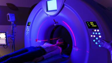
The Elekta Versa HD is the linear accelerator on which Biruni Hospital delivers external beam radiation therapy, with a 160-leaf collimator, high dose rate beams and cone beam CT imaging before every session. This page covers the techniques, from VMAT to radiosurgery, the cancers treated, what large trials show about shorter courses for breast, prostate and metastases, the path from first visit to last session, side effects, and how patients from abroad arrange treatment.
Glioblastoma Radiation Therapy
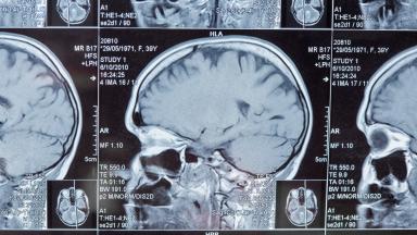
A glioblastoma has no edge, so surgery alone is never enough. Radiation covers the cavity and a margin of brain around it, together with a daily temozolomide capsule, over six weeks or three depending on age and fitness.