
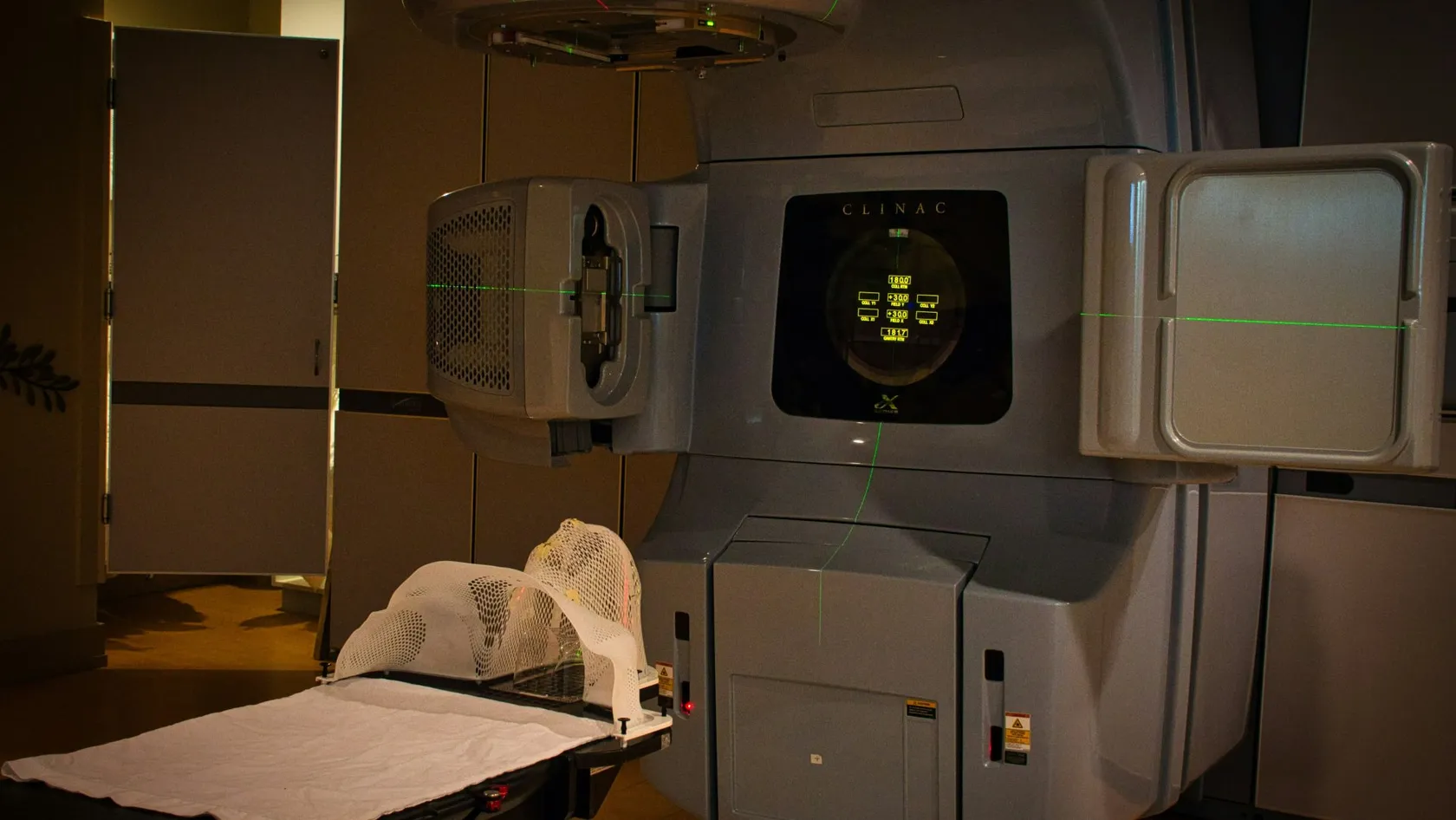
Brachytherapy - Internal Radiation Therapy
A beam from outside has to cross healthy tissue to reach a tumor. Brachytherapy avoids that by placing a rice-sized radioactive source inside the tumor for a few minutes, then taking it out again. It is an essential part of curing cervical cancer and a tested option for womb, prostate and early breast cancer.
About This Department
The radiation source stays inside for minutes and leaves with the machine. Women with cervical cancer who receive brachytherapy live longer.
Brachytherapy places a small radioactive source inside the tumor or right against it. The cancer receives a dose that no beam from outside the body could deliver without harming what lies in its path. Doctors treat it as a fixed part of curative treatment for cervical cancer and a well-tested option in cancers of the womb, prostate and breast. The evidence is strong. In a United States analysis of 7,359 women with cervical cancer, four-year survival reached 58.2 percent when brachytherapy was part of the treatment and 46.2 percent when it was left out.
Doctors call it brachytherapy, from the Greek word for short, and patient leaflets call it internal radiation, so this page uses both in its title, Brachytherapy - Internal Radiation Therapy. The short distance is the whole idea.
What brachytherapy is
Brachytherapy means radiotherapy from a distance of millimeters. A sealed radioactive source, smaller than a grain of rice, is brought into the tumor or a body cavity beside it through an applicator, a needle or a thin plastic tube. Physics does the rest. Radiation weakens with the square of the distance from its source, so tissue one centimeter away receives a very high dose and tissue three centimeters away receives a ninth of it, which spares the bladder, the bowel or the skin just beyond the target in a way that external beams cannot match.
This table scrolls sideways on a narrow screen. Swipe or drag to see every column.
| Form | How it works | Where it is used most |
|---|---|---|
| High dose rate, HDR | A single iridium source on a wire travels from a shielded machine into the applicator, stops at programmed points for 5 to 20 minutes in all, and returns to the machine | Cervix, womb, vagina, prostate boost, breast, some skin and esophageal tumors |
| Low dose rate, temporary | Sources stay in place for one to several days while the patient remains in a shielded single room | Replaced by HDR in most centers |
| Permanent seeds | Dozens of iodine seeds are placed through needles and left in the organ, where their radiation fades over months | Early prostate cancer |
| Intracavitary or interstitial | The applicator either sits in a natural cavity or consists of needles passed through the tissue itself | Cavity for cervix and vagina, needles for prostate, breast and bulky cervical tumors |
Will I be radioactive?
The answer depends on the form. For nearly everyone it is no.
- After an HDR session the source is back in its safe. Nothing radioactive remains in the body, and you can hold a baby that evening
- During temporary low dose rate treatment you stay in a shielded room and visits are limited. Once the sources are removed you are free of radiation
- With permanent prostate seeds a weak radiation persists for some months. Men are advised for the first two months not to hold children on the lap for long periods and to keep a little distance from pregnant women
- Seeds can set off radiation detectors at airports for several months. Carry the implant card that states the isotope, the activity and the date
What it is used for, and what the studies found
Four cancers account for nearly all brachytherapy.
Cervical cancer
For cervical cancer that has grown beyond the cervix, the standard is five to six weeks of external radiotherapy with weekly chemotherapy, followed by brachytherapy as the final boost. No center should skip that boost. Han and colleagues examined 7,359 American women treated between 1988 and 2009 and found that the share who received brachytherapy had fallen from 83 to 58 percent, after which they matched patients who did and did not receive it. Survival differed.
58.2% vs 46.2%Overall survival at four years with and without brachytherapy in stage IB2 to IVA cervical cancer. Cancer-specific survival was 64.3 against 51.5 percent. Han and colleagues, Int J Radiat Oncol Biol Phys, 2013.
Cancer of the womb lining
After a hysterectomy for endometrial cancer with some risk features, the commonest site of relapse is the top of the vagina. The Dutch PORTEC-2 trial asked whether treating that area alone, with a cylinder placed in the vagina for a few minutes on three occasions, protects as well as five weeks of radiotherapy to the whole pelvis.
1.8% vs 1.6%Vaginal recurrence at five years after vaginal brachytherapy and after pelvic external radiotherapy among 427 women. Survival did not differ. Nout and colleagues, Lancet, 2010.
Prostate cancer
In the prostate, doctors use brachytherapy either alone, as permanent seeds for early disease, or as a boost after external radiotherapy for higher-risk disease. The Canadian ASCENDE-RT trial tested the boost by randomizing 398 men with intermediate or high-risk cancer, all on hormone therapy and pelvic radiotherapy, to a boost with external beams to 78 gray or a boost with seeds.
83% vs 62% at nine yearsMen free of a rising PSA after a seed boost and after an external beam boost. Men given the external boost were twice as likely to relapse. Morris and colleagues, Int J Radiat Oncol Biol Phys, 2017.
Early breast cancer
After breast-conserving surgery, radiotherapy can be limited to the area around the scar in selected low-risk patients. Thin catheters do it. Several are passed through the breast and stay for four to five days.
1.44% vs 0.92%Local recurrence at five years after partial breast brachytherapy and after whole-breast radiotherapy in 1,184 women, a difference inside the margin set for non-inferiority. Strnad and colleagues, Lancet, 2016.
Who it suits, and who it does not
Reachable tumors of limited size, sitting beside organs worth protecting, suit brachytherapy best. A woman finishing chemoradiation for cervical cancer is the clearest case, and if her own hospital cannot offer the boost, traveling for it is reasonable and at times lifesaving. The clock matters. Guidance from the American Brachytherapy Society asks for the whole course, external treatment and brachytherapy together, to be completed within eight weeks, so the referral has to be made while external radiotherapy is still running.
Others gain nothing from it. Tumors that have spread widely are not helped by a local boost. A very large prostate, severe urinary obstruction or a previous prostate operation makes seed implantation difficult, and patients who cannot have anesthesia, or who cannot lie still for hours with an applicator in place, need another plan. A man with low-risk prostate cancer who qualifies for active surveillance needs no radiation of any kind.
The procedure
An HDR session for cervical cancer, step by step
- Preparation. Fasting from midnight, blood tests and an anesthesia review. A bowel preparation the evening before.
- Placement. Under spinal or general anesthesia the doctor examines the tumor, places a thin tube into the womb and a ring or two small holders against the cervix, and adds needles if the tumor extends sideways. Gauze packing pushes the bladder and rectum away.
- Imaging. An MRI or CT scan with the applicator in place. The doctor outlines the remaining tumor and the organs, and the physicist calculates where the source must stop and for how long.
- Treatment. In the shielded room the applicator is connected to the machine, and staff leave, watch by camera and talk by intercom. The source travels in, pauses at each position and returns. This takes 10 to 20 minutes and cannot be felt.
- Removal. The applicator is taken out, with pain relief. Cramping and light bleeding for a day are common.
- Repeat. Three to five sessions are given over one to two weeks, some centers keeping the applicator in overnight to give two sessions from one placement.
Placement and imaging take far longer than the radiation itself.
Prostate and breast implants
Doctors place prostate seeds in a single procedure of one to two hours under anesthesia, through needles passed through the skin behind the scrotum under ultrasound guidance. The man goes home the same day or the next, with a catheter for a short time. Breast catheters go in under local or general anesthesia. They stay for four to five days, with two short treatments a day, and come out in the clinic.
Side effects and risks
Side effects stay close to the treated organ. That is the point of the method. After gynecological brachytherapy, expect cramping, discharge, burning on passing urine and looser stools. They last one to three weeks. In the longer term the vagina can become dry, narrower and shorter, and regular use of a vaginal dilator or regular intercourse from a few weeks after treatment keeps it open, which matters for comfort and for the examinations that check for recurrence. After prostate brachytherapy the urine stream weakens and frequency rises for weeks to months. A few men need a catheter for a time. Erections decline gradually in a proportion of men over the following years. Breast catheters leave small marks and sometimes cause infection or firmness at the site. The procedure itself carries the risks of anesthesia, bleeding and infection, and on rare occasions a needle or applicator injures the womb, the bladder or the bowel.
If the plan changes
The examination under anesthesia sometimes shows that the tumor has shrunk less than hoped, and the plan moves from a simple applicator to one with added needles, which lengthens the procedure and may mean a night in hospital. Low blood counts or an infection can postpone a session. The delay is a few days. When a womb cannot be entered safely, the team may finish with an external boost. They will say plainly that this is second best. Chemotherapy and external radiotherapy are both delivered at Biruni Hospital on the same site. A change on one side is coordinated with the other. No records move.
Keep the calendar tightIn cervical cancer every week beyond eight lowers the chance of control. If you are coming for the brachytherapy part only, send the dates and doses of your external radiotherapy before you book, so that the first placement falls in the final week of external treatment or directly after it.
Time in Istanbul, cost and the trip
Your stay depends on one choice, the whole course or brachytherapy alone.
This table scrolls sideways on a narrow screen. Swipe or drag to see every column.
| Treatment | Schedule | Time in Istanbul |
|---|---|---|
| Cervical cancer, whole course | Five to six weeks of external radiotherapy with weekly chemotherapy, then three to five HDR sessions | Eight to ten weeks, planning included |
| Cervical cancer, brachytherapy only | Three to five HDR sessions over one to two weeks | Two to three weeks |
| Womb cancer after surgery | Three vaginal HDR sessions, as in PORTEC-2 | Two weeks |
| Prostate seeds | Planning scan and one implant procedure | One week |
| Partial breast | Catheters for four to five days | One to two weeks |
The first two rows reflect programs that Turkish hospitals and medical travel agencies publish, and the rest follow the schedules used in the trials above. Five things set the cost. They are the form of brachytherapy, the number of placements, the type of anesthesia, whether MRI is used for planning, and whether needles are added. Seeds carry a price per implant and form the costliest single item. External radiotherapy and chemotherapy, where they are part of the plan, are quoted separately. Your own health enters through anesthesia. Heart or lung disease, blood thinners and diabetes all change the preparation. Published packages cover the consultation, planning imaging, the placements and sessions, anesthesia, a short hospital stay where needed, an interpreter and transfers, and leave out flights, the hotel, chemotherapy drugs and the treatment of a complication. Send the reports first. The review that comes before any quote is free, and it tells you which of these items apply.
A coordinator from the international patients office handles the case from the first message to the day of discharge, and the team works in English, Arabic, French, Russian, Serbian, Romanian and Spanish, with interpreters for other languages. For a treatment this intimate, the interpreter in the consent conversation can be a woman, and a female doctor can be requested in the first message and is arranged by the department wherever the rota allows. The office sends the appointment confirmation and an invitation letter naming the hospital and the treating doctor ten days before travel. Patient rooms have a companion bed. Halal, vegetarian and diabetic meals are available, and a prayer room is on site.
After you fly home
You can fly the day after the last HDR session if you feel well. After a seed implant, wait a few days until the catheter is out and urine flows. The treatment summary lists the isotope, the dose per session, the number of sessions, the total dose once combined with any external radiotherapy, and the doses received by the bladder, the rectum and the bowel. Your oncologist at home needs it for follow-up. Any future radiotherapy team needs it even more.
Follow-up after cervical cancer means an examination every three months at first and a scan at three months. Prostate follow-up means a PSA test every six months. The PSA often bounces upward once in the first two years. One rise does not mean relapse. Once you are back home the coordinator stays reachable on the same WhatsApp number and passes clinical questions to the treating doctor.
Brachytherapy - Internal Radiation Therapy FAQ
Is brachytherapy painful?
Am I radioactive after brachytherapy?
Can I have external radiotherapy at home and come to Istanbul only for the brachytherapy?
How many sessions will I need?
Will prostate seeds set off airport alarms?
Can I ask for a female doctor and interpreter?
References
- Han K, Milosevic M, Fyles A, Pintilie M, Viswanathan AN. Trends in the utilization of brachytherapy in cervical cancer in the United States. Int J Radiat Oncol Biol Phys. 2013;87(1):111-9.
- Potter R, Tanderup K, Schmid MP, et al. MRI-guided adaptive brachytherapy in locally advanced cervical cancer (EMBRACE-I): a multicentre prospective cohort study. Lancet Oncol. 2021;22(4):538-547.
- Nout RA, Smit VT, Putter H, et al. Vaginal brachytherapy versus pelvic external beam radiotherapy for patients with endometrial cancer of high-intermediate risk (PORTEC-2): an open-label, non-inferiority, randomised trial. Lancet. 2010;375(9717):816-23.
- Morris WJ, Tyldesley S, Rodda S, et al. Androgen Suppression Combined with Elective Nodal and Dose Escalated Radiation Therapy (the ASCENDE-RT Trial): An Analysis of Survival Endpoints for a Randomized Trial Comparing a Low-Dose-Rate Brachytherapy Boost to a Dose-Escalated External Beam Boost for High- and Intermediate-risk Prostate Cancer. Int J Radiat Oncol Biol Phys. 2017;98(2):275-285.
- Strnad V, Ott OJ, Hildebrandt G, et al. 5-year results of accelerated partial breast irradiation using sole interstitial multicatheter brachytherapy versus whole-breast irradiation with boost after breast-conserving surgery for low-risk invasive and in-situ carcinoma of the female breast: a randomised, phase 3, non-inferiority trial. Lancet. 2016;387(10015):229-38.
Editor's note
Written by the Biruni Hospital medical editorial team. Reviewed by Prof. Dr. Metin GÜDEN, Radiation Oncology.
Medically reviewed by
Prof. Dr. Metin GÜDEN
Radiation Oncology
Related Treatments
View All
Brain Tumor Radiation Therapy
The plan for a brain tumor starts with what must be protected, the optic nerves, the brainstem, the hippocampus, and then shapes the dose around them. Radiosurgery in one to five sessions now treats most metastases, and six weeks of radiotherapy with temozolomide remains the standard after surgery for glioblastoma.
Chemoradiation Therapy - Concurrent Chemoradiotherapy
Blood test on Monday, a drip on Tuesday, radiotherapy every weekday, and the same again for six or seven weeks. Chemoradiation asks more of a patient than either treatment alone and, for several cancers, cures more. See the survival gains trial by trial, the side effects they came with, who is fit for it and how a long stay is organized.
Elekta Versa HD Radiation Therapy
The Elekta Versa HD is the linear accelerator on which Biruni Hospital delivers external beam radiation therapy, with a 160-leaf collimator, high dose rate beams and cone beam CT imaging before every session. This page covers the techniques, from VMAT to radiosurgery, the cancers treated, what large trials show about shorter courses for breast, prostate and metastases, the path from first visit to last session, side effects, and how patients from abroad arrange treatment.
Glioblastoma Radiation Therapy
A glioblastoma has no edge, so surgery alone is never enough. Radiation covers the cavity and a margin of brain around it, together with a daily temozolomide capsule, over six weeks or three depending on age and fitness.
IGRT Radiation Therapy - Image Guided Radiation Therapy
IGRT is how each radiotherapy session is aimed. A scan taken on the treatment machine, with you already in position, is matched to the planning CT, and the table shifts by millimeters before the beam turns on.