
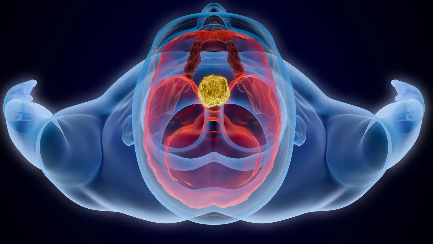
Brain Tumor Radiation Therapy
The plan for a brain tumor starts with what must be protected, the optic nerves, the brainstem, the hippocampus, and then shapes the dose around them. Radiosurgery in one to five sessions now treats most metastases, and six weeks of radiotherapy with temozolomide remains the standard after surgery for glioblastoma.
About This Department
Radiotherapy for a brain tumor is planned around what the brain has to keep doing. Memory, sight and speech are drawn on the plan as areas to protect.
Radiation forms part of treatment for nearly every kind of brain tumor, whether it began in the brain or spread there from another organ. The way of giving it has changed. For one to a few metastases, focused radiosurgery has replaced whole-brain treatment, and in a trial in JAMA thinking skills declined in 63.5 percent of patients after radiosurgery alone against 91.7 percent when whole-brain radiotherapy was added, with no difference in survival. For glioblastoma, adding temozolomide tablets to six weeks of radiotherapy raised two-year survival from 10.4 to 26.5 percent.
This page covers brain tumor radiation therapy for adults, from glioblastoma to metastases to benign tumors such as meningioma. The numbers in it are averages from trials. Some of them are hard to read. They appear here because families plan better with real figures than with reassurance, and because no average describes one person.
How radiation treats a brain tumor
Radiation damages the DNA of cells that are dividing. Tumor cells divide often and repair badly, and healthy brain cells divide rarely, so a dose split into daily portions harms the tumor more than the tissue around it. A six-week course rests on that logic. Radiosurgery works differently. It aims so many narrow beams at a small target from so many directions that the dose at the crossing point destroys the tissue outright in one to five sessions, while each beam on its way in is too weak to matter.
Surgery removes what can be seen, and radiation treats what cannotGliomas send cells into normal-looking brain well beyond the edge visible on MRI. After even the best operation, radiotherapy is aimed at the cavity plus a margin of one to two centimeters, which is where most regrowth occurs.
Which tumor, which radiotherapy
The schedule follows the diagnosis, and the diagnosis now includes molecular tests on the tumor tissue.
This table scrolls sideways on a narrow screen. Swipe or drag to see every column.
| Tumor | Usual radiotherapy | Sessions |
|---|---|---|
| Glioblastoma, under 65 to 70 and fit | Focal radiotherapy to 60 gray with daily temozolomide, then six monthly cycles of temozolomide | 30 over 6 weeks |
| Glioblastoma, age 65 and over | Shorter course to 40 gray with temozolomide | 15 over 3 weeks |
| Lower-grade gliomas | Focal radiotherapy, timing and chemotherapy decided by molecular type | 28 to 33 over 6 weeks |
| One to four metastases, in selected cases up to ten | Stereotactic radiosurgery to each lesion | 1 to 5 |
| Many metastases, or spread along the brain lining | Whole-brain radiotherapy, with the hippocampus spared and memantine tablets where suitable | 10 to 12 over 2 to 3 weeks |
| Meningioma, vestibular schwannoma, pituitary adenoma | Radiosurgery for small tumors, a six-week course for larger ones or those against the optic nerves | 1 to 5, or 25 to 30 |
What the trials found
Glioblastoma
Glioblastoma, the commonest malignant brain tumor in adults, is also the hardest to treat. One trial set the schedule now used around the world, and Stupp and colleagues published it in the New England Journal of Medicine in 2005.
14.6 vs 12.1 months, and 26.5% vs 10.4% at two yearsMedian survival and two-year survival with and without temozolomide. Severe effects on the blood count occurred in 7 percent during the combined phase. Stupp and colleagues, N Engl J Med, 2005.
Glioblastoma after the age of 65
Six weeks of daily treatment is a large share of the time an older patient has. A shorter course was tested for that reason.
9.3 vs 7.6 monthsMedian survival with and without temozolomide added to the three-week course. In patients whose tumor carried a methylated MGMT gene, the figures were 13.5 and 7.7 months. Perry and colleagues, N Engl J Med, 2017.
One to a few brain metastases
For decades every patient with brain metastases received radiotherapy to the whole brain. Radiosurgery treats only the visible lesions, and doctors had to find out whether leaving the rest of the brain untreated costs lives.
63.5% vs 91.7%Patients whose thinking had declined at three months after radiosurgery alone and after radiosurgery plus whole-brain radiotherapy. New metastases appeared sooner without whole-brain treatment, and survival was the same, 10.4 against 7.4 months with no significant difference. Brown and colleagues, JAMA, 2016.
When the whole brain has to be treated
Some patients have too many metastases for radiosurgery. Whole-brain radiotherapy still works for them, and its cost to memory falls when the dose is shaped away from the hippocampus, the small paired structure deep in each temporal lobe where new memories form.
26 percent lower risk of cognitive failureWith hippocampal sparing, a hazard ratio of 0.74. At four months executive function had worsened in 23.3 against 40.4 percent, and patients reported less fatigue and less trouble remembering and speaking. Brown and colleagues, J Clin Oncol, 2020.
How many metastases are too many for radiosurgery?
A Japanese study of 1,194 patients, published in The Lancet Oncology in 2014, found that survival after radiosurgery alone was the same with five to ten metastases as with two to four, 10.8 months in both groups, and that side effects of any grade occurred in 9 percent of each. Since then many centers offer radiosurgery for up to ten small lesions. Total volume matters more than the count.
Who should travel, and who should not
Timing drives the decision. After surgery for glioblastoma, radiotherapy should start within six weeks, so a family that wants treatment abroad has to send the scans and the pathology in the first fortnight after the operation, while the patient is still recovering. Patients with a benign tumor or a slow-growing glioma have time to plan. Patients with metastases depend on how fast the disease is moving elsewhere in the body.
- A patient who is drowsy, confused or largely in bed should be treated close to home, with a short course or with supportive care alone
- Seizures that are not yet controlled, or headaches with vomiting from pressure in the skull, have to be stabilized before any flight
- Children and teenagers with brain tumors belong in a pediatric center, and some of them are better served by proton therapy
- A patient whose local hospital offers the same schedule on modern equipment gains little from six weeks away from family
From the mask to the last session
Every course follows the same steps, whether it lasts six weeks or a single session.
- Consultation. The radiation oncologist examines you, reviews the MRI and pathology, and explains the aim, the schedule and the side effects. Steroid and seizure medication are checked.
- Mask. A sheet of warm plastic mesh is molded over the face and hardens in a few minutes. It clips to the table and holds the head still to within a millimeter or two. You can breathe and see through it. Patients who dread it are offered a trial run and, if needed, a mild sedative.
- Planning scans. A CT in the mask and a fresh MRI. The doctor outlines the target, the optic nerves, the brainstem, the hippocampi and the inner ears, and the physicist builds the plan. This takes three to seven working days, less for radiosurgery.
- Sessions. Monday to Friday, 15 to 20 minutes in the room with the beam on for a minute or two. Radiosurgery sessions last longer. Nothing is felt. Some people see blue flashes or notice a smell, which are harmless.
- Tablets. In glioblastoma, temozolomide is swallowed every day of the course, weekends included, with an anti-sickness tablet, and blood counts are taken weekly.
- Weekly review. The doctor asks about headache, tiredness and seizures and adjusts the steroid dose.
- Completion. A treatment summary, a medication plan and the date of the first MRI.
Side effects, early and late
During a course, tiredness leads the list of complaints. It builds from the third week, peaks one to two weeks after the last session, and returns in some patients as a wave of sleepiness a month or two later, which is normal and passes. Hair falls out in the path of the beams from the third week. The scalp reddens and itches. Headache and nausea come from swelling around the tumor and respond to a steroid such as dexamethasone, which brings its own problems of appetite, sleep, mood and blood sugar when taken for long, so the dose is kept as low as symptoms allow. Temozolomide adds nausea, constipation and a fall in platelets and white cells, which explains the weekly blood test. Radiosurgery causes little hair loss or fatigue. Its risks lie in swelling around the target in the following weeks and a small chance of a seizure in the first days, and the team gives a short steroid course when the target is large or sits in a sensitive area.
Hair grows back within three to four months in most patientsRegrowth can be thinner or a different texture, and after high doses a patch may stay bare. Netherlands Cancer Institute patient information.
Late effects are a separate matter.
They appear months to years afterward and depend on dose and location. Slower thinking and weaker short-term memory are the most common, more so after whole-brain treatment and in older patients. Radiation necrosis, an area of damaged tissue that swells and mimics tumor regrowth on MRI, follows a minority of radiosurgery treatments, and doctors manage it with steroids, sometimes other drugs, and occasionally surgery to remove the damaged area. Treatment near the pituitary gland lowers hormone levels in some patients years later. A blood test detects it. Hearing and vision are at risk only when the tumor sits beside the inner ear or the optic nerves, and those doses are kept within known limits.
If the scan or the plan changes
Families should know about one trap in advance.
The first MRI after chemoradiation for glioblastoma sometimes looks worse than the scan before it, with more swelling and more contrast uptake, without the tumor having grown. Doctors call this pseudoprogression. The treatment causes it, and it settles over the following months. Telling it apart from true growth takes repeat scans, special MRI sequences and patience, and a change of treatment on the strength of that first scan alone is a mistake that experienced teams take care to avoid. Other changes come from practical causes. Low platelets or white cells pause temozolomide while radiotherapy continues. A rise in pressure calls for a higher steroid dose or a neurosurgical opinion. A planning MRI for radiosurgery sometimes reveals more metastases than the referring scan showed, since thinner slices find smaller lesions, and the plan then shifts to more targets or to whole-brain treatment with hippocampal sparing. Chemotherapy and radiotherapy are both delivered at Biruni Hospital on the same site, so such changes are made in one meeting.
Weeks in Istanbul, flying and cost
The length of stay follows the schedule. A six-week course means seven to eight weeks in Istanbul once the planning days at the start are counted, the three-week course four to five, radiosurgery one to two, and whole-brain treatment three. Check your visa against the longest of these. The international patients office sends an invitation letter with the appointment confirmation ten days before travel, naming the hospital and the treating doctor.
Flying with a brain tumorAirlines ask for a medical information form after recent brain surgery, and air trapped in the skull after an operation has to be gone before a flight, which the surgeon confirms from a scan. Seizures should be controlled and the steroid dose stable. Carry all tablets in hand luggage, with a letter listing them.
Cost follows the schedule too. Hospitals price radiosurgery per treatment and by the number of lesions, and a fractionated course by technique and number of sessions. The planning MRI, the mask, daily image guidance, temozolomide, anti-sickness and steroid tablets, blood tests and any hospital admission appear as separate items on some quotes and inside the total on others, so a family comparing two quotes has to line the items up. Published packages in Istanbul cover consultation, planning, all sessions, weekly reviews, an interpreter and transfers. They leave out the hotel, flights and chemotherapy drugs. Age, fitness, seizures, diabetes made worse by steroids, and previous radiotherapy to the head all change the plan and the figure.
Send the images first. Reading them costs you nothing.
One coordinator handles the case from first contact to departure. The team works in English, Arabic, French, Russian, Serbian, Romanian or Spanish, or through an interpreter. A patient with a brain tumor should not travel or stay alone. Two family members may attend consultations, and if an admission is needed the room has a companion bed. Halal, vegetarian and diabetic meals are available in hospital, a prayer room is on site, and a female doctor can be requested when you first write.
After you fly home
Treatment for glioblastoma continues at home. Four weeks after radiotherapy ends, temozolomide restarts for five days in every 28, for six cycles, with a blood count before each one. Your oncologist prescribes it from the plan in the discharge file. The first MRI is due four weeks after radiotherapy, then every two to three months. After radiosurgery the first MRI comes at six to eight weeks and then every three months, since new metastases elsewhere in the brain are common and respond to a further session of radiosurgery when they are found small.
Once you are back home the coordinator stays reachable on the same WhatsApp number. Clinical questions go to the treating doctor. Driving rules after a seizure or brain surgery differ by country, so ask your local doctor before you drive.
Brain Tumor Radiation Therapy FAQ
Will I lose my hair?
How many sessions are needed?
Is radiosurgery better than whole-brain radiotherapy for metastases?
How soon after surgery should radiotherapy start?
Can I fly to Istanbul with a brain tumor?
Can the chemotherapy continue in my own country?
References
- Stupp R, Mason WP, van den Bent MJ, et al. Radiotherapy plus concomitant and adjuvant temozolomide for glioblastoma. N Engl J Med. 2005;352(10):987-96.
- Perry JR, Laperriere N, O'Callaghan CJ, et al. Short-Course Radiation plus Temozolomide in Elderly Patients with Glioblastoma. N Engl J Med. 2017;376(11):1027-1037.
- Brown PD, Jaeckle K, Ballman KV, et al. Effect of Radiosurgery Alone vs Radiosurgery With Whole Brain Radiation Therapy on Cognitive Function in Patients With 1 to 3 Brain Metastases: A Randomized Clinical Trial. JAMA. 2016;316(4):401-409.
- Brown PD, Gondi V, Pugh S, et al. Hippocampal Avoidance During Whole-Brain Radiotherapy Plus Memantine for Patients With Brain Metastases: Phase III Trial NRG Oncology CC001. J Clin Oncol. 2020;38(10):1019-1029.
- Yamamoto M, Serizawa T, Shuto T, et al. Stereotactic radiosurgery for patients with multiple brain metastases (JLGK0901): a multi-institutional prospective observational study. Lancet Oncol. 2014;15(4):387-95.
Editor's note
Written by the Biruni Hospital medical editorial team. Reviewed by Prof. Dr. Metin GÜDEN, Radiation Oncology.
Medically reviewed by
Prof. Dr. Metin GÜDEN
Radiation Oncology
Related Treatments
View All
Brachytherapy - Internal Radiation Therapy
A beam from outside has to cross healthy tissue to reach a tumor. Brachytherapy avoids that by placing a rice-sized radioactive source inside the tumor for a few minutes, then taking it out again. It is an essential part of curing cervical cancer and a tested option for womb, prostate and early breast cancer.
Chemoradiation Therapy - Concurrent Chemoradiotherapy
Blood test on Monday, a drip on Tuesday, radiotherapy every weekday, and the same again for six or seven weeks. Chemoradiation asks more of a patient than either treatment alone and, for several cancers, cures more. See the survival gains trial by trial, the side effects they came with, who is fit for it and how a long stay is organized.
Elekta Versa HD Radiation Therapy
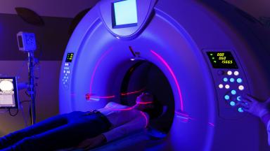
The Elekta Versa HD is the linear accelerator on which Biruni Hospital delivers external beam radiation therapy, with a 160-leaf collimator, high dose rate beams and cone beam CT imaging before every session. This page covers the techniques, from VMAT to radiosurgery, the cancers treated, what large trials show about shorter courses for breast, prostate and metastases, the path from first visit to last session, side effects, and how patients from abroad arrange treatment.
Glioblastoma Radiation Therapy
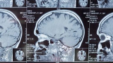
A glioblastoma has no edge, so surgery alone is never enough. Radiation covers the cavity and a margin of brain around it, together with a daily temozolomide capsule, over six weeks or three depending on age and fitness.
IGRT Radiation Therapy - Image Guided Radiation Therapy
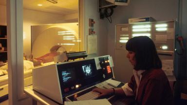
IGRT is how each radiotherapy session is aimed. A scan taken on the treatment machine, with you already in position, is matched to the planning CT, and the table shifts by millimeters before the beam turns on.