
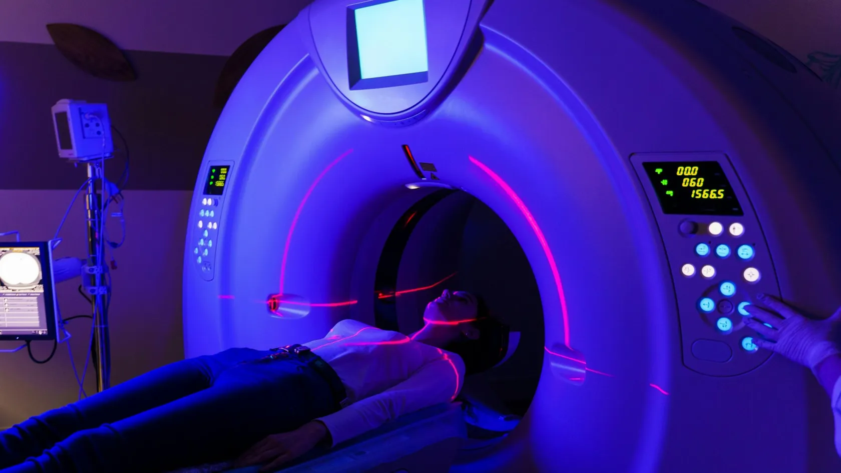
Elekta Versa HD Radiation Therapy
The Elekta Versa HD is the linear accelerator on which Biruni Hospital delivers external beam radiation therapy, with a 160-leaf collimator, high dose rate beams and cone beam CT imaging before every session. This page covers the techniques, from VMAT to radiosurgery, the cancers treated, what large trials show about shorter courses for breast, prostate and metastases, the path from first visit to last session, side effects, and how patients from abroad arrange treatment.
About This Department
The linear accelerator at Biruni Hospital shapes each beam with 160 tungsten leaves and holds its aim to within a millimeter. The machine is the tool. The plan is the treatment.
The Elekta Versa HD is a linear accelerator, the standard machine for external beam radiation therapy, fitted with a 160-leaf collimator that sculpts the beam to the outline of a tumor from every angle, high dose rate beams for short treatments of small targets, and a built-in CT scanner that checks the patient's position before each session. Physicists who commissioned one measured the rotation of its gantry, collimator and couch to within one millimeter of a single point (Narayanasamy and colleagues, Journal of Applied Clinical Medical Physics, 2016). That precision is what allows the treatments described on this page, from a five-session course for early breast cancer to a single high dose to a metastasis in the lung or spine. This page explains what the machine does, which cancers it treats and by which techniques, what the trials say about shorter courses, what the path from first visit to last session looks like, the side effects, and how patients from abroad arrange radiation therapy at Biruni Hospital in Istanbul.
What the Versa HD is
A linear accelerator makes X-rays of far higher energy than a diagnostic machine, points them at a tumor from a rotating arm, and shapes the beam so that the tumor receives the prescribed dose while the tissue around it receives as little as the geometry allows. Every modern radiation therapy course, whatever the cancer, is delivered on a machine of this kind. The Versa HD is Elekta's current design, and Biruni Hospital treats on one.
What the hardware does
- The collimator, called Agility, carries 160 tungsten leaves that slide in and out under computer control as the arm rotates, so that the beam's outline changes continuously to match the shape of the tumor from each direction. Leaf transmission was measured at about half a percent, meaning almost nothing leaks through the closed leaves.
- Flattening filter free beams remove a metal cone from the beam path and deliver dose at up to four times the ordinary rate. Forty minutes becomes ten. That matters when a patient must hold still and hold a breath.
- An X-ray panel and a cone beam CT scanner mounted on the arm image the patient on the treatment couch before each session. The picture is matched to the planning scan and the couch moves to correct any difference, in six directions, before the beam starts.
- A surface camera system and breath-hold monitoring, where fitted, track the patient's chest during treatment of the left breast and the lung, so that the heart stays out of the field.
Techniques and cancers
One machine. Several techniques, which differ in how finely the dose is shaped, how many sessions it is split into and how much imaging is done at each.
What is the difference between IMRT, VMAT and SBRT?
Is it the same as CyberKnife or Gamma Knife?
Gantry, collimator and couch isocentricity measurements were within 1 mm, 0.7 mm and 0.7 mm diameter respectively (Narayanasamy and colleagues, 2016).
Cancers treated
- Breast. After lumpectomy or mastectomy, to the whole breast or chest wall and, where needed, the lymph nodes, with breath-hold to protect the heart on the left side.
- Prostate. Moderate hypofractionation over four weeks, or SBRT in five sessions for selected patients, with markers or a spacer gel where indicated.
- Lung. SBRT for early tumors in patients who cannot or prefer not to have surgery, and conventional courses with chemotherapy for locally advanced disease.
- Head and neck. Six to seven week VMAT courses that spare the salivary glands and the swallowing muscles as far as the tumor allows.
- Brain. Radiosurgery for metastases and benign tumors in a frameless mask, and whole brain or partial brain courses where needed.
- Metastases in bone, liver, lymph nodes and adrenal. SBRT for a limited number of metastases, and short palliative courses for pain.
- Rectum, cervix, esophagus, bladder, lymphoma, sarcoma and skin. Conventional and hypofractionated courses, with chemotherapy where the protocol calls for it.
What it does not do
Protons. Brachytherapy, in which a source is placed inside the body, is a separate service and a separate question. And a linear accelerator treats what can be seen and outlined on a scan, so a cancer that has spread widely is treated with drugs, with radiation reserved for the sites that hurt or threaten.
Fewer sessions, same result
Not a machine. The most important change in radiation therapy over the past fifteen years has been the finding, in large randomized trials, that many cancers can be treated in far fewer sessions with a higher dose at each, without loss of control and without more side effects, provided the delivery is precise enough. The Versa HD was built for that kind of delivery.
26 Gy in five fractions over 1 week is non-inferior to the standard of 40 Gy in 15 fractions over 3 weeks for local tumor control, and is as safe in terms of normal tissue effects up to 5 years (Murray Brunt and colleagues, Lancet, 2020).
From first visit to first session
Seven to ten days, for most courses.
The steps
First, the radiation oncologist reviews the pathology, the scans and any previous treatment, examines the patient and confirms the intent, whether cure, control or relief of symptoms, and the technique and schedule. The patient then has a planning CT in the treatment position, lying on a mold or a mask made that day, with the scan sometimes fused to an MRI or a PET. Over the following days the oncologist outlines the tumor and every organ near it on each slice of that scan, a physicist and a dosimetrist build a plan that meets the prescribed dose to the target and the limits set for each organ, and the plan is checked by a second physician and by measurement on the machine itself before the patient is called. The first session runs long. It includes a verification image. A patient who has already had the planning scan and the outlining done at home cannot reuse them, because the plan belongs to the machine it was made for, but the scans and the previous dose record are essential and shorten the review.
What a session is like
Painless, and shorter than the wait for it.
- The radiation therapists position the patient on the couch using the mold or mask and the skin marks from planning, then leave the room and watch on camera and intercom.
- The cone beam CT runs, the image is matched to the plan, and the couch moves. This takes two to three minutes and is the part that makes the precision possible.
- The arm rotates and the beam runs. A conventional VMAT session lasts two to four minutes of beam time. A stereotactic session on the high dose rate beam lasts five to fifteen minutes. The patient feels nothing and hears a hum.
Side effects
Where the beam goes decides them.
Late effects
Coming from abroad
Radiation therapy is one of the treatments patients travel for most often, because the schedule is predictable, the cost is a known quantity, and a modern machine with an experienced team is not available everywhere.
Combining treatment
Patients who need chemotherapy alongside radiation receive it in the hospital's medical oncology unit on the same days, and those who need surgery first are scheduled so that radiation follows in the window the protocol requires.
Cost
An estimate follows the file review and depends on the technique, the number of sessions, whether imaging is done at every session and whether chemotherapy runs alongside. Hospitals in this market quote radiation therapy as a package for the course, with planning, imaging and weekly reviews included and accommodation listed separately.
Confirm what the figure includes and whether the follow-up scan is priced with it.
References
- Narayanasamy G, Saenz D, Cruz W, Ha CS, Papanikolaou N, Stathakis S. Commissioning an Elekta Versa HD linear accelerator. J Appl Clin Med Phys. 2016;17(1):179-191.
- Murray Brunt A, Haviland JS, Wheatley DA, et al. Hypofractionated breast radiotherapy for 1 week versus 3 weeks (FAST-Forward). 5-year efficacy and late normal tissue effects results from a multicentre, non-inferiority, randomised, phase 3 trial. Lancet. 2020;395(10237):1613-1626.
- Dearnaley D, Syndikus I, Mossop H, et al. Conventional versus hypofractionated high-dose intensity-modulated radiotherapy for prostate cancer. 5-year outcomes of the randomised, non-inferiority, phase 3 CHHiP trial. Lancet Oncol. 2016;17(8):1047-1060.
- Palma DA, Olson R, Harrow S, et al. Stereotactic ablative radiotherapy versus standard of care palliative treatment in patients with oligometastatic cancers (SABR-COMET). A randomised, phase 2, open-label trial. Lancet. 2019;393(10185):2051-2058.
Editor's note
Written by the Biruni Hospital medical editorial team. Reviewed by Prof. Dr. Özcan YILDIZ, Medical Oncology.
Medically reviewed by

Prof. Dr. Özcan YILDIZ
Medical Oncology
Related Treatments
View All
Brachytherapy - Internal Radiation Therapy
A beam from outside has to cross healthy tissue to reach a tumor. Brachytherapy avoids that by placing a rice-sized radioactive source inside the tumor for a few minutes, then taking it out again. It is an essential part of curing cervical cancer and a tested option for womb, prostate and early breast cancer.
Brain Tumor Radiation Therapy
The plan for a brain tumor starts with what must be protected, the optic nerves, the brainstem, the hippocampus, and then shapes the dose around them. Radiosurgery in one to five sessions now treats most metastases, and six weeks of radiotherapy with temozolomide remains the standard after surgery for glioblastoma.
Chemoradiation Therapy - Concurrent Chemoradiotherapy
Blood test on Monday, a drip on Tuesday, radiotherapy every weekday, and the same again for six or seven weeks. Chemoradiation asks more of a patient than either treatment alone and, for several cancers, cures more. See the survival gains trial by trial, the side effects they came with, who is fit for it and how a long stay is organized.
Glioblastoma Radiation Therapy
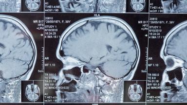
A glioblastoma has no edge, so surgery alone is never enough. Radiation covers the cavity and a margin of brain around it, together with a daily temozolomide capsule, over six weeks or three depending on age and fitness.
IGRT Radiation Therapy - Image Guided Radiation Therapy
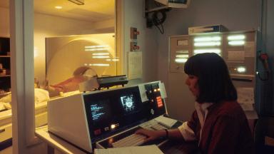
IGRT is how each radiotherapy session is aimed. A scan taken on the treatment machine, with you already in position, is matched to the planning CT, and the table shifts by millimeters before the beam turns on.