
Glioblastoma Radiation Therapy
A glioblastoma has no edge, so surgery alone is never enough. Radiation covers the cavity and a margin of brain around it, together with a daily temozolomide capsule, over six weeks or three depending on age and fitness.
About This Department
Surgery cannot remove every glioblastoma cell. Radiation with temozolomide is the treatment that reaches the rest.
Someone you love, or you yourself, has had a brain operation or a biopsy. The pathology report says glioblastoma. The next treatment is already decided in outline, and it is radiotherapy to the area of the tumor with a daily chemotherapy capsule called temozolomide. That combination became the worldwide standard in 2005, when a trial of 573 patients showed that adding temozolomide to six weeks of radiation raised the share of patients alive at two years from 10.4 percent to 26.5 percent. Glioblastoma remains a serious diagnosis. Treatment does not cure it in most people. It does add time, often good time. How much depends on age, on how much tumor the surgeon removed, and on a marker called MGMT. What follows explains the course, the choices inside it, the timing after surgery, which matters more than families are told, and what a stay of several weeks in Istanbul involves for the patient and for whoever comes along.
Why radiation follows surgery
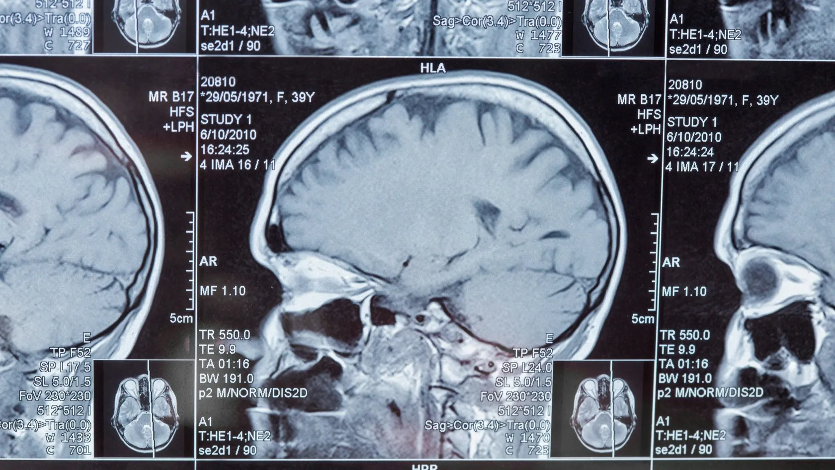
Glioblastoma Radiation Therapy exists because of how this tumor grows. A glioblastoma has no edge. The bright mass on the MRI is its densest part, and around it tumor cells travel along nerve fibers into brain that looks normal on any scan, two centimeters and more from the visible margin.
A neurosurgeon can remove what is visible. Nobody can remove what is not, because those cells sit inside working brain.
Surgery takes the mass. Radiation treats the ground it grew in. The radiation field covers the cavity, any tumor left behind, and a margin of brain around them where the scattered cells are most likely to be.
That explains two things families ask about. Doctors recommend radiation even after a surgeon reports a complete removal, since complete refers to the scan and not to the cells. And focused single-session treatments such as Gamma Knife have no place in a newly diagnosed glioblastoma, because a beam sharp enough to spare everything outside the visible tumor spares the tumor cells out there too.
The trials behind the standard treatment
Four randomized trials define current practice, and each one answers a question a family will meet.
| Trial | Who and what | Result |
|---|---|---|
| Stupp and colleagues, New England Journal of Medicine 2005 | 573 adults, median age 56. Radiation of 60 Gy alone, or with daily temozolomide followed by six monthly cycles | Median survival 14.6 months against 12.1. Alive at two years, 26.5 percent against 10.4 percent |
| Hegi and colleagues, same journal, same trial | 206 tumors from that trial tested for MGMT methylation, found in 45 percent | With methylated MGMT, 21.7 months with temozolomide against 15.3 without. Little benefit from the drug in unmethylated tumors |
| Perry and colleagues, New England Journal of Medicine 2017 | 562 patients aged 65 to 90. Three weeks of radiation, 40 Gy in 15 sessions, alone or with temozolomide | 9.3 months against 7.6. With methylated MGMT, 13.5 against 7.7. Quality of life the same in both groups |
| Stupp and colleagues, JAMA 2017 | 695 patients who had finished chemoradiation. Maintenance temozolomide alone, or with tumor-treating fields worn on the scalp 18 hours a day | 20.9 months against 16.0 from randomization. Skin irritation under the pads in 52 percent |
Read the numbers for what they are. A median marks the point that half of patients pass, and some pass it by years. The figures come from trial patients, who are fitter than average, yet the direction has not changed in twenty years, with radiation and temozolomide as the backbone and everything newer added on top of it.
What the pathology report decides
Two laboratory results on the tumor shape the plan more than the scan does.
| Marker | What it means | Effect on radiotherapy |
|---|---|---|
| MGMT promoter methylation | The tumor has switched off a gene that repairs the DNA damage temozolomide causes. Found in a little under half of glioblastomas | Temozolomide works much better. In an older or frail patient with an unmethylated tumor, radiation alone is a reasonable choice |
| IDH mutation | Since the 2021 WHO classification, a tumor with this mutation is no longer called glioblastoma. It is an astrocytoma, grade 4 at most, and behaves more slowly | Radiation and temozolomide are still used. Survival is counted in years more often than months, and late effects of radiation matter more |
If your report lacks either result, ask the laboratory that holds the tissue to run it. Both run on the stored block. We arrange it in Istanbul on slides you bring.
Six weeks, three weeks or one
Doctors use three schedules, and choosing between them means judging the person as much as the tumor.
| Schedule | For whom | Temozolomide |
|---|---|---|
| 60 Gy in 30 sessions over six weeks | Adults up to 70 or so in good general condition | Daily during radiation, then six monthly cycles |
| 40 Gy in 15 sessions over three weeks | Patients of 65 to 70 and over, and younger patients who are frail | Daily during radiation and afterwards, above all when MGMT is methylated |
| 25 Gy in 5 sessions over one week | Very frail or very elderly patients for whom three weeks of daily travel is too much | Usually none |
Between 65 and 70
The two main trials overlap in this age band, and fitness decides. A 68-year-old who walked into the hospital, lives independently and had most of the tumor removed is offered six weeks. A 68-year-old who needs help to dress is better served by three, since the Perry trial showed, in a group whose median age was 73, that the shorter course with temozolomide extends life without lowering its quality.
Higher doses have been tried repeatedly. None has improved survival, and all have increased damage to normal brain.
The clock after surgery
Radiation should start three to six weeks after surgery. Earlier than that the wound has not healed. Much later, and a fast-growing tumor has had time to regrow. Planning MRI scans show exactly that in a share of patients who waited.
Flying after a craniotomy needs the surgeon's agreement. Air trapped inside the skull at operation expands at altitude, and neurosurgeons want it absorbed before a flight, which takes a few weeks and shows on a CT scan done shortly before the planned date of travel. Tell us the date of surgery and we plan around it.
How the target is drawn
Radiotherapy at Biruni Hospital is delivered on an Elekta Versa HD linear accelerator with cone-beam CT imaging before treatment. Planning takes five to seven working days from the mask.
Living through the weeks of treatment
Each session lasts fifteen minutes and cannot be felt. The treatment as a whole is felt, gradually, from the third week.
- Temozolomide capsules are taken every day including weekends, on an empty stomach, an hour before radiation or at bedtime, with an anti-sickness tablet half an hour before.
- Blood counts are checked every week, because the drug lowers platelets and white cells. In the 2005 trial this reached a serious level in 7 percent of patients.
- An antibiotic to prevent a particular lung infection is prescribed for the weeks of combined treatment, since steroids and temozolomide together weaken that part of immunity.
- Dexamethasone, the steroid that controls brain swelling, is kept at the lowest dose that controls symptoms and reduced when possible.
- Anti-seizure medicine continues unchanged. Driving is off limits in nearly every country after a seizure or a craniotomy, for a period set by local law.
The radiation oncologist sees you every week. Say what has changed, however small. A new headache in the morning, more weakness on one side, or sleepiness that is out of proportion can mean swelling, and a change of steroid dose fixes it within a day or two.
Side effects, early and late
During treatment and the month after
Tiredness affects nearly everyone. It peaks in the last two weeks and the two weeks that follow. Hair falls out over the treated area from week three, in a patch that matches the beam paths, and regrows within three to six months for the majority, thinner where the dose was highest and sometimes with a different texture. The scalp reddens and itches. Nausea comes from the capsule more than from the radiation. A few weeks after the course ends, some people go through a spell of heavy sleepiness and low mood that lasts two weeks and lifts by itself.
Months to years later
Slower thinking, poorer short-term memory and difficulty doing two things at once affect a share of longer-term survivors. The tumor, the surgery, the seizure medicines and the radiation all contribute. Nobody can divide the blame. Radiation necrosis, an area of treated brain that breaks down and swells, appears in a small minority from six months onwards. Steroids treat it, sometimes with the drug bevacizumab added, and occasionally surgery is needed. Hormone deficiency follows in some patients when the pituitary region lies in the field. A yearly blood test finds it.
Honesty requires one more sentence. For most patients with glioblastoma the late effects of radiation are a problem they would be glad to live long enough to have.
The first scan afterwards, and the months that follow
The first MRI can mislead
The first scan is done four weeks after the last session. In a fifth to a third of patients it looks worse. More contrast uptake, more swelling. In many of them the cause is the treatment working, a reaction called pseudoprogression, which is commoner in MGMT-methylated tumors and settles over the following months. For that reason neuro-oncologists avoid declaring failure on any scan within twelve weeks of radiation unless new tumor appears outside the treated area, and they continue temozolomide while watching, repeating the MRI after a further four to eight weeks.
Six cycles of temozolomide
After a four-week break the drug restarts at a higher dose. Five days in every twenty-eight. Six cycles. A capsule, nothing more. Any oncologist at home can prescribe and monitor it with a blood count before each cycle and an MRI every two to three months, and that is how nearly all our international patients continue.
Tumor-treating fields
Marketed as Optune, this is a set of adhesive pads worn on a shaved scalp and connected to a battery pack, delivering a weak alternating electric field that disturbs dividing cells. In the 2017 trial it added nearly five months to median survival, 20.9 against 16.0, when worn 18 hours a day alongside maintenance temozolomide, at the price of skin irritation under the pads in half of users. Availability and reimbursement differ greatly between countries, and the device is supplied by its manufacturer, so the practical question is whether it can be obtained where you live.
If the tumor comes back
Most glioblastomas return. Most return within two centimeters of where they started, inside the area already irradiated. Reading that sentence is hard, and families planning ahead need it.
No single standard exists for recurrence, and the choice depends on how long the first treatment held, on where the new growth sits and on how the patient is. Operating again helps when the tumor is reachable and causing pressure, and it also provides fresh tissue for the pathologist. Restarting temozolomide makes sense if it worked the first time and was stopped more than a few months ago. Lomustine and bevacizumab are the usual second-line drugs. A second, shorter course of radiation to a small volume, 35 Gy in 10 sessions being a common schedule, is an option when at least six months have passed since the first and the new growth is small enough to treat without overlapping the optic nerves or the brainstem. Clinical trials deserve a serious look at this stage, at home or abroad. And for some patients the right decision is to stop treating the tumor and treat the symptoms well.
How long to stay in Istanbul, and flying
| Schedule | Planning | Treatment | Total |
|---|---|---|---|
| 60 Gy in 30 sessions | 1 to 1.5 weeks | 6 weeks | 7 to 8 weeks |
| 40 Gy in 15 sessions | 1 to 1.5 weeks | 3 weeks | 4 to 5 weeks |
| 25 Gy in 5 sessions | 1 week | 1 week | 2 weeks |
| Surgery at Biruni Hospital, then the six-week course | 3 to 4 weeks of recovery, pathology and planning | 6 weeks | 10 to 11 weeks |
You attend as an outpatient, five days a week. Weekends stay free.
Radiation sets no limit on flying, and you are not radioactive. Brain swelling causes the real concern. Anyone whose symptoms are controlled on a stable steroid dose can fly home in the days after the last session, with the steroid and anti-seizure tablets in hand luggage, a letter describing the diagnosis and treatment, and a companion in the next seat. Break up a long flight. If the last week brought new headaches or drowsiness, we settle that first.
What the quote is made of
No price appears here, because a glioblastoma quote is a sum of parts that differ from one patient to the next, and the parts are these. The schedule decides the largest share, thirty sessions costing more than fifteen. Hospitals charge planning once, and it covers the mask, the planning CT, the MRI and the physics work. Pharmacies price temozolomide by body surface area and by the number of days, and whether the quote includes the capsules or expects you to buy them matters a great deal. Weekly blood tests, supporting medicines and consultations are small lines. Over six weeks they add up. An admission for swelling or a seizure is never inside a package. Published packages from Turkish hospitals and medical travel agencies for brain radiotherapy cover consultation, planning, all sessions and transfers, and leave out drugs, MRI scans during follow-up, flights, and seven weeks of accommodation for two people, which is a large sum in its own right.
A quote for surgery is separate and depends on the position of the tumor, the use of navigation, monitoring or awake mapping, and the days in intensive care, so send the file first, and the figure you receive will name the schedule it assumes and say whether temozolomide is included.
For the person traveling with the patient
A patient with glioblastoma should not travel or stay alone. The companion carries more here than in almost any other treatment. You will manage the tablets, notice the changes the patient does not, and be the memory for every conversation with the doctors.
Biruni Hospital assigns one coordinator from the first message, and the international patients team works in English, Arabic, French, Russian, Serbian, Romanian and Spanish, with interpreters arranged for other languages, which matters most when a tired patient with word-finding difficulty is trying to follow a consultation. The office arranges accommodation for both of you for the whole course, close enough that the daily trip stays short, along with airport transfers and the daily car. It issues the appointment confirmation and invitation letter for a visa, naming the hospital and the treating doctor, ten days or so before travel. After any admission, the room has a bed for you. The kitchen provides halal, vegetarian and diabetic meals, the last of these being relevant because steroids raise blood sugar. A prayer room is on site. Request a female physician in your first message if you want one. Bring a month of the patient's regular medicines from home, in their boxes, so that doses can be checked against what we prescribe.
Look after yourself as well. Seven weeks wears anyone down. Another family member who takes over for two weeks in the middle makes a difference.
Care at home
Once you get back home, three people need the handover file. The oncologist who will prescribe temozolomide, the neurologist or family doctor who manages seizures and steroids, and whoever reads the MRI scans. The file contains the radiation summary with dose, sessions and the doses to the optic nerves and brainstem, the planning images, the pathology with markers, the schedule for the six cycles, and the name and direct contact of the doctor who treated you here.
- MRI four weeks after radiation, then every two to three months
- Blood count before each temozolomide cycle, and on day 21 of the cycle
- Steroid dose reviewed at every visit, with the aim of stopping
- A plan, written down, for what to do if a seizure lasts more than five minutes
Your coordinator stays on the same WhatsApp number. Send each new MRI, and the radiation oncologist will compare it with the treatment plan, which is the only reliable way to tell regrowth from radiation effect when the change lies inside the treated area.
Glioblastoma Radiation Therapy FAQ
How long does radiation therapy for glioblastoma take?
Does radiation cure glioblastoma?
Is Gamma Knife or CyberKnife better than ordinary radiotherapy for glioblastoma?
How soon after surgery should radiation start?
Can we fly home straight after the last session?
Will the staff be able to talk to us in our language?
Who continues the treatment when we are home?
References
- Stupp R, Mason WP, van den Bent MJ, et al. Radiotherapy plus concomitant and adjuvant temozolomide for glioblastoma. The New England Journal of Medicine. 2005;352(10):987-996.
- Hegi ME, Diserens AC, Gorlia T, et al. MGMT gene silencing and benefit from temozolomide in glioblastoma. The New England Journal of Medicine. 2005;352(10):997-1003.
- Perry JR, Laperriere N, O'Callaghan CJ, et al. Short-Course Radiation plus Temozolomide in Elderly Patients with Glioblastoma. The New England Journal of Medicine. 2017;376(11):1027-1037.
- Niyazi M, Andratschke N, Bendszus M, et al. ESTRO-EANO guideline on target delineation and radiotherapy details for glioblastoma. Radiotherapy and Oncology. 2023;184:109663.
- Stupp R, Taillibert S, Kanner A, et al. Effect of Tumor-Treating Fields Plus Maintenance Temozolomide vs Maintenance Temozolomide Alone on Survival in Patients With Glioblastoma: A Randomized Clinical Trial. JAMA. 2017;318(23):2306-2316.
Editor's note
Written by the Biruni Hospital medical editorial team. Reviewed by Prof. Dr. Mustafa Teoman YANMAZ, Medical Oncology.
Medically reviewed by

Prof. Dr. Mustafa Teoman YANMAZ
Medical Oncology
Related Treatments
View All
Brachytherapy - Internal Radiation Therapy
A beam from outside has to cross healthy tissue to reach a tumor. Brachytherapy avoids that by placing a rice-sized radioactive source inside the tumor for a few minutes, then taking it out again. It is an essential part of curing cervical cancer and a tested option for womb, prostate and early breast cancer.
Brain Tumor Radiation Therapy
The plan for a brain tumor starts with what must be protected, the optic nerves, the brainstem, the hippocampus, and then shapes the dose around them. Radiosurgery in one to five sessions now treats most metastases, and six weeks of radiotherapy with temozolomide remains the standard after surgery for glioblastoma.
Chemoradiation Therapy - Concurrent Chemoradiotherapy
Blood test on Monday, a drip on Tuesday, radiotherapy every weekday, and the same again for six or seven weeks. Chemoradiation asks more of a patient than either treatment alone and, for several cancers, cures more. See the survival gains trial by trial, the side effects they came with, who is fit for it and how a long stay is organized.
Elekta Versa HD Radiation Therapy
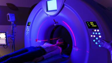
The Elekta Versa HD is the linear accelerator on which Biruni Hospital delivers external beam radiation therapy, with a 160-leaf collimator, high dose rate beams and cone beam CT imaging before every session. This page covers the techniques, from VMAT to radiosurgery, the cancers treated, what large trials show about shorter courses for breast, prostate and metastases, the path from first visit to last session, side effects, and how patients from abroad arrange treatment.
IGRT Radiation Therapy - Image Guided Radiation Therapy
IGRT is how each radiotherapy session is aimed. A scan taken on the treatment machine, with you already in position, is matched to the planning CT, and the table shifts by millimeters before the beam turns on.