
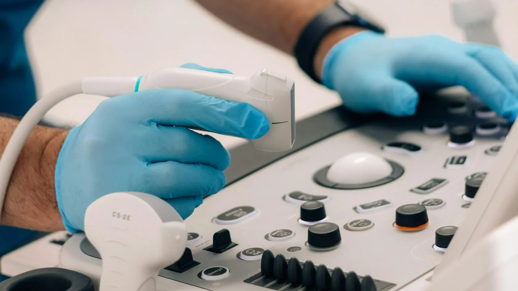
EUS - Endoscopic Ultrasound
EUS, or endoscopic ultrasound, carries a small ultrasound probe through the mouth or rectum to examine the pancreas, bile duct, lymph nodes and the layers of the gut wall from a few millimeters away, and guides a needle to sample what it finds. Across 33 studies it confirmed pancreatic cancer with 85 percent sensitivity and 98 percent specificity. This page explains what EUS shows, the needle, its uses, accuracy, the day of the test, risks, pancreatic cysts and care from abroad.
About This Department
Across 33 studies and 4,984 patients, a needle guided by endoscopic ultrasound confirmed pancreatic cancer with 85 percent sensitivity and 98 percent specificity. No scan can do that, because no scan takes a sample.
Those figures come from a meta-analysis of EUS-guided fine needle aspiration for solid pancreatic masses (Hewitt and colleagues, Gastrointestinal Endoscopy, 2012). EUS, or endoscopic ultrasound, combines two instruments in one. An endoscope carries a small ultrasound probe through the mouth to the stomach and duodenum, or through the rectum, and from there the probe looks through the wall of the gut at the organs beside it, a few millimeters away instead of through the whole abdomen. That closeness gives pictures of the pancreas, bile duct, gallbladder, lymph nodes and the layers of the gut wall that CT and MRI cannot match, and a needle passed under ultrasound guidance turns a picture into a diagnosis. This page explains what EUS is, what it is used for, how accurate it is, what the day involves, what the risks are, and how patients from abroad can arrange it at Biruni Hospital in Istanbul.
What EUS is
Ordinary ultrasound, the kind used in pregnancy, looks into the body from the skin, and it works beautifully wherever the sound can travel through water and tissue without interruption. Sound waves have to cross fat, muscle and, worst of all, gas in the bowel, which scatters them. The pancreas lies behind the stomach, surrounded by exactly that gas, and it is the organ that abdominal ultrasound sees least well.
EUS solves the problem by moving the probe to the other side of the gas. From inside the stomach the pancreas is a few millimeters away, and the picture is as clear as the pictures of a baby.
- The instrument. An endoscope like the one used for a gastroscopy, with an ultrasound transducer built into the tip, either radial, which scans a full circle, or linear, which scans a wedge and lets a needle be watched as it advances.
- The view. The layers of the gut wall, each one visible as a separate line, and the organs beyond it. The pancreas, bile duct, gallbladder, liver edge, spleen, adrenal gland, kidney, lymph nodes and large vessels of the abdomen from above. The prostate, rectum and pelvic nodes from below.
- The needle. A fine needle passed through the scope and into a lesion under direct ultrasound view, to draw cells or a core of tissue.
A CT scan finds a shadow on the pancreas. EUS says what the shadow is.
Where it fits with other tests
Most patients reach EUS after a CT or MRI has raised a question. The scan surveys the whole abdomen and shows spread to the liver and lungs. EUS excels at small lesions, at telling a cyst from a tumor, at the layers of the gut wall, at nearby lymph nodes, and above all at obtaining tissue safely. Doctors use the two together. A patient who arrives with a good scan saves a step.
The needle
Does the needle hurt?
What it is used for
Three questions bring most patients to EUS. What is this lesion. How far has this cancer gone. Can a diagnosis be made without an operation.
| Situation | What EUS adds | What may follow |
|---|---|---|
| A mass in the pancreas on CT | Confirms or excludes cancer by sampling, and shows whether the main vessels are involved | Surgery, chemotherapy, or reassurance |
| A pancreatic cyst | Size, wall, nodules and fluid analysis to judge the risk of malignancy | Surveillance or surgery |
| Esophageal, gastric or rectal cancer | Depth of invasion through the wall and nearby nodes, the T and N stage | Endoscopic removal, surgery, or chemotherapy and radiotherapy first |
| A lump under the lining of the stomach or esophagus | Which layer it arises from and what it is likely to be, with sampling | Surveillance, endoscopic or surgical removal |
| Bile duct stones or narrowing | Finds small stones and tumors that other tests miss | Removal of stones by ERCP, sampling of a narrowing |
| Enlarged lymph nodes in the chest or abdomen | Sampling without surgery, for lung cancer, lymphoma and infection | Diagnosis and staging |
| Chronic pancreatitis | Fine changes in the gland and its duct | Diagnosis, and nerve block for pain in selected cases |
| Unexplained pancreatitis | A hidden stone, a small tumor or a duct abnormality | Treatment of the cause |
The list is long because the probe sits next to more organs than any other single instrument reaches.
- EUS also guides treatment. Fluid collections after pancreatitis are drained into the stomach through a stent placed under EUS, which has largely replaced surgery for this problem.
- The nerves that carry pain from the pancreas can be blocked with alcohol or steroid under EUS guidance in cancer and chronic pancreatitis.
- A blocked bile duct can be drained through the stomach wall when the usual route fails.
How accurate it is
Numbers matter here, because the reason to have EUS is a decision that depends on the answer.
For solid masses of the pancreas, needle sampling under EUS had a pooled sensitivity of 85 percent and a specificity of 98 percent across 33 studies. Counting suspicious and atypical results as positive raised sensitivity to 91 percent at the cost of some specificity, and accuracy was higher in prospective multicenter studies (Hewitt and colleagues, 2012).
| Question | Result | Source |
|---|---|---|
| Is a pancreatic mass cancer | Sensitivity 85 percent, specificity 98 percent | Hewitt 2012, 4,984 patients |
| Has esophageal cancer invaded through the wall, T4 | Sensitivity 92.4 percent, specificity 97.4 percent | Puli 2008, 49 studies, 2,558 patients |
| Is esophageal cancer confined to the surface, T1 | Sensitivity 81.6 percent, specificity 99.4 percent | Puli 2008 |
| Are the lymph nodes involved in esophageal cancer | Sensitivity 84.7 percent by imaging alone, 96.7 percent with needle sampling | Puli 2008 |
Two things follow from the table. A negative needle result does not fully exclude cancer, since 15 percent of cancers gave a benign sample in the pooled data, so a suspicious mass with a negative sample is sampled again or followed closely. And the needle changes the answer for lymph nodes far more than the picture alone. EUS without sampling is the weaker test.
The day of the test
- Preparation. Nothing to eat for six hours and nothing to drink for two. Blood thinners and antiplatelet drugs are managed in advance because a needle is involved, and a recent blood count and clotting test are checked. For rectal EUS an enema is given.
- Sedation. Deep sedation with propofol supervised by an anesthesiologist for most EUS, since the examination is longer and more detailed than a gastroscopy and stillness improves the pictures.
- The examination. The scope passes with a swallow. The probe is placed against the wall at each station, the organs are examined and measured, and the needle is passed where sampling is planned. Twenty to sixty minutes.
- Recovery. An hour or two in the recovery area. A sore throat, bloating and a dull ache are usual. Eating resumes that day unless the pancreas was sampled, when a light diet is advised for 24 hours.
- Results. The imaging report is given the same day with pictures. Cytology takes two to four days and tissue core results up to a week, with molecular tests longer. The results are explained in a consultation.
Risks
Without a needle, EUS carries the risks of a gastroscopy. Small ones. The needle adds a little.
| Complication | Rate | Comment |
|---|---|---|
| Any complication | 0.98 percent | Most mild or moderate |
| Pancreatitis after sampling the pancreas | 0.44 percent | Usually settles with a day or two of rest and fluids |
| Pain after the procedure | 0.34 percent | Short-lived |
| Bleeding, infection, perforation | Each rare | Infection risk is higher for cysts, which are given antibiotic cover |
| Death attributable to the procedure | 0.02 percent | Two patients in the whole series |
Complication rates in these studies were higher in prospective series than in retrospective ones, which means the true rates sit nearer the higher figures and the honest number for pancreatic mass sampling is around 2 percent, nearly all of it minor (Wang and colleagues, Gastrointestinal Endoscopy, 2011).
| Symptom | Possible cause | Action |
|---|---|---|
| Severe abdominal pain, especially spreading to the back | Pancreatitis | Same-day assessment and blood tests |
| Fever or chills | Infection, especially after a cyst is sampled | Same-day assessment |
| Black stools or vomiting blood | Bleeding | Emergency department |
| Chest pain, breathlessness, pain on swallowing that worsens | Perforation, rare | Emergency department |
Needle track seeding
A theoretical concern with any needle biopsy of a cancer is that cells could be carried along the track. For EUS of the pancreas this has been reported only in a handful of cases worldwide, and because the track through the stomach or duodenum is removed with the tumor at surgery for most head-of-pancreas cancers, it has not changed practice. For cancers of the body and tail the track stays behind. There the question is weighed case by case.
Coming from abroad
Pancreatic cysts, the commonest dilemma
Scans find them by accident. A CT or MRI ordered for something else shows a small fluid-filled space in the pancreas, and the patient, who felt nothing, is told it might be nothing or might one day be cancer. Cysts of this kind are found in a few percent of adults and in more than one in ten over seventy, and the great majority never cause harm. A small minority are precancerous, and a few are already malignant. Telling them apart from the outside is unreliable. EUS looks at the cyst wall for thickening and nodules, measures the duct it may connect to, and draws fluid through a needle for analysis of a protein marker, glucose, enzymes, cells and, increasingly, DNA changes that separate the cyst types with growing accuracy. The result places the patient in one of three groups. Those who can be reassured and discharged. Those who need a scan or an EUS at intervals for years. And those who should have the cyst removed, which is a major operation and is not undertaken on the strength of a scan alone.
For a patient carrying this diagnosis across a border, EUS with fluid analysis tends to be the single test that ends months of uncertainty. One hour.
Who does the test, and why it matters
No examination in gastroenterology depends more on the operator. Interpretation happens in real time, stations are found by feel and experience, and the yield of the needle depends on the hand that guides it and the pathologist who reads what it brings back. Training bodies set minimum numbers of supervised procedures before a doctor practices independently, and the accuracy figures in the published studies climb with the volume a center performs, so that the same needle in the same pancreas gives a different answer in a unit that does two hundred a year and a unit that does twenty. Volume matters. So does the pathologist. Three questions belong to any patient choosing where to have EUS, and none of them is rude. How many EUS procedures does the doctor perform each year. Is a cytopathologist available to check the sample during the procedure. And will the same team see the patient afterward to act on the result, so that the diagnosis does not become a report in search of a doctor.
Good answers exist, and a patient may want them in writing.
Cost
The estimate follows the file review and depends on whether sampling is done, the number of needle passes, cytology and molecular tests, and anesthesia. Hospitals in this market quote EUS as a procedure fee with pathology and anesthesia listed separately.
Confirm what the figure includes before you travel.
References
- Hewitt MJ, McPhail MJ, Possamai L, Dhar A, Vlavianos P, Monahan KJ. EUS-guided FNA for diagnosis of solid pancreatic neoplasms, a meta-analysis. Gastrointest Endosc. 2012;75(2):319-331.
- Puli SR, Reddy JB, Bechtold ML, Antillon D, Ibdah JA, Antillon MR. Staging accuracy of esophageal cancer by endoscopic ultrasound, a meta-analysis and systematic review. World J Gastroenterol. 2008;14(10):1479-1490.
- Wang KX, Ben QW, Jin ZD, et al. Assessment of morbidity and mortality associated with EUS-guided FNA, a systematic review. Gastrointest Endosc. 2011;73(2):283-290.
Editor's note
Written by the Biruni Hospital medical editorial team. Reviewed by Assistant Professor Koray KOÇHAN, Gastroenterology.
Medically reviewed by
Assistant Professor Koray KOÇHAN
Gastroenterology
Related Treatments
View All
Barrett's Esophagus Treatment
Most people told they have Barrett's esophagus have been told only half the story. The yearly cancer risk is 0.12 percent, and the grade of dysplasia in the biopsies decides whether the right plan is a daily tablet with endoscopy every few years, or radiofrequency ablation to remove the lining.
Capsule Endoscopy - Wireless Capsule Endoscopy
When a gastroscopy and a colonoscopy are both normal and the blood count keeps falling, the search moves to the small bowel, which neither instrument reaches. A swallowed capsule camera films that stretch in eight hours without sedation and finds a cause in six of ten patients examined for bleeding.
Colonoscopy
Ask one question before choosing where to have a colonoscopy, which is how often the doctor finds polyps. In a study of 314,872 examinations that figure ranged from 7 to 53 percent between doctors, and it predicted who later developed bowel cancer. This guide covers preparation in a hotel room, sedation, findings and the flight home.
Colorectal Polyp Removal
A large polyp in the colon does not automatically mean bowel surgery. Nearly all polyps, including flat ones wider than 20 millimeters, come out through the colonoscope, and the technique is chosen by size and shape.
Endoscopic Mucosal Resection - EMR
Endoscopic mucosal resection lifts a precancerous growth or an early cancer off the muscle wall with an injection and removes it with a snare through an ordinary endoscope, in the colon, esophagus, stomach or duodenum, without an operation. In a study of 479 patients with large colon polyps, 83.7 percent avoided surgery and none died of the procedure. This page explains what EMR removes, which lesions qualify, how it compares with ESD and surgery, evidence, risks, follow-up and care from abroad.