
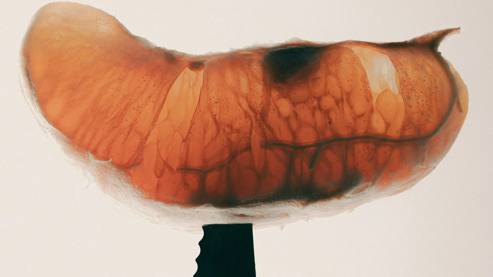
Endoscopic Submucosal Dissection - ESD
Removed in one piece, an early tumor can be measured at its edges and at its deepest point, and that measurement decides whether you are cured or need surgery. ESD is the endoscopic technique that makes it possible in the stomach, esophagus and bowel. See which lesions qualify, what recovery involves and how to judge an operator.
About This Department
An early tumor of the stomach, esophagus or bowel can come out in one piece through the endoscope. In a Japanese trial of 470 patients, five-year survival was 97 percent.
That trial, run by the Japan Clinical Oncology Group at 29 hospitals, treated early stomach cancers larger than the old size limits with endoscopic submucosal dissection. The tumor came out in a single piece in 99 percent of patients. Seven in ten were cured by the endoscopy alone. They kept their stomach. The remainder went on to surgery once the pathologist had examined the specimen. Send us your endoscopy report, photographs and biopsy result, and a gastroenterologist will tell you whether your lesion can be removed this way.
Your endoscopist has found something that a wire loop cannot safely take out, perhaps a flat polyp four centimeters across, perhaps an early cancer, and the word surgery has been mentioned. Between the loop and the operating room lies a third option.
Endoscopic submucosal dissection was developed in Japan for early stomach cancer and is now used along the whole digestive tract, and for the right lesion it removes the disease and leaves the organ. Which lesions are right, how good the operator has to be, what the recovery involves and what the pathology report will decide are all covered here.
What is endoscopic submucosal dissection?
Endoscopic submucosal dissection, ESD, removes a flat or early tumor from the inner lining of the digestive tract in a single intact piece, working entirely through an endoscope passed by the mouth or the back passage. The wall of the gut has layers. Tumors start in the innermost one, the mucosa, and ESD peels that layer off together with the loose layer beneath it, the submucosa, leaving the muscle wall whole. The doctor first marks a ring of dots around the lesion. Fluid injected underneath lifts the lesion away from the muscle, a fine electrosurgical knife cuts around the ring, and the same knife then frees the underside millimeter by millimeter until the whole disc comes away. A disc of eight or ten centimeters can be removed like this.
Size does not limit the technique. Depth does.
Once a cancer has grown deep into the submucosa it can reach lymph nodes, and no endoscope can remove a lymph node, so everything in the selection of patients turns on estimating depth before the procedure and measuring it afterward.
ESD, EMR or surgery
Endoscopic mucosal resection, EMR, the older technique, lifts the lesion the same way and takes it with a wire loop. A loop holds two centimeters. Anything larger comes out in several pieces, with two consequences. The pathologist cannot reassemble the pieces to check the edges or measure the deepest point of a cancer, and tissue left between the bites regrows.
This table scrolls sideways on a narrow screen. Swipe or drag to see every column.
| EMR, loop | ESD, knife | Surgery | |
|---|---|---|---|
| How the lesion comes out | Whole up to about 2 cm, in pieces above that | Whole, at any size | With a segment of the organ and its lymph nodes |
| What pathology can say | Edges and depth uncertain when in pieces | Edges and depth measured exactly | Everything, including lymph nodes |
| Regrowth at the site | 5.1 percent at six months in a French randomized trial of large colon polyps | 0.6 percent in that trial, and none when the edges were clear | Rare |
| Complications | 24.5 percent in that trial, mostly minor | 35.6 percent in that trial, mostly minor | Higher, with the risks of an operation and its anesthetic |
| Time and stay | Under an hour, same day | One to three hours, one or two nights | Several hours, a week or so |
| Operator | Most endoscopists | A small number of trained specialists | Surgeon |
After ESD, regrowth at six months occurred in 1 of 161 patients. After piecemeal EMR it occurred in 8 of 157. Complications were more frequent with ESD.RESECT-COLON randomized trial, Annals of Internal Medicine, 2024
Both halves of that result belong in the decision. For a large benign polyp of the colon, piecemeal EMR with a check colonoscopy at six months remains a sound choice, any regrowth is treated then, and the patient has avoided a longer procedure with a higher complication rate. Cancer changes the sum. ESD earns its extra time and risk when there is a real chance of cancer in the lesion, because only an intact specimen can show whether the cancer has been cured.
Which lesions are suitable?
The European Society of Gastrointestinal Endoscopy updated its guideline in 2022, and its recommendations differ by organ. For superficial squamous cancers of the esophagus and for superficial lesions of the stomach, ESD is the treatment of choice. That is settled. For Barrett's esophagus it is suggested for lesions that look as though they may have entered the submucosa, for cancers larger than 20 mm and for lesions in scarred areas, with EMR used for the others. In the colon and above all the rectum, ESD should be considered when the surface pattern raises suspicion of shallow invasion, particularly above 20 mm, or when a lesion cannot be removed completely with a loop. The duodenum is the exception. There the guideline advises against routine ESD, because the wall is thin and the complication rate high, and the few lesions that need it should go to the handful of units that publish their duodenal results.
Appearance decides. An experienced endoscopist examines the lesion with high-definition imaging and dye or virtual staining, reading the pit and vessel patterns that separate a surface lesion from one that has gone deep. The same guideline advises against routine CT, MRI or endoscopic ultrasound before resection, since for a lesion that looks superficial the specimen itself is the staging test.
Some lesions are wrong for ESD. A tumor that does not lift on injection, an ulcerated cancer with deep features, a poorly differentiated or signet-ring cancer on biopsy outside strict size limits, and anything with enlarged lymph nodes on a scan belong with a surgeon. So does a patient who could not tolerate an operation if a perforation needed one.
- The endoscopy or colonoscopy report with its photographs, and video if any was recorded.
- The biopsy pathology report.
- Any CT, MRI or endoscopic ultrasound report already done.
- Whether removal has been attempted before, since scarring makes a second attempt harder.
- A full medication list with blood thinners marked, and your main medical conditions.
A gastroenterologist replies with one of three opinions, namely suitable for ESD, better served by EMR, or a case for surgery.
What happens during the procedure and the hospital stay?
Among 692 patients treated at ten North American centers, lesions came out in one piece in 91.5 percent and with clear margins in 84.2 percent. Bleeding occurred in 2.3 percent and perforation in 2.9 percent, and one patient needed surgery for a complication.Prospective multicenter study, Gastroenterology, 2021
Risks, and why the operator matters
No endoscopic procedure is harder to learn, and results vary with experience more than with equipment. A meta-analysis of 97 studies of colorectal ESD found clear-margin removal in 85.6 percent of procedures in Asian centers and 71.3 percent elsewhere, and surgery for complications in 0.8 percent against 3.1 percent. The reviewers were blunt. Outside Asia, they wrote, the standard technique was still failing to reach acceptable performance. The North American figures above, published four years later, show the gap closing in dedicated units. Ask the endoscopist how many ESDs he or she has performed in the organ concerned, and for personal rates of one-piece removal, clear margins and perforation, because a specialist who does this work every week has those numbers to hand and one who does it a few times a year does not.
What does recovery involve, and what will the pathology report decide?
- The first day. Clear fluids once the team is satisfied, then milk, soup and yogurt.
- Days two to seven. Soft, cool or lukewarm food. No alcohol, no anti-inflammatory painkillers such as ibuprofen. After stomach or esophageal ESD an acid-suppressing tablet is taken for four to eight weeks while the wound heals.
- The first two weeks. No heavy lifting, straining or vigorous exercise. Blood thinners restart on the day the endoscopist specifies.
- Three to twelve months. A check endoscopy of the scar, timed according to the pathology.
Pathology arrives in five to seven working days. The report matters more than anything said on the day, and it answers four questions, how deep the lesion went, whether the edges are clear, how aggressive the cells look, and whether tumor has entered small blood or lymph vessels.
This table scrolls sideways on a narrow screen. Swipe or drag to see every column.
| Result | What it means | What follows |
|---|---|---|
| Very low or low risk, often called curative | Removed in one piece with clear edges, confined to the mucosa or the shallowest submucosa, well or moderately differentiated, no vessel invasion. | No further treatment. Scheduled check endoscopies. |
| Local risk | A side edge is positive or the lesion came out in pieces, with no invasion and no other high-risk feature. | Endoscopic surveillance or a repeat endoscopic treatment. Surgery is not advised. |
| High risk, non-curative | Deeper submucosal invasion, a positive deep edge, poorly differentiated cells, vessel invasion, or in the bowel a high grade of tumor budding. | Full staging, then discussion at a tumor board, where surgery with lymph node removal is considered case by case. |
A non-curative result counts as a finding. It does not mean the procedure failed. In the Japanese gastric trial, 28 percent of patients had surgery added after the specimen was read, and the group as a whole reached 97 percent survival at five years. Of the 317 patients whose removal met the curative criteria, none had a recurrence. Not one. For those who do go on to surgery, the ESD has cost a few weeks and has spared everyone else an operation they did not need.
- Black or bloody stools, or vomiting blood.
- Severe or worsening pain in the chest or abdomen, or a hard swollen abdomen.
- Fever above 38 degrees, chills or shortness of breath.
- Food sticking on the way down after esophageal ESD, which needs a prompt call and not an ambulance.
How many days do I need in Istanbul?
Plan on seven to ten days. One day for the consultation and a repeat look at the lesion where the referring images are not good enough, one or two nights in hospital, then several days in a hotel nearby while the first week of bleeding risk passes and the pathology is read. Staying for that report has a point. If it says curative, you fly home finished, and if it says high risk, the tumor board meets while you are still here and you leave with a plan, whether surgery is done in Istanbul, where surgery, chemotherapy and radiotherapy share one site, or at home.
Delayed bleeding can occur for up to two weeks. Short flights after the first week are reasonable once the endoscopist agrees in writing, while long-haul flights and destinations far from a hospital with an endoscopy service are better left until the two weeks are over.
What decides the cost?
Time, devices and nights set the price of ESD. A two-centimeter lesion in the lower stomach and a seven-centimeter lesion wrapped around a fold of the colon are different jobs, and scarring from a previous attempt can double the time. The knife, the injection solution, the hemostatic forceps and every clip are single-use items charged to the case. General anesthesia costs more than deep sedation. Nights count. Each one is a line on the bill, and a complication adds nights. Pathology for an ESD specimen takes more work than for a biopsy, because the disc is sliced at two-millimeter intervals and every slice is read under the microscope. A preliminary endoscopy with staining, where the outside images are inadequate, is a separate procedure. Your own health counts as well, since blood thinners, heart or lung disease and diabetes add cardiology and anesthetic checks before anyone will put you to sleep for three hours.
Packages published in this market for endoscopic removal of large lesions bundle the consultation, the procedure, one day of hospitalization with medications, a companion's stay, interpreter services and airport transfers, and some add hotel nights. Four details separate one quote from another. Find out whether the pathology is inside the figure, how many hospital nights are covered, what a night beyond that costs, and whether a repeat endoscopy for bleeding would be charged.
Your own figure follows the free review of your report and images.
What about language, companions and going home?
One coordinator from the international patients team handles your case from the first message to the pathology result. The team works in English, Arabic, French, Russian, Serbian, Romanian and Spanish, arranges interpreting in other languages on request, and provides an interpreter for the consent conversation and again when the pathology is explained. Your room has a companion bed, so one relative can stay both nights, and a second pair of ears is useful when the pathology is explained. Airport transfers and the hotel for the days after discharge are arranged by the international patients office, and it helps to ask for a hotel within a short drive of the emergency entrance. Halal, vegetarian and diabetic meals come from the hospital kitchen, which also prepares the soft diet of the first days. A prayer room is on site, and requests for a female doctor are passed to the department and met wherever the rota allows.
Back home, your coordinator stays reachable on the same WhatsApp number. You leave with the procedure report, photographs, the pathology report in English and a letter stating when the check endoscopy is due, which any gastroenterologist can perform. Glass slides are released on request for a second opinion.
ESD FAQ
Is ESD a surgery?
What is the difference between ESD and EMR?
Is ESD a cure for early cancer?
How long will I be in hospital?
When can I eat normally?
Can my relative stay with me?
References
- Pimentel-Nunes P, Libanio D, Bastiaansen BAJ, et al. Endoscopic submucosal dissection for superficial gastrointestinal lesions: European Society of Gastrointestinal Endoscopy (ESGE) Guideline, Update 2022. Endoscopy. 2022;54(6):591-622.
- Hasuike N, Ono H, Boku N, et al. A non-randomized confirmatory trial of an expanded indication for endoscopic submucosal dissection for intestinal-type gastric cancer (cT1a): the Japan Clinical Oncology Group study (JCOG0607). Gastric Cancer. 2018;21(1):114-123.
- Jacques J, Schaefer M, Wallenhorst T, et al. Endoscopic En Bloc Versus Piecemeal Resection of Large Nonpedunculated Colonic Adenomas: A Randomized Comparative Trial. Annals of Internal Medicine. 2024;177(1):29-38.
- Draganov PV, Aihara H, Karasik MS, et al. Endoscopic Submucosal Dissection in North America: A Large Prospective Multicenter Study. Gastroenterology. 2021;160(7):2317-2327.
- Fuccio L, Hassan C, Ponchon T, et al. Clinical outcomes after endoscopic submucosal dissection for colorectal neoplasia: a systematic review and meta-analysis. Gastrointestinal Endoscopy. 2017;86(1):74-86.
Editor's note
Written by the Biruni Hospital medical editorial team. Reviewed by Prof. Dr. Oğuzhan ÖZTÜRK, Gastroenterology.
Medically reviewed by
Prof. Dr. Oğuzhan ÖZTÜRK
Gastroenterology
Related Treatments
View All
Barrett's Esophagus Treatment
Most people told they have Barrett's esophagus have been told only half the story. The yearly cancer risk is 0.12 percent, and the grade of dysplasia in the biopsies decides whether the right plan is a daily tablet with endoscopy every few years, or radiofrequency ablation to remove the lining.
Capsule Endoscopy - Wireless Capsule Endoscopy
When a gastroscopy and a colonoscopy are both normal and the blood count keeps falling, the search moves to the small bowel, which neither instrument reaches. A swallowed capsule camera films that stretch in eight hours without sedation and finds a cause in six of ten patients examined for bleeding.
Colonoscopy
Ask one question before choosing where to have a colonoscopy, which is how often the doctor finds polyps. In a study of 314,872 examinations that figure ranged from 7 to 53 percent between doctors, and it predicted who later developed bowel cancer. This guide covers preparation in a hotel room, sedation, findings and the flight home.
Colorectal Polyp Removal
A large polyp in the colon does not automatically mean bowel surgery. Nearly all polyps, including flat ones wider than 20 millimeters, come out through the colonoscope, and the technique is chosen by size and shape.
Endoscopic Mucosal Resection - EMR
Endoscopic mucosal resection lifts a precancerous growth or an early cancer off the muscle wall with an injection and removes it with a snare through an ordinary endoscope, in the colon, esophagus, stomach or duodenum, without an operation. In a study of 479 patients with large colon polyps, 83.7 percent avoided surgery and none died of the procedure. This page explains what EMR removes, which lesions qualify, how it compares with ESD and surgery, evidence, risks, follow-up and care from abroad.