
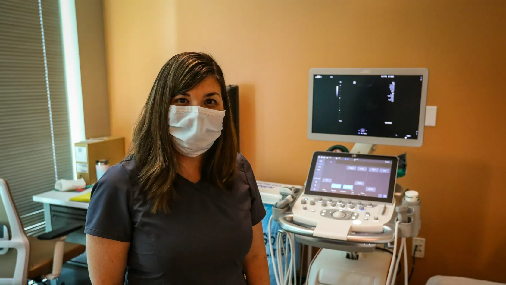
Ultrasound-Guided Biopsy
Patients ask whether a biopsy is dangerous. The two questions that decide the outcome are whether to put a needle in at all, and which needle to use.
About This Department
Across 145,928 thyroid aspirations the biggest problem was not a wrong answer. The indeterminate ones carried a 27 percent cancer risk.
A meta-analysis pooling 51 studies and 145,928 fine needle aspirations of the thyroid examined the two result categories that settle nothing. Among the specimens that went on to surgery, 27 percent of those reported as atypia of undetermined significance turned out to be cancer, along with 31 percent of those called a follicular neoplasm. The authors concluded that the published framework had understated the risk in the first group.
Two decisions come before the needle, and patients ask about neither
Almost everyone sent for an ultrasound-guided biopsy asks the same question first. Is it dangerous. That question has a reassuring answer, and it ranks third in importance among the things being decided in that room, behind two others that nobody raises.
Order matters here, because the first two decisions get made before you arrive, by somebody reading your scan, and by the time you are lying on the couch they have already been settled. A patient who understands them can raise them. A patient who only asks whether it will hurt gets a true answer to a small question.
The order this page takes them in
The three questions come in that order below.
Whether a needle should go in at all
Ultrasound finds things. Finding things is the point of it and also the problem, because the great majority of what turns up in a thyroid, a breast or a neck does no harm to anybody.
How radiologists decide who gets a needle
When the American College of Radiology set out its thyroid system in 2017 it stated the difficulty plainly in the opening lines. Thyroid nodules turn up often on neck ultrasound, most of them prove benign, and many get biopsied in order to identify the small number that are malignant or need surgery for a definitive answer. The committee built a scoring system from the appearance of the nodule, covering its composition, echogenicity, shape, margin and any bright foci inside it, then paired the resulting score with a size threshold, so that the two numbers together decide who gets a needle. A nodule that looks highly suspicious earns one at a centimeter, a nodule that looks mildly suspicious waits until one and a half centimeters, and the gap between those two thresholds is entirely a matter of appearance. One that looks benign may need nothing at all, whatever its size, and that last case is the one patients find hardest to accept. Nothing is a legitimate recommendation.
Size alone is the worst possible reason to biopsy something, and it remains the commonest one patients are given. A large nodule with a reassuring appearance is safer than a small one with a suspicious pattern.
Breast imaging works the same way through the BI-RADS categories, where a lesion assessed as probably benign gets followed instead of punctured while a lesion assessed as suspicious gets biopsied, and neck lymph nodes are judged on shape, on the loss of their normal internal architecture and on the pattern of blood flow through them. Different organ, identical logic.
The question to put to whoever ordered the biopsy
Establish first which category your lesion was placed in and what that category means. If nobody can tell you, the decision to biopsy has been made on something other than the imaging.
Cytology or histology, and why the two are not interchangeable
Two instruments, two different tests, and the difference is not the thickness of the needle.
Drag the table sideways on a narrow screen.
| Fine needle aspiration | Core needle biopsy | |
|---|---|---|
| What comes out | Loose cells drawn through a thin needle, smeared on slides | An intact cylinder of tissue with its architecture preserved |
| What the pathologist sees | Cell shape and nuclear detail, with no information about how the cells are arranged | Cells in context, so invasion, growth pattern and tissue structure are visible |
| Extra tests possible | Limited, though molecular testing on aspirated material is increasingly available | Receptor status, immunohistochemistry and most molecular panels |
| Typical use | Thyroid nodules, simple cysts, confirming a known cancer has spread to a node | Breast lesions, lymphoma, sarcoma, liver and kidney lesions, anything needing a subtype |
The number that settles the breast question
Twelve prospective studies compared the two tests head to head in the same patients, pooled in one systematic review covering 1,802 people with suspicious breast lesions. Core needle biopsy reached a sensitivity of 87 percent, with a confidence interval of 84 to 88 percent. Fine needle aspiration cytology reached 74 percent, interval 72 to 77. Specificity came out close, 98 percent against 96 percent. Both tests performed better on lumps that could be felt than on lesions found only on imaging, and that detail matters a great deal here, since the lesions biopsied under ultrasound are precisely the ones nobody can feel. So the setting in which the gap between the two needles is widest happens to be the setting in which ultrasound guidance is used, which is an awkward fact and not a reason to distrust either test at all. Ask which one you are being given.
Thirteen percentage points of sensitivity does not count as a technicality. Call it one cancer in eight handed a falsely reassuring answer.
Find out which needle is planned and what the pathologist will be able to do with what comes out.
How dangerous it actually is
Now the question everyone opens with, which has the most reassuring answer on this page.
One systematic review and meta-analysis gathered 51 cohort studies covering 12,481 patients who had percutaneous ultrasound-guided liver biopsy, which is the procedure people fear most, since the liver bleeds and sits near the lung. Pooled major complication rates came out at zero. So did pooled minor complication rates. The specific rates for bleeding, pain, pneumothorax, vasovagal reactions and death were each pooled at zero as well. Needle gauge made a small difference. The thickest needles carried the only measurable minor complication rates in the subgroup analysis, and the authors concluded that the procedure is safe to perform.
Bleeding, pain, pneumothorax, vasovagal reactions and death all pooled at zero across 12,481 patients. That is the answer to the question everybody asks first.
The fear about spreading the cancer
Needle tract seeding means a tumor growing along the path the needle took, and it is the objection patients raise most often after they have read something online. A review examining it in sarcoma, where the concern has been debated longest, found that pooled data from four large referral centers put the risk of needle tract seeding in retroperitoneal sarcoma at 0.37 percent. Only a few cases have been reported in extremity sarcoma. Cutting out the needle track along with the tumor, which some surgeons do routinely, has not been shown to improve outcomes, and the review described the evidence for doing it as poor. The authors concluded that the benefits of getting a diagnosis before treatment outweigh the risk of seeding. Gastrointestinal stromal tumors carry a theoretical risk of spread into the abdominal cavity after a percutaneous approach, which is why endoscopic ultrasound is preferred for them where it is available, and the review noted that even this theoretical risk remains unproven.
Less than one case in two hundred and fifty, in the tumor type where the worry is oldest, set against the certainty of treating a cancer without knowing what it is. That is the trade being made.
Genuine risks are smaller and more ordinary. Bruising. Soreness for a day or two. A small chance of bleeding that needs watching. A pneumothorax where the needle passes near a lung. Chest wall and upper abdominal biopsies get planned with exactly that in mind.
What the appointment involves
Twenty to forty minutes in the room. The needle accounts for two of them.
- The radiologist scans the area again to find the target and choose a path that avoids vessels, nerves and anything containing air.
- The skin is cleaned and local anesthetic is injected, first into the skin and then along the planned track. This is the part that stings and it takes a few seconds.
- The needle goes in under live ultrasound, so it is visible on the screen throughout and the radiologist watches the tip reach the target.
- Samples are taken. A fine needle aspiration involves several passes with a to and fro motion, and a core biopsy makes a loud click each time the device fires, which is worth knowing in advance because the noise startles people more than the sensation does.
- Pressure is applied, a dressing goes on, and you are observed for a short period before going home.
What it feels like
Anesthetic stings going in. After that most people report pressure and a dull push instead of sharp pain, and the core biopsy click is louder than it is uncomfortable. Thyroid biopsies produce an urge to swallow or cough that patients are asked to resist for a few seconds at a time, and most name that as the worst part. Deep biopsies of the liver or kidney can ache afterwards, sometimes referred to the shoulder. Simple pain relief covers it, and anything worse than that is a reason to call rather than to wait it out.
When the biopsy answers nothing
Nobody prepares patients for this outcome, and it happens often enough to deserve its own section.
That discordant case is the dangerous one. A benign biopsy result from a lesion that looked suspicious on imaging does not close the question, because the needle may have missed, and the accepted response is to act on the higher of the two levels of suspicion and not on the more comfortable one. Anyone told their biopsy was benign should ask whether the result matches what the scan predicted. A no there needs a plan attached to it.
Blood thinners and the rest of the preparation
Most ultrasound-guided biopsies need very little preparation. Medication that affects clotting is the one exception, and it needs sorting out days in advance and not on the morning itself.
Scroll the table sideways if your screen is narrow.
| Item | What it usually involves |
|---|---|
| Warfarin, apixaban, rivaroxaban and similar | Stopping for a defined number of days, decided by the drug, your kidney function and why you take it. Never stopped on your own initiative. |
| Aspirin and clopidogrel | Frequently continued for superficial biopsies and paused for deep ones, so the target decides this more than the drug does. |
| Blood tests | A clotting profile and platelet count for deep organ biopsies. Superficial thyroid and breast biopsies frequently need none. |
| Eating and drinking | Usually no restriction for superficial biopsies. Fasting for several hours where sedation is planned or the target is deep. |
| Getting home | Driving yourself is fine after a superficial biopsy and not after sedation. Arrange this before the day. |
| What to bring | Previous imaging, previous pathology reports if any, and a current medication list including anything bought without a prescription. |
The two items that go wrong most
Two questions get settled at booking. Whether to stop a blood thinner and for how long, and whether any blood tests are needed first. Both go wrong more than the biopsy does.
Reading the report when it arrives
Pathology reports get written for clinicians and read by patients, a mismatch nobody has solved.
The structure underneath the language
Most reports have three parts. A description of what was received and seen down the microscope, a diagnosis, and sometimes a comment that explains the reasoning or flags a limitation. That comment is the part patients skip and frequently the part that matters, because it is where a pathologist writes that the material was scanty, or that a particular diagnosis cannot be excluded, or that correlation with the imaging is advised. Read the comment first and the diagnosis second.
Take a copy of the report itself and not a summary of it. This matters most for anyone who will show it to a second doctor later, because the phrasing in the comment is the part a specialist reads first.
How long the results take, and why
Why histology takes longer than cytology
Cytology slides get read within a day or two. Core biopsy tissue has to be fixed, processed, embedded, cut and stained before anyone looks at it, which takes a working day or two on its own, and the diagnosis then needs additional stains that take another day or more each. So the real ranges run two to four working days for straightforward cytology, three to seven for routine histology, and one to three weeks where immunohistochemistry, molecular testing or a second opinion from another pathologist comes into it. Lymphoma and sarcoma sit at the long end, since the diagnosis there depends on panels of stains and not on appearance alone. A case that goes to a multidisciplinary meeting waits for the next meeting, which is a scheduling delay and never a signal of bad news. Knowing which of those three tracks your case is on turns an open-ended wait into a finite one, and that single piece of information does more for how the fortnight feels than any reassurance anybody can offer.
Get the expected date at the time of the biopsy, and get the name of the person who will call you. Results that go to a referring doctor who has not been told to expect them are the commonest reason a patient waits three weeks for a report that was finished in four days.
What to be skeptical of
Five things that should prompt a question
And one that runs the other way. Be equally wary of anyone who describes a biopsy as nothing, because a needle into an organ is a procedure with a small complication rate and a real chance of an answer that creates more questions.
Having a biopsy while away from home
Biopsy means a short appointment with a long tail, and the tail needs arranging when you are away from where you live.
How long it actually ties you down
Length of stay here is measured in hours and not nights. Under an hour covers the procedure including observation, and superficial biopsies allow normal activity the same day with flying permitted almost immediately. The result is what holds you in place. Cytology can come back before you fly. Core biopsy histology rarely does, and anything needing immunohistochemistry or a second pathology opinion certainly will not, so the question to settle is whether you intend to be here when the answer arrives. A diagnosis of cancer delivered by email to somebody who has already flown home is a bad way to receive it and a worse way to begin planning treatment, and rearranging a return flight after the fact costs more than staying an extra week would have. Decide this before you book the outbound flight rather than in the days after the needle, and build a week of slack into whatever you book, because the difference between a benign result and a malignant one is the difference between going home and starting a conversation about treatment.
Once you are back home
Three things travel home with you. The report, the images on a disc or a shared link, and the contact details of the pathologist who signed it, since anyone reviewing your case later will want all three and the pathologist contact is the item nobody thinks to request while it is easy to get. Ask for it at discharge, because obtaining it later from another country is far harder.
How a quotation for a biopsy is built
No prices appear on this page. The figure depends on what gets sampled and on what the pathologist is then asked to do with it, and those two things are separate line items quoted as one more often than not.
Five lines, and the one people forget
Swipe the table sideways on a small screen.
| Line on the quotation | What sits behind it |
|---|---|
| The ultrasound and the radiologist | Scanning time to locate and plan, then the procedure itself under live imaging. |
| Consumables | The biopsy device, which for a core needle system is a real cost, along with the anesthetic and dressings. |
| Basic pathology | Processing the specimen and a routine stain, producing the diagnosis in most straightforward cases. |
| Additional pathology | Immunohistochemistry panels, molecular testing and receptor status. Each is priced separately, and each can cost more than the biopsy did. |
| Sedation or an overnight stay | Needed for deep biopsies in some patients and for anxious ones, and not needed for most superficial procedures. |
One line people forget. Additional pathology moves a quotation more than any other line and gets mentioned least when one is prepared. Establish whether immunohistochemistry and molecular testing are included or billed afterwards, because for a lymphoma or a sarcoma they are not optional extras but the part of the test that produces the diagnosis.
Ultrasound-guided biopsy FAQ
Does it hurt?
Local anesthetic stings for a few seconds. After that most people describe pressure instead of pain. A core biopsy device makes a loud click that startles people more than it hurts. Thyroid biopsies bring an urge to swallow that you are asked to hold back briefly, and patients name that as the worst part.
Can a biopsy spread the cancer?
This ranks as the commonest fear and the evidence does not support it as a meaningful risk. In retroperitoneal sarcoma, the tumor type where the concern has been debated longest, pooled data from four large referral centers put needle tract seeding at 0.37 percent, and cutting out the needle track has not been shown to improve outcomes. Treating a cancer without knowing its type is the larger risk by a wide margin.
How safe is the procedure itself?
Very. A meta-analysis of 51 cohort studies covering 12,481 patients who had percutaneous ultrasound-guided liver biopsy found pooled major and minor complication rates of zero, with bleeding, pain, pneumothorax, vasovagal reactions and death all pooled at zero. Liver biopsy ranks among the riskier sites, which puts superficial thyroid and breast biopsies below even that.
Why does the result say indeterminate?
Because the material showed something abnormal that cannot be classified from what was collected. It is a genuine category and not a mistake, and it happens a great deal. Across 51 studies and 145,928 thyroid aspirations, specimens reported as atypia of undetermined significance turned out to be cancer in 27 percent of cases that went on to surgery, and those called a follicular neoplasm in 31 percent. The next step is repeat sampling, molecular testing or removal, depending on the imaging.
Should I ask for a core biopsy instead of a fine needle?
For a suspicious breast lesion, the evidence favors it. Pooled across twelve studies and 1,802 patients, core needle sensitivity was 87 percent against 74 percent for cytology, with similar specificity. For suspected lymphoma or sarcoma it is not a preference but a requirement. For a thyroid nodule, fine needle aspiration remains the established first test.
How long until I know?
Two to four working days for straightforward cytology, three to seven for routine histology, and one to three weeks where extra stains, molecular testing or a second pathology opinion come into it, which means the single most useful thing to leave the appointment with is the expected date and the name of the person who will contact you. Get both in writing.
Can I fly afterwards?
After a superficial thyroid, breast or lymph node biopsy, yes, essentially straight away. After a deep liver or kidney biopsy, allow a day or two and get it confirmed, since the small bleeding risk sits highest in the first hours and a long flight is a poor place to discover it, which makes the more useful question whether you want to be in the country when the result arrives at all.
References
- Straccia P, Rossi ED, Bizzarro T, Brunelli C, Cianfrini F, Damiani D, Fadda G. A meta-analytic review of the Bethesda System for Reporting Thyroid Cytopathology. Has the rate of malignancy in indeterminate lesions been underestimated? Cancer Cytopathology. 2015;123(12):713-722.
- Wang M, He X, Chang Y, Sun G, Thabane L. A sensitivity and specificity comparison of fine needle aspiration cytology and core needle biopsy in evaluation of suspicious breast lesions. A systematic review and meta-analysis. Breast. 2016;31:157-166.
- Tessler FN, Middleton WD, Grant EG, Hoang JK, Berland LL, Teefey SA, et al. ACR Thyroid Imaging, Reporting and Data System (TI-RADS). White paper of the ACR TI-RADS Committee. Journal of the American College of Radiology. 2017;14(5):587-595.
- Tian G, Kong D, Jiang T, Li L. Complications after percutaneous ultrasound-guided liver biopsy. A systematic review and meta-analysis of a population of more than 12,000 patients from 51 cohort studies. Journal of Ultrasound in Medicine. 2020;39(7):1355-1365.
- Berger-Richardson D, Swallow CJ. Needle tract seeding after percutaneous biopsy of sarcoma. Risk and benefit considerations. Cancer. 2017;123(4):560-567.
Editor's note
Written by the Biruni Hospital medical editorial team. Reviewed by Prof. Dr. Birnur YILMAZ, Radiology.
Medically reviewed by

Prof. Dr. Birnur YILMAZ
Radiology
Related Treatments
View All
Biliary Stenting - Bile Duct Stent Placement
Draining the bile before a pancreatic cancer operation doubles the complication rate, metal stents beat plastic ones by months, and both sides of a blocked liver do no better than one. Each of those findings came from trials, and each overturned what doctors assumed. This guide gives them plainly, along with the routes, the risks and the days to plan.
Bone Marrow Biopsy
Collecting the marrow takes 10 to 30 minutes, while the genetic tests on it can take three weeks, and that wait decides how long a trip to Istanbul should last.
Cryoablation - Tumor Freezing Surgery
Cryoablation destroys a tumor with a ball of ice grown around it through needles the width of a pencil lead, with no incision and one night in hospital. For small kidney tumors it matched surgery in the Mayo Clinic series, and it treats lung metastases and painful bone metastases.
CT-Guided Biopsy - Computed Tomography Guided Biopsy
A CT-guided biopsy uses the scanner to steer a needle into a lung nodule, a deep abdominal mass, a bone lesion or a lymph node and remove cores of tissue for diagnosis and molecular testing, under local anesthetic in about twenty minutes. This page explains when CT guidance is chosen, why a core of tissue matters, what the day involves, the risks of lung biopsy and of other sites with figures from large studies, what the report means, and how patients from abroad arrange it in Istanbul.
Irreversible Electroporation - Nanoknife Surgery
Irreversible electroporation, known by the brand name NanoKnife, destroys a tumor with high-voltage electrical pulses between needle electrodes while sparing the blood vessels, bile ducts and nerves around it. It covers how it works, which pancreatic, liver, prostate and kidney tumors it treats, what the registry and trial data show and what they do not, the procedure, the risks, the follow-up, and how patients from abroad arrange it in Istanbul.