A CT-guided biopsy uses the scanner to steer a needle into a lung nodule, a deep abdominal mass, a bone lesion or a lymph node and remove cores of tissue for diagnosis and molecular testing, under local anesthetic in about twenty minutes. This page explains when CT guidance is chosen, why a core of tissue matters, what the day involves, the risks of lung biopsy and of other sites with figures from large studies, what the report means, and how patients from abroad arrange it in Istanbul.
About This Department
Interventional radiology
A needle guided by a CT scanner reaches a lump in the lung, the liver, the bone or a lymph node and brings back a piece of it. Twenty minutes. Local anesthetic. The piece decides everything that follows.
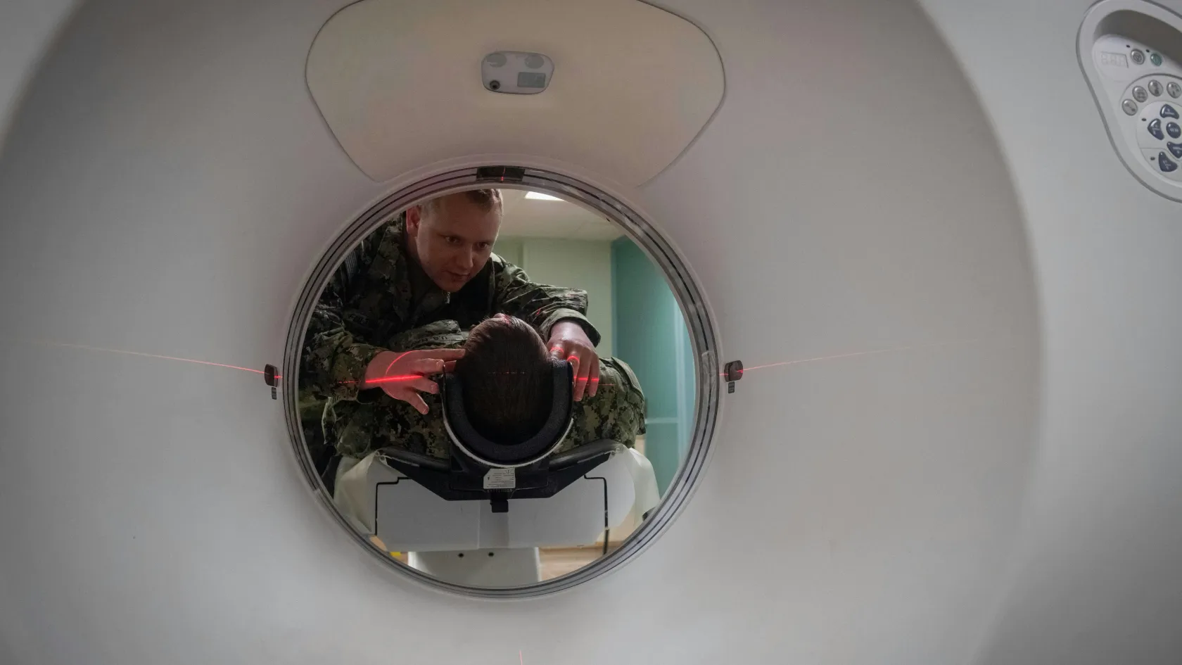
A CT-guided biopsy uses the scanner to place a needle in a target that cannot be seen by ultrasound or reached by an endoscope, and removes a core of tissue for the pathologist. For a lung nodule, a deep abdominal mass, a bone lesion or a lymph node in the chest or the back of the abdomen, it has become the standard route to a diagnosis. In an analysis of 15,865 lung biopsies across four American states, any collapse of the lung occurred in 15.0 percent, a chest tube was needed in 6.6 percent, and bleeding complicated 1.0 percent (Wiener and colleagues, Annals of Internal Medicine, 2011). A meta-analysis of 8,133 core biopsies found major complications in 5.7 percent (Heerink and colleagues, European Radiology, 2017). Outside the lung the risks are lower. This page explains what the procedure is and when it is chosen, why a core of tissue matters more than it used to, what the day involves, the risks organ by organ, what the report means, and how patients from abroad arrange it at Biruni Hospital in Istanbul.
15.0
Percent of lung biopsies followed by any air leak around the lung, most of them needing no treatment
6.6
Percent of lung biopsies needing a chest tube for the air leak, in 15,865 procedures
1.0
Percent of lung biopsies complicated by bleeding
Free
Review of your CT or PET and the question the biopsy must answer
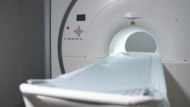
First, a picture. The radiologist marks the skin, numbs it, advances a needle a few centimeters, and takes another picture to see where the tip has reached. Adjust, scan, adjust again, until the tip sits inside the target. A cutting needle then passes through the guide and removes a cylinder of tissue a millimeter or two wide and a centimeter long, two to four times. The whole thing takes fifteen to thirty minutes, and the patient lies still on the scanner table throughout, breathing quietly or holding a breath on request.
Why CT rather than ultrasound?
Ultrasound guides most biopsies of the liver, the thyroid, the breast and the neck, because it shows the needle in real time and uses no radiation. It cannot see through air or bone. A lung nodule, a lesion inside a rib or a vertebra, a lymph node behind the lung or at the back of the abdomen, an adrenal mass and a deep pelvic lesion are hidden from ultrasound and visible to CT. CT is also chosen when the target is small, when it lies behind bowel that must be avoided, or when an earlier ultrasound-guided attempt failed.
Targets reached under CT guidance
Region
Typical target
Route
Chest
Lung nodule or mass, pleural thickening, mediastinal node
Through the abdominal wall, sometimes through the liver
Kidney
Renal mass before ablation or when surgery is in doubt
Through the flank
Bone
Vertebral, pelvic or rib lesion, suspected metastasis or infection
Through the cortex with a drill needle
Pelvis
Deep pelvic mass or node
Through the buttock or the abdominal wall
Soft tissue
Deep muscle or retroperitoneal mass
Direct
Why a core of tissue matters
A biopsy used to answer one question. Cancer or not. Today it answers four, and the fourth has changed what the radiologist must bring back.
Guidelines from the Society of Interventional Radiology define percutaneous needle biopsy as a procedure with established thresholds for diagnostic yield and complications, and set out the indications, the patient selection and the quality measures a service is held to (Gupta and colleagues, Journal of Vascular and Interventional Radiology, 2010).
1
Diagnosis
Whether the lesion is cancer, infection, inflammation or something benign. A core of tissue shows the pathologist the architecture, which a few cells from a fine needle often cannot.
2
Type and origin
Which cancer, and where it started. A lung nodule may be a lung cancer or a metastasis from the bowel, and the treatment differs entirely. Immunostains on the core settle it.
3
Molecular profile
The mutations and markers that decide whether a targeted drug or immunotherapy will work. These tests need more tissue than a diagnosis alone, and a biopsy that yields too little sends the patient back for another.
The fourth question is the reason the modern biopsy takes three or four cores rather than one, uses a slightly larger needle, and is planned with the oncologist before the needle goes in, so that the pathologist receives what the tests will need. A patient who has had a biopsy elsewhere that gave a diagnosis but not enough tissue for molecular testing is one of the commonest reasons for a repeat.
The core is the product. Everything else on this page exists to obtain it safely.
The day of the procedure
Outpatient for most. One night in hospital for a lung biopsy in some centers and for patients who live far away.
1
Before
Fasting for four to six hours. Blood tests for clotting and platelets. Blood thinners paused on a schedule agreed in advance, from a day for some to five days for others. The radiologist reviews the scans and chooses the route that crosses the least lung, avoids vessels and bowel, and reaches the most solid part of the lesion.
2
Positioning
On the back, the front or the side, depending on the route, with a pillow and straps to hold the position for twenty minutes. A planning scan is taken and the entry point marked on the skin.
3
The needle
Local anesthetic to the skin and the deeper tissues, which is the part that stings. The guide needle advances in steps with a scan after each. Patients feel pressure and, in the lung or the liver, sometimes a brief sharp sensation as the needle crosses the surface. Then the cores are taken, each with a click.
4
After
A final scan checks for bleeding or air. A dressing goes on. The patient is observed for one to four hours, lying on the biopsied side for a lung biopsy so that the puncture seals, with a chest X-ray before going home.
Nobody is put to sleep for an ordinary CT-guided biopsy. Sedation through a vein is offered to anxious patients and for bone biopsies, which take longer and hurt more. General anesthesia is reserved for children and for the rare deep biopsy that needs the patient perfectly still.
1
The same evening
Rest. No driving after sedation. Simple painkillers for a sore puncture site. A patient from abroad stays in the hotel near the hospital rather than flying, and a lung biopsy patient does not fly for 48 hours.
Does it hurt?
The anesthetic injection stings for a few seconds. The rest is pressure. A bone biopsy through the cortex is felt as a deep ache during the drilling and for a day or two afterward. Pain that grows in the hours after a biopsy, breathlessness, coughing blood beyond a few streaks, or faintness are reasons to call the number given at discharge.
Bring the list of medicines, and say so at booking if any are blood thinners, including aspirin, clopidogrel, warfarin, apixaban, rivaroxaban or enoxaparin.
Bring the outside CT or PET on disk or as a link, because the radiologist plans the route on the pictures rather than the report.
Arrange for someone to accompany you if sedation is planned.
Lung biopsy and its risks
The lung draws the most needles and the most complications. Its surface leaks air when punctured, and everything about the technique follows from that.
What is a pneumothorax and how often does it happen?
Air escaping from the punctured lung into the space around it, which lets the lung sag away from the chest wall. In the four-state analysis of 15,865 biopsies it followed 15 percent of procedures, and 6.6 percent of all biopsies needed a chest tube to drain the air, with smokers, patients with emphysema and patients in their sixties at higher risk (Wiener and colleagues, 2011). Most small air leaks cause no symptoms, are seen only on the check X-ray, and seal on their own within a day. A larger one causes breathlessness and is treated by a thin tube through the chest wall, left in for one to three days.
1
What reduces the risk
A route that crosses as little lung as possible, a small nodule reached without passing through a fissure, breath-holding at the moment of puncture, lying on the biopsied side afterward, and an experienced radiologist. Emphysema on the scan raises the risk and is taken into account when the radiologist decides whether to biopsy at all or to propose bronchoscopy or surgery instead.
2
Bleeding
A few streaks of blood in the sputum after a lung biopsy in a proportion of patients, settling within hours. Bleeding heavy enough to matter complicated 1.0 percent of biopsies in the population study, and about one in six of those needed a transfusion.
Complications of CT-guided lung biopsy
Complication
How often
What happens
Any air leak around the lung
15 to 25 percent
Watched, X-ray repeated, most settle unaided
Air leak needing a chest tube
5 to 7 percent
Tube for one to three days, sometimes an overnight stay
Blood-streaked sputum
5 to 10 percent
Settles within hours
Bleeding into the lung or chest
About 1 percent
Observation, rarely transfusion or a procedure
Air entering a blood vessel
Under 0.1 percent
Rare and serious, treated at once with oxygen and positioning
Death
Under 0.1 percent
Reported in large series, almost always in patients with severe lung disease
Core or fine needle
The meta-analysis of 49 studies found overall complications in 38.8 percent of core biopsies and 24.0 percent of fine-needle aspirations, most of them minor, with major complications in 5.7 percent and 4.4 percent, a difference that was not significant (Heerink and colleagues, 2017). The core gives more tissue for the same major risk, and it serves as the default for a lung nodule that may need molecular testing. A fine needle is chosen for a small nodule in a patient with emphysema, where every millimeter of needle counts, and for a lesion that is almost certainly a metastasis of a known cancer.
Liver, bone, kidney, adrenal and lymph nodes
Lower risk, different questions.
1
Liver
CT takes over from ultrasound for lesions high under the dome of the diaphragm, small lesions seen only on contrast scans, and patients whose bowel gas or body shape blocks the ultrasound window. The risk is bleeding, in under one percent, higher in cirrhosis and on blood thinners, and the puncture is planned through a cuff of normal liver that tamponades the track.
2
Bone
A drill needle through the cortex of a vertebra, the pelvis or a rib, to tell a metastasis from infection, myeloma or a primary bone tumor. Longer, more uncomfortable, done with sedation, and the sample often goes for culture as well as pathology.
3
Kidney and adrenal
A kidney mass is biopsied before ablation, when the scan cannot say whether it is cancer, or when surgery would be risky. An adrenal mass is biopsied only after a blood test has excluded pheochromocytoma, because a needle into that tumor can trigger a dangerous surge in blood pressure.
Lymph nodes in the chest between the lungs are reached by endobronchial ultrasound through the airway, and nodes around the stomach and pancreas by EUS through the gut, both with lower risk than a needle through the chest or the abdomen. CT is used for the nodes those scopes cannot reach, along the aorta at the back of the abdomen, in the pelvis and in the neck root.
Seeding
Cancer cells carried along the needle track and growing there later. Rare for most tumors. Well under one percent, and the reason for the coaxial technique, in which the cutting needle passes through an outer guide so that the track is protected. For a few tumors, above all hepatocellular carcinoma and mesothelioma, seeding happens frequently enough that the biopsy is avoided when the scan is diagnostic, or the track is planned so that surgery removes it.
A lesion that the scans have already characterized with confidence, a liver cancer with typical contrast behavior in a cirrhotic liver for example, does not need a biopsy, and the tumor board will say so. The needle answers a question. Where there is none, it stays in the drawer.
The report and what it means
Three to seven working days for a diagnosis. Two to three weeks for a full molecular profile.
The pathologist reports the tissue type, whether cancer is present, its grade, the immunostains that identify its origin, and, when requested, the mutations and markers relevant to treatment. The result goes to the doctor who asked the question, and a patient from abroad receives it in writing with a consultation to explain it.
What if the biopsy does not give an answer?
It happens in 5 to 10 percent of biopsies, when the needle samples dead tissue in the center of a tumor, scar around it, or too little for the stains. The report says non-diagnostic rather than benign, which is a different thing, and the options are a repeat biopsy aimed at the edge of the lesion, a different route such as bronchoscopy or EUS, or surgical removal of a lesion that is small and suspicious enough to justify it. A non-diagnostic result never means the lesion is safe to ignore.
Day of the biopsy. The cores go into fixative and, for suspected lymphoma or infection, some go fresh for flow cytometry or culture.
Days two to four. Sections are cut, stained and examined. A preliminary result of cancer or no cancer is available for most.
Days four to seven. Immunostains identify the type and origin. The written report is issued.
Weeks two to three. Molecular tests, where ordered, are added to the report. The oncologist plans treatment on the complete result.
Second opinions on the slides
Blocks and slides belong to the patient. A patient who returns home with the report can have the slides sent to a pathologist there for a second reading, and a patient who arrives with slides from home can have them reviewed here without a new biopsy, which is often the faster and safer route to a treatment decision.
Coming from abroad
Few patients travel for a biopsy alone.
It comes first when a patient arrives for cancer treatment without a tissue diagnosis, or with one that lacks the molecular results the treatment needs, and the hospital schedules it in the first days so that the tumor board can meet on a complete file rather than on a scan and a guess, which saves the week that would otherwise pass between the first consultation and the first decision. The length of stay for the biopsy alone runs to three or four days. Two days after a lung biopsy, and the day after most others, patients are fit to fly, and a patient staying on for treatment simply continues. The one arrangement worth making before travel is to ask the doctor at home whether the slides from any earlier biopsy can be released, since a review of existing slides can replace a new needle altogether and the answer to that question decides whether the first days in Istanbul are spent in the scanner or in the clinic.
One coordinator from the international patients office handles arrangements from the first message through discharge and answers the same WhatsApp number once you are back home. The office works in English, Arabic, French, Russian, Serbian, Romanian and Spanish and books interpreters for other languages. It arranges the visa invitation letter, airport transfers, daily transport and accommodation near the hospital. The kitchen prepares halal, vegetarian and diabetic meals, a prayer room is on site, and a request for a female physician is met wherever the rota allows.
What to send
Send the CT or PET as image files rather than a report alone, any earlier biopsy report and, if possible, the slides or blocks, the list of medicines with blood thinners marked, and the question the treating doctor wants answered. Within a few working days the interventional radiology team replies. Whether a biopsy is needed at all, which route, whether sedation is planned, the stay, and a cost estimate.
Cost
An estimate follows the file review and covers the procedure, the CT time, local anesthetic or sedation, the observation period, the chest X-ray and the pathology with immunostains. Hospitals in this market quote CT-guided biopsy as a procedure fee with pathology and molecular testing listed separately, since the molecular panel depends on the cancer found.
Confirm what the figure includes and whether the molecular tests are priced with it.
References
Wiener RS, Schwartz LM, Woloshin S, Welch HG. Population-based risk for complications after transthoracic needle lung biopsy of a pulmonary nodule. An analysis of discharge records. Ann Intern Med. 2011;155(3):137-144.
Heerink WJ, de Bock GH, de Jonge GJ, Groen HJ, Vliegenthart R, Oudkerk M. Complication rates of CT-guided transthoracic lung biopsy. Meta-analysis. Eur Radiol. 2017;27(1):138-148.
Gupta S, Wallace MJ, Cardella JF, Kundu S, Miller DL, Rose SC. Quality improvement guidelines for percutaneous needle biopsy. J Vasc Interv Radiol. 2010;21(7):969-975.
Editor's note
Written by the Biruni Hospital medical editorial team. Reviewed by Specialist Dr. Onur Aksoy, Radiology; GETAT (Traditional and Complementary Medicine).