Irreversible electroporation, known by the brand name NanoKnife, destroys a tumor with high-voltage electrical pulses between needle electrodes while sparing the blood vessels, bile ducts and nerves around it. It covers how it works, which pancreatic, liver, prostate and kidney tumors it treats, what the registry and trial data show and what they do not, the procedure, the risks, the follow-up, and how patients from abroad arrange it in Istanbul.
About This Department
Interventional oncology
Some tumors wrap around the vessels and ducts that keep a patient alive. Heat would destroy both. Irreversible electroporation kills the tumor and spares the pipes. Electricity does it. Nothing is cut.
Irreversible electroporation, or IRE, marketed as NanoKnife, destroys a tumor with short, high-voltage electrical pulses delivered between thin needle electrodes placed around it. The pulses open permanent holes in the membranes of the cells, which die over the following days, while the collagen framework of blood vessels, bile ducts and nerves survives. Despite the name, no cutting takes place. In a registry of 200 patients with locally advanced pancreatic cancer treated after chemotherapy, median survival was 24.9 months and local recurrence occurred in 3 percent (Martin and colleagues, Annals of Surgery, 2015). A prospective phase II trial of the percutaneous technique reached a median of 17 months from diagnosis (Ruarus and colleagues, Radiology, 2020). This page explains what IRE is and how it differs from heat ablation, which tumors it treats, what the studies show and what they do not, the procedure itself, the risks, the follow-up, and how patients from abroad arrange it at Biruni Hospital in Istanbul.
24.9
Months median survival in 200 patients with locally advanced pancreatic cancer treated with chemotherapy and IRE
3
Percent local recurrence at a median follow-up of 29 months in the same registry
77
Percent of men with prostate cancer recurring after radiation who needed no further treatment four years after IRE
Free
Review of your CT or MRI and the tumor board's view on whether IRE fits
Every ablation method on the market before this one killed tumors with temperature. Radiofrequency and microwave probes heat tissue past 60 degrees. Cryoablation freezes it past minus 40. Both work, and both have the same limit, which is that heat and cold do not know the difference between a tumor and the artery running through it. A tumor pressed against the portal vein, the bile duct, the ureter or the nerves of the pelvis could not be ablated without risking the structure beside it, and a tumor surrounding those structures could not be ablated at all.
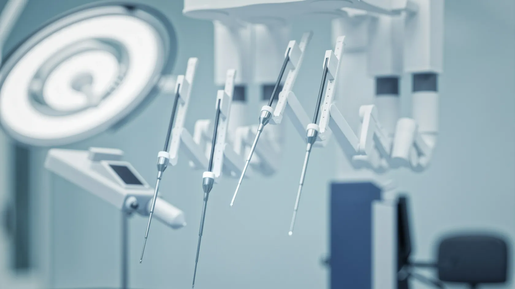
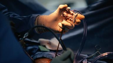
IRE uses no temperature. Two to six needle electrodes are placed around the tumor under CT or ultrasound guidance, and a generator delivers about ninety pulses of up to 3,000 volts between each pair, each pulse lasting a hundred microseconds. The electric field punches nanometer-scale holes in the cell membranes. Above a threshold the holes do not close, the cell cannot hold its contents, and it dies over hours to days. Vessels, ducts and nerves are made largely of collagen and elastin rather than cells, and they keep their structure. Blood keeps flowing through a treated artery. Bile keeps draining through a treated duct.
Is NanoKnife the same as IRE?
Yes. NanoKnife is the brand name of the generator and electrodes made by one manufacturer, and it is the system in clinical use for this procedure. Patients search for either name. The word knife is marketing. Nothing is cut, and the procedure is done through needles placed through the skin or, in some pancreatic cases, during an open operation.
What it needs
Four things.
General anesthesia with complete muscle relaxation, because each pulse contracts every muscle in its path. Every pulse timed to the heartbeat, since one delivered at the wrong moment of the cardiac cycle can trigger an arrhythmia. Electrodes parallel to one another and within millimeters of the plan. And a team that has done it before, since the learning curve is steep and the organs involved forgive little.
Which tumors it treats
The ones heat cannot reach safely. That is the whole indication.
By organ
Pancreas. Locally advanced cancer that has grown around the celiac or superior mesenteric arteries and cannot be removed, in a patient who has responded to or at least not progressed on several months of chemotherapy. The largest use and the one with the most data. Also used during surgery to treat the margin where the tumor cannot be fully separated from a vessel.
Liver. Tumors, primary or metastatic, within a centimeter of the main bile ducts or the major veins, where a heat probe would cause a stricture or a thrombosis. IRE treats them and leaves the ducts open.
Prostate. Focal treatment of a cancer confined to one part of the gland, and salvage treatment of cancer that has returned after radiation, where surgery and further radiation carry high rates of incontinence and rectal injury.
Kidney. Tumors at the hilum, where the vessels and the collecting system enter, that partial nephrectomy and heat ablation would both endanger.
Other. Selected tumors of the pelvis near nerves, tumors of the adrenal near the vena cava, and lung tumors near the hilum, in specialist centers and small numbers.
IRE does not replace surgery for a tumor that can be removed, does not replace heat ablation for a tumor away from vital structures, and does not treat a cancer that has spread beyond the organ. It treats the tumor between the needles and nothing else. A patient with pancreatic cancer and metastases in the liver gains nothing from it, which is the reason every candidate has a fresh scan and a tumor board review before the date is set.
What the studies show
No randomized trial has compared IRE with anything. What exists is one large registry, one prospective phase II trial and several single-center series, and a patient should read the numbers with that in mind.
The multicenter registry followed 200 patients with stage III pancreatic cancer, all of whom had received chemotherapy for a median of six months and half of whom had also received chemoradiation. IRE was performed alone in 150 and combined with resection in 50. Complications occurred in 37 percent, mostly minor. Median hospital stay was six days. At a median follow-up of 29 months, local recurrence had occurred in 3 percent and median overall survival was 24.9 months, against 12 to 14 months expected with chemotherapy alone (Martin and colleagues, 2015).
PANFIRE-2, the prospective trial. Fifty patients, 40 with locally advanced and 10 with recurrent pancreatic cancer, treated by CT-guided percutaneous IRE at Amsterdam. Median survival was 17 months from diagnosis and 10 months from the procedure. Local recurrence developed in 46 percent. Major complications occurred in 21 patients, and one death was probably related to the procedure. The trial met its survival target and the authors noted that a tumor volume over 37 milliliters and a very high CA 19-9 level predicted worse outcomes (Ruarus and colleagues, 2020).
Salvage prostate IRE. Seventy-four men whose prostate cancer had returned after radiation were treated with focal IRE in Sydney. At a median of four years, 77 percent had needed no further treatment, 91 percent were free of metastases, one rectal fistula had occurred, and 93 percent of those who answered questionnaires remained continent (Geboers and colleagues, BJU International, 2023).
The gap between them. The registry included patients selected by surgeons and treated in the operating room. The trial treated sicker patients through the skin. The difference in survival, 24.9 against 17 months, reflects selection as much as technique, and the honest range for a patient with locally advanced pancreatic cancer who responds to chemotherapy and then has IRE is somewhere between the two.
Does IRE cure pancreatic cancer?
No, and any page that says otherwise should be read with suspicion. Locally advanced pancreatic cancer is not cured by any local treatment, because the disease is systemic in nearly every patient by the time it is found. What the studies show is that in patients who have already responded to chemotherapy, destroying the primary tumor adds months, in the registry roughly a year over historical figures, and relieves the pain that the tumor causes by invading the nerves behind the pancreas. A real gain, for the patient who qualifies. Not a cure. The team here says so at the first consultation.
Reading the numbers as a patient
Three conditions. No spread beyond the pancreas on a scan taken within the last month. At least three to four months of chemotherapy completed, with the tumor no larger than before it started. And a tumor smaller than about four centimeters, since larger volumes were the strongest predictor of failure in the trial. Meeting all three gives a reasonable expectation of local control and of a survival measured in a year or two rather than months. Meeting none of them means chemotherapy should continue instead, and the file review will say so plainly.
The procedure
Ninety minutes to three hours. General anesthesia. Two to six nights.
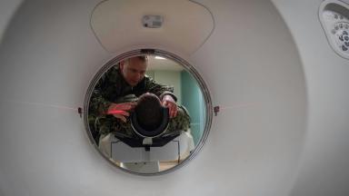
The electrodes are placed one at a time under CT, each checked against the plan, and the generator delivers its pulses between every pair in turn while the anesthesiologist watches the heart rhythm on the monitor and the muscle relaxant keeps the patient still. The whole delivery takes minutes. The placement takes most of the time.
1
Before
A contrast CT within the month, blood tests including CA 19-9 for pancreatic cancer, an ECG and an echocardiogram, because a patient with an irregular heart rhythm or a pacemaker may not be suitable. Chemotherapy is paused for two weeks before and restarted two to four weeks after. A stent in the bile duct, if one is present, is changed from metal to plastic beforehand, because a metal stent between the electrodes conducts the current.
2
Placement
Under general anesthesia, on the CT table for the percutaneous route or in the operating room for the open route, two to six electrodes are advanced into position around the tumor, spaced 1.5 to 2 centimeters apart and parallel, with the active tips exposed to a length matched to the tumor. A scan confirms the geometry before any pulse is delivered.
3
Delivery
Ten test pulses, then the full sequence between each pair of electrodes, each pulse timed to the heartbeat. The electrodes are pulled back and the sequence repeated if the tumor is longer than the exposed tips. A contrast scan at the end confirms that the vessels are open.
4
After
Intensive care or a monitored bed for the first night after pancreatic IRE, because of the risk to the heart rhythm and the bowel. Pain, which peaks on the second day, is controlled with a pump. Eating resumes on the first or second day. Discharge follows on day three to six.
Risks
Higher than for heat ablation of a simple tumor. The tumors IRE treats sit against the most dangerous structures in the abdomen.
In the registry, complications occurred in 37 percent of patients with a median grade of 2, meaning most were treated with medicines or observation. In the prospective trial, 21 of 50 patients had a major complication and one death was probably related to the procedure. The complications specific to IRE are an irregular heart rhythm during delivery, prevented by synchronization and treated at once by the anesthesiologist, and pancreatitis, a leak from the pancreas or a bile leak in the days afterward. Bleeding, portal vein thrombosis, bowel injury and infection of the treated area occur as they do after any ablation. For prostate IRE the specific risks are a rectal fistula, seen once in 74 men, temporary urinary retention, and erectile dysfunction in a proportion of men who had function before.
What lowers the risk
A team that does the procedure regularly. Tumors under four centimeters. Recovery from chemotherapy, with a good blood count and no infection. A plastic rather than a metal stent. And restraint. The commonest serious harm from IRE in the published series came from treating patients who should not have been treated, whose disease had already spread or whose tumors were too large, and the file review exists to prevent that.
After the procedure
Two things happen in the first weeks. Pain eases. The scan looks worse before it looks better.
IRE does not shrink a tumor at once. The treated zone swells in the first days, shows as a larger area on the scan at one month, and settles slowly over three to six months. The pathologist, when tissue is available, finds dead cells inside an intact scaffold. The radiologist judges success by the absence of enhancement inside the zone and by the tumor marker, CA 19-9, which should fall by half within three months. A marker that does not fall predicts trouble, and the trial identified it as one of the three strongest signs of a poor outcome.
1
Chemotherapy resumes
Two to four weeks after IRE, on the oncologist's schedule, since the systemic disease is what determines survival and the local treatment does not replace it.
2
Scans
Contrast CT at one month, three months and every three months thereafter, with the images from each visit kept for comparison with the next.
Local recurrence developed in 23 of 50 participants, 46 percent, after percutaneous IRE (Ruarus and colleagues, 2020). Local recurrence occurred in 3 percent in the surgical registry (Martin and colleagues, 2015). Both numbers are true, for different patients treated by different routes, and a patient should ask which group resembles his own case.
Repeat treatment
Recurrence at the edge of the treated zone can be treated again, by IRE or by radiation, if the disease elsewhere remains controlled. Spread in the meantime sends the patient back to drug treatment, and the IRE has bought the months in between.
Coming from abroad
IRE is a treatment patients travel for because few centers offer it and fewer offer it with the surgical and interventional experience the pancreas demands. The length of stay is ten to fourteen days, which covers the tumor board, the scan, the cardiac assessment, the procedure, three to six nights in hospital and a week of recovery near the hospital before the flight, since the risk of a delayed pancreatic leak makes an early departure unwise. Patients are fit to fly when the surgeon or the interventional radiologist confirms that the drain, if one was placed, is out and the blood tests are stable, in the second week for most. What the team needs before travel is the CT or MRI as image files, the pathology, the chemotherapy record with dates and response, the most recent CA 19-9, the list of medicines, an ECG and a note of any heart condition or implanted device. The tumor board reviews the file without charge and replies with whether IRE is appropriate, by which route, or whether continued chemotherapy, radiation or surgery would serve the patient better.
One coordinator from the international patients office handles arrangements from the first message through discharge and answers the same WhatsApp number once you are back home. The office works in English, Arabic, French, Russian, Serbian, Romanian and Spanish and books interpreters for other languages, and it arranges the visa invitation letter, airport transfers, daily transport and accommodation near the hospital for the patient and a companion for the full stay. Halal, vegetarian and diabetic meals, a prayer room on site, and a female physician on request wherever the rota allows. The follow-up scans are reviewed by the same team wherever they are taken, and the oncologist at home receives the procedure report and the plan for restarting chemotherapy before the patient lands.
Cost
One figure, after the file review.
It depends on the route, percutaneous or surgical, the number of electrodes, the anesthesia, the monitored bed and the hospital stay. Hospitals in this market quote IRE as a package for the procedure with the single-use electrodes, which are a large part of the cost, included, and the pre-procedure scans, chemotherapy and any longer stay for complications priced separately.
Confirm what the figure includes. Confirm, too, what a longer stay would add, since the stay after IRE varies more than after any other ablation, from three nights for an uncomplicated percutaneous case to two weeks or more for a patient who develops a pancreatic leak and needs a drain, antibiotics and time.
References
Martin RC 2nd, Kwon D, Chalikonda S, et al. Treatment of 200 locally advanced (stage III) pancreatic adenocarcinoma patients with irreversible electroporation. Safety and efficacy. Ann Surg. 2015;262(3):486-494.
Ruarus AH, Vroomen LGPH, Geboers B, et al. Percutaneous irreversible electroporation in locally advanced and recurrent pancreatic cancer (PANFIRE-2). A multicenter, prospective, single-arm, phase II study. Radiology. 2020;294(1):212-220.
Geboers B, Scheltema MJ, Blazevski A, et al. Median 4-year outcomes of salvage irreversible electroporation for localized radio-recurrent prostate cancer. BJU Int. 2023;131 Suppl 4:14-22.
Editor's note
Written by the Biruni Hospital medical editorial team. Reviewed by Specialist Dr. Onur Aksoy, Radiology; GETAT (Traditional and Complementary Medicine).