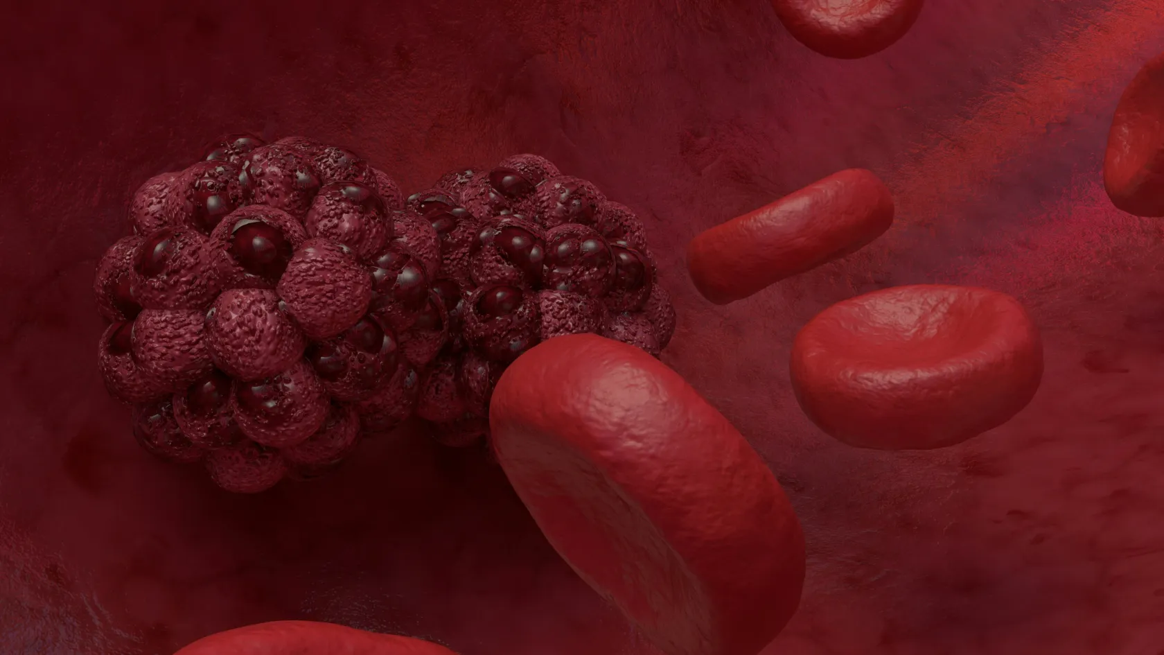
Tumor embolization blocks the artery feeding a tumor from inside, through a catheter from the wrist or groin, with particles, chemotherapy beads or yttrium-90 microspheres. For liver cancer that cannot be removed, chemoembolization raised two-year survival from 27 to 63 percent in the Barcelona trial.
About This Department
Interventional Radiology
Every tumor needs an artery. Embolization blocks it from the inside, through a catheter the width of a strand of spaghetti.
A liver tumor the surgeon cannot remove. A kidney tumor that will bleed heavily on the operating table. A metastasis in the spine that has to come out, and a cancer that is bleeding and will not stop. Each of these has an artery feeding it, and embolization is the treatment that reaches that artery from inside the body and blocks it, with particles, with chemotherapy-loaded beads, or with radioactive microspheres, delivered through a catheter threaded from the wrist or the groin under X-ray guidance. For liver cancer the evidence is old and firm. In the Barcelona trial published in the Lancet in 2002, chemoembolization raised two-year survival from 27 percent to 63 percent, and a Hong Kong trial the same year found the same direction. For single liver tumors up to 8 centimeters, radioembolization with yttrium-90 produced a response in 88 percent of patients in the LEGACY study, and a third of them went on to a transplant or an operation. This page explains the four kinds of embolization, which tumors they suit, what the week after feels like, and how to plan a stay of a few days in Istanbul.
63% vs 27%
Alive at two years, chemoembolization against supportive care, liver cancer, Lancet 2002
88%
Single liver tumors responding to yttrium-90 radioembolization, LEGACY study
1 to 2 nights
In hospital after most embolizations
Free
Review of your scans and liver tests
Free consultation
The review of your file is free and commits you to nothing. Send the CT or MRI with its images and report, the liver blood tests including bilirubin and albumin, any biopsy result, the treatments given so far, your medicine list, and a note of any viral hepatitis or cirrhosis. An interventional radiologist and a liver specialist read them together and tell you which form of embolization fits, or whether ablation, surgery or drug treatment would serve you better, and a coordinator writes to you in your own language.
Tumor Embolization is a treatment in which the blood supply to a tumor is blocked from within the artery that feeds it. No cut anywhere. A catheter enters an artery at the wrist or the groin, is steered under X-ray to the branch that supplies the tumor, and delivers something that stops the flow. Tumor tissue dies within days for want of oxygen, while the organ around it, fed by other branches, carries on.
Three reasons make it work in the liver above all. A healthy liver takes three quarters of its blood from the portal vein and only a quarter from the hepatic artery, while a liver tumor draws nearly all of its supply from the artery. Blocking the artery therefore hits the tumor and spares the liver. And the same catheter that blocks the artery can leave chemotherapy or radiation behind, concentrated in the tumor at doses the rest of the body never sees.
The tumor lives on the artery. The liver lives on the vein. That asymmetry underlies the whole of liver embolization. It explains why the treatment has held its place for forty years.
Four kinds of embolization
Phone screens show this table in part. Drag it left for the rest.
Four kinds of embolization
Kind
What goes in
What it is for
Bland embolization
Particles or gelatin sponge, and no drug
Cutting off blood before an operation, stopping a bleeding tumor, and some liver tumors
Chemoembolization, TACE
Chemotherapy mixed with an oily contrast agent, or loaded onto tiny beads that release it slowly
Liver cancer that cannot be removed, liver tumors while a patient waits for a transplant, and some liver metastases
Radioembolization, TARE or SIRT
Glass or resin microspheres carrying yttrium-90, a radioactive isotope with a range of a few millimeters
Liver cancer, including tumors that have grown into the portal vein, and liver metastases from bowel and other cancers
Preoperative embolization
Particles, coils or glue, hours to days before surgery
Kidney tumors, bone and spine metastases, meningiomas and other tumors that would bleed heavily at operation
Three treatments and one preparation
The first three are treatments in their own right. The fourth makes another treatment safer, and it appears on this page because the same team, the same room and the same catheter do the work.
Liver cancer and chemoembolization
Hepatocellular carcinoma, the cancer that arises in a liver already damaged by hepatitis or cirrhosis, is the disease that made embolization standard. Two randomized trials, both published in 2002, set the figures.
Barcelona, Lancet 2002
112 patients with liver cancer that could not be removed, in a liver still working well, were assigned to repeated bland embolization, repeated chemoembolization with doxorubicin, or supportive care. The trial was stopped early because chemoembolization was clearly winning. Survival at one and two years was 82 and 63 percent with chemoembolization, 75 and 50 percent with bland embolization, and 63 and 27 percent with supportive care.
Hong Kong, Hepatology 2002
80 patients assigned to chemoembolization with cisplatin and iodized oil, repeated every two to three months, or supportive care. Survival at one, two and three years was 57, 31 and 26 percent with treatment and 32, 11 and 3 percent without. Patients received a median of four and a half sessions.
Two facts in those trials still shape practice. Both selected patients whose liver was working well, and in the Hong Kong trial death from liver failure was commoner in the treated group, which is why the liver tests decide eligibility as much as the scan does. And both repeated the treatment, since a single session rarely finishes the job.
Chemoembolization today uses drug-eluting beads in most centers, which release the drug over days and put less of it into the bloodstream. Guidelines make it the standard for intermediate-stage liver cancer, several tumors confined to the liver in a patient who is well. It also holds a tumor still while a patient waits on the transplant list.
Radioembolization with yttrium-90
Radioembolization delivers radiation from inside the tumor. Millions of microspheres, each thinner than a hair and carrying yttrium-90, lodge in the smallest arteries of the tumor and irradiate it from a range of a few millimeters for about two weeks. The dose reaching the tumor can be many times what an external beam could safely give.
Phone screens show this table in part. Drag it left for the rest.
Two studies of yttrium-90 in liver cancer
SARAH, Lancet Oncology 2017
LEGACY, Hepatology 2021
Patients
459 with advanced or intermediate liver cancer after failed chemoembolization, at 25 French centers
162 with a single liver tumor of up to 8 cm and good liver function, at three American centers
Design
Randomized against the drug sorafenib
Consecutive patients, response judged by blinded central review
Main result
Median survival 8.0 months against 9.9, no significant difference
Response in 88.3 percent, lasting six months or more in 62.2 percent
Survival
No better than the drug
86.6 percent at three years for all, 92.8 percent for those who went on to transplant or resection
Side effects
Fewer serious effects than the drug. Severe diarrhea 1 against 14 percent, severe hand-foot skin reaction under 1 against 6 percent, severe fatigue 9 against 19 percent
Not the focus of the study
Bridge to another treatment
21 percent went on to transplant and 7 percent to resection
Read the two together
In advanced disease, radioembolization did not lengthen life beyond what a tablet achieved, and it did make the months better. In a single, well-selected tumor, it controlled the disease in nearly nine patients out of ten and carried a third of them to a curative operation. The review tells you which of those two patients you are.
A radioembolization has two visits a week or two apart. On the first, a mapping angiogram studies the arteries, blocks with coils any branch that leads to the stomach or bowel, and injects a test dose of harmless particles that a scan then traces to measure how much would leak to the lungs. On the second comes the treatment itself. The microsphere dose is calculated from that scan.
Before an operation, and for bleeding tumors
Cutting off blood before an operation
Some tumors are so richly supplied that removing them means removing them through a field of blood. Large kidney tumors. Metastases in the spine and pelvis from kidney or thyroid cancer. Meningiomas fed by the arteries of the skull, and bone tumors of the pelvis. Embolization the day before, or the same morning, turns the operation from a race against bleeding into a controlled one, with fewer transfusions and a clearer view for the surgeon. The neurosurgery, orthopedic and urology teams at Biruni Hospital work on the same site as interventional radiology, and the two procedures are scheduled together.
A tumor that is bleeding
A cancer in the kidney, bladder, uterus, bowel or lung can bleed enough to need transfusion, and embolization stops it within the hour for nearly all of them. It is done as an emergency for a patient already in the hospital, and it is the reason a patient with a bleeding tumor should be in a hospital with an interventional radiologist on call rather than on a plane.
Who is a candidate
For liver embolization the answer has three parts, and the scan answers only one of them.
How far has the cancer spread
Chemoembolization suits cancer confined to the liver, several tumors or one large one, where surgery and ablation are ruled out. Radioembolization reaches the same patients and also those whose tumor has grown into a branch of the portal vein, and some with limited spread outside the liver.
How well does the liver work
Child-Pugh class A, or B with a low score. Bilirubin below about 2 mg per deciliter and no fluid in the abdomen for chemoembolization. Radioembolization tolerates portal vein involvement better and poor liver function worse.
How well is the patient
Up and about most of the day. A patient in bed for more than half the day is not helped by either.
Reasons to say no
For chemoembolization, tumor in the main portal vein, liver failure, bile duct obstruction, a previous bile duct operation, poor kidney function. For radioembolization, too much flow to the lungs on the mapping scan, arteries to the bowel that cannot be blocked, liver failure.
Metastases in the liver from bowel, breast, neuroendocrine and other cancers are treated with embolization when drugs have stopped working or as an addition to them, and the choice between chemoembolization, radioembolization and ablation is made by the tumor board from the number, size and position of the deposits.
Preparation
Preparation fills a week with small tasks. The blood tests carry the most weight.
Blood tests within a week
Liver function including bilirubin and albumin, clotting, a blood count, kidney function, and for liver cancer the tumor marker alpha-fetoprotein.
Scans
A contrast CT or MRI within six weeks, showing the arteries of the liver and any portal vein involvement. For radioembolization, the mapping visit one to two weeks before treatment.
Medicines
Blood thinners paused for three to seven days on the advice of the doctor who prescribed them, arranged before travel. Metformin stopped on the day of the procedure and for two days afterwards, because of the contrast dye.
Fasting and anesthesia
Six hours without food, clear fluids until two hours before. Nearly every embolization is done under sedation with local anesthetic at the puncture site, and general anesthesia is kept for children, for long procedures and for anyone who asks for it.
The procedure
Five steps, one to two hours in the room.
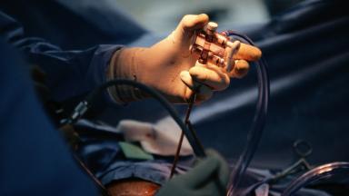
Access. A local anesthetic at the wrist or the groin, and a sheath the width of a drinking straw placed in the artery.
Mapping. A catheter is steered to the liver, the kidney or the tumor in question, and contrast is injected to show every artery feeding it and every branch that must be protected.
Selection. A microcatheter, thinner than a strand of spaghetti, is advanced into the branch or branches that feed the tumor alone, as far from healthy tissue as the anatomy allows.
Delivery. Particles, drug-loaded beads or microspheres are injected slowly until the flow to the tumor stops or the planned dose has gone in. For radioembolization the injection takes minutes, and the room is cleared of everyone not needed.
Closure. The catheter and sheath come out, and the artery is sealed with a closure device or with pressure. At the wrist a small band does it, and at the groin you lie flat for two to four hours while the puncture seals.
One to two nights in hospital for liver embolization. Same-day discharge for many preoperative and radioembolization procedures.
The first week
Patients feel the treatment more than they felt the procedure. What they feel has a name.
Post-embolization syndrome
Pain under the right ribs, fever, nausea and tiredness that begin within hours of a liver embolization, peak on the second or third day and settle within a week. It comes from the dying tumor and the inflammation around it, and it affects the majority after chemoembolization, a minority after radioembolization. Pain relief, anti-sickness medicine and fluids cover it. Everyone expects it, and nobody counts it as a complication.
Radioembolization gives a quieter first week, and then the second and third weeks bring tiredness that lasts a month, with loss of appetite for a few weeks. Preoperative embolization has no week of its own. Its symptoms belong to the operation that follows.
Risks
Six things can go wrong, and the first is the one the blood tests exist to prevent.
Liver failure after chemoembolization
Uncommon when the liver tests were good beforehand. The Hong Kong trial recorded more deaths from liver failure in the treated group, all in patients with borderline function, and that is the reason the tests decide eligibility.
Liver abscess
A few percent, higher after a previous bile duct operation. Antibiotics and drainage.
Particles reaching the wrong organ
Rare. Ulcers in the stomach or bowel, or inflammation of the gallbladder, when a branch was missed, which careful mapping prevents and which radioembolization guards against by coiling the branches to the gut beforehand.
Radiation reaching the lungs
Rare. The test scan before radioembolization exists to measure it, and treatment is withheld when the calculated lung dose is too high.
Bleeding or a bruise at the puncture
A few percent, fewer at the wrist than at the groin, treated with pressure and only rarely with a small procedure.
Kidney injury from contrast dye
Uncommon, higher in diabetics and those with kidney disease. Fluids before and after, and a smaller contrast volume.
Death within 30 days of a liver embolization done in a patient who met the criteria is rare, in the low single figures per hundred. Nearly all of it comes from liver failure in patients whose function was borderline.
Results, and repeating the treatment
The scan tells the story.
Four to six weeks after chemoembolization it shows how much of the tumor has died. Dead tumor takes up no contrast. Living tumor at the edge is treated again, and chemoembolization is repeated as needed, every two to three months, until the tumor no longer responds or the liver can no longer tolerate it. Two to four sessions in a year is the usual count.
With radioembolization the scan waits three months, since the response develops slowly, and a treated segment of liver shrinks over the following year while the rest grows to compensate. A second radioembolization to the other lobe is possible when the first was tolerated.
A tumor that shrinks enough may become removable, or small enough for ablation, or stable enough to keep a patient on the transplant list. Liver surgery, ablation, radiotherapy on an Elekta Versa HD linear accelerator and medical oncology are all delivered at Biruni Hospital on the same site, so the next step after a good response is a change of room rather than a change of hospital.
Days in Istanbul
A chemoembolization session takes four to five days in Istanbul. Consultation and blood tests on the first day, the procedure and one to two nights in hospital on the second and third, rest on the fourth, and the flight on the fifth. Radioembolization takes two visits of three days each, one to two weeks apart, or a single stay of ten to twelve days that covers the mapping and the treatment. Preoperative embolization sits inside the surgical stay and adds one day before the operation.
Flying home after chemoembolization waits until the fever and pain of the first days have settled, which for the majority is three to four days after the procedure.
Radioembolization sets no such limit. The radiation in the liver is too weak and too short-ranged to affect anyone nearby, so no precautions apply on the flight, and a patient who wants the second lobe treated later or a chemoembolization repeated can have it on a later visit or from an interventional radiologist at home on the protocol we write.
What a quote contains
No figure appears on this page, because the four kinds of embolization differ from each other more than most treatments differ from their alternatives.
Phone screens show this table in part. Drag it left for the rest.
What moves an embolization quote
Kind
Main lines
Notes
Bland or preoperative
Particles or coils, the catheter set, the angiography suite
The least expensive of the four
Chemoembolization
Drug-eluting beads, the chemotherapy, one to two hospital nights
Multiplied by the number of sessions over the year
Radioembolization
Yttrium-90 microspheres priced per dose, the mapping angiogram, the nuclear medicine scan
Far above the others, and the mapping is a separate procedure on a separate day
Every kind
Consultation, blood tests, a follow-up scan, an interpreter and transfers
Inside most published packages
Packages published by Turkish hospitals and medical travel agencies for liver embolization cover the consultation, one session, one to two nights in hospital, an interpreter and transfers, and leave out repeat sessions, flights, insurance, accommodation and the treatment of any complication.
A quote you can compare names the kind of embolization, the number of sessions it assumes, whether the mapping visit is inside the figure for radioembolization, and the number of hospital nights. It follows the free review.
Coordinator, language, companion
Language
Biruni Hospital gives every international patient one coordinator from the first message, reachable on the same WhatsApp number afterwards, and the international patients team works in English, Arabic, French, Russian, Serbian, Romanian and Spanish with interpreters arranged for other languages. For this treatment the interpreter matters at two points, the consultation where the kind of embolization is chosen and the reasons explained, and the discharge, when the symptoms of the week ahead are gone through so that a fever on day two does not send the family to an emergency department.
Companion and paperwork
Bring a companion for the days after the procedure. The office books accommodation close to the hospital, arranges airport transfers and the car to the hospital, and issues an appointment confirmation and invitation letter for a visa ten days or so before travel. Patient rooms have a companion bed. The hospital kitchen prepares halal, vegetarian and diabetic meals, and follows the low-salt diet a liver patient is prescribed. A prayer room is on site, and a female physician is arranged where the rota allows for anyone who asks in the first message.
Follow-up at home
Once you get back home, the procedure report with the arteries treated and the dose given, the images, the blood tests at discharge and the follow-up calendar go to your own oncologist or liver specialist.
Liver blood tests and alpha-fetoprotein at four weeks, then every three months
Contrast CT or MRI at four to six weeks after chemoembolization, at three months after radioembolization, then every three months in the first year
Blood thinners restarted on the date written on the discharge note
Any scan sent to the coordinator for comparison with the treatment images at no charge, and a recommendation on whether another session is due
Fever above 38 degrees beyond the first week, pain that gets worse instead of better, yellowing of the skin or eyes, confusion, swelling of the abdomen, or black stools go to a local emergency department the same day, with the discharge note in hand.
Tumor Embolization FAQ
What is the difference between chemoembolization and radioembolization?
Both block the artery feeding a liver tumor through a catheter. Chemoembolization leaves chemotherapy behind on beads. It is repeated every two to three months. Radioembolization leaves radioactive yttrium-90 microspheres behind, needs a mapping visit first, and is given once per lobe. Chemoembolization is the standard for intermediate-stage liver cancer, and radioembolization is chosen for single large tumors, for tumors that have entered the portal vein, and for patients in whom chemoembolization has failed.
Does tumor embolization cure cancer?
Rarely on its own. It controls a liver tumor, extends life, and can shrink a tumor enough for surgery, ablation or a transplant. In the LEGACY study, 28 percent of patients with a single tumor went on to a transplant or an operation after radioembolization.
How long is the hospital stay?
One to two nights after chemoembolization, for the fever and pain of the first days. Same-day discharge or one night after radioembolization and after preoperative embolization.
Is the procedure painful?
The procedure itself is done under sedation and local anesthetic and is not painful. The days after a liver embolization bring pain under the ribs, fever and nausea in the majority, which is expected and treated with medicines, and settles within a week.
How long do I need to stay in Istanbul?
Four to five days for a chemoembolization session. Two visits of three days each, or a single stay of ten to twelve days, for radioembolization, since the mapping and the treatment are one to two weeks apart.
Is it safe to be near family after radioembolization?
Yes. The radiation from yttrium-90 travels a few millimeters in tissue and does not leave the body in any meaningful amount, so no isolation and no precautions on the flight home are needed.
Will the staff be able to speak my language?
The international patients team works in English, Arabic, French, Russian, Serbian, Romanian and Spanish and arranges interpreters for other languages, including for the consultation and the discharge instructions.
References
Llovet JM, Real MI, Montana X, et al. Arterial embolisation or chemoembolisation versus symptomatic treatment in patients with unresectable hepatocellular carcinoma: a randomised controlled trial. The Lancet. 2002;359(9319):1734-1739.
Lo CM, Ngan H, Tso WK, et al. Randomized controlled trial of transarterial lipiodol chemoembolization for unresectable hepatocellular carcinoma. Hepatology. 2002;35(5):1164-1171.
Vilgrain V, Pereira H, Assenat E, et al. Efficacy and safety of selective internal radiotherapy with yttrium-90 resin microspheres compared with sorafenib in locally advanced and inoperable hepatocellular carcinoma (SARAH): an open-label randomised controlled phase 3 trial. The Lancet Oncology. 2017;18(12):1624-1636.
Salem R, Johnson GE, Kim E, et al. Yttrium-90 Radioembolization for the Treatment of Solitary, Unresectable HCC: The LEGACY Study. Hepatology. 2021;74(5):2342-2352.
Editor's note
Written by the Biruni Hospital medical editorial team. Reviewed by Prof. Dr. Birnur YILMAZ, Radiology.