
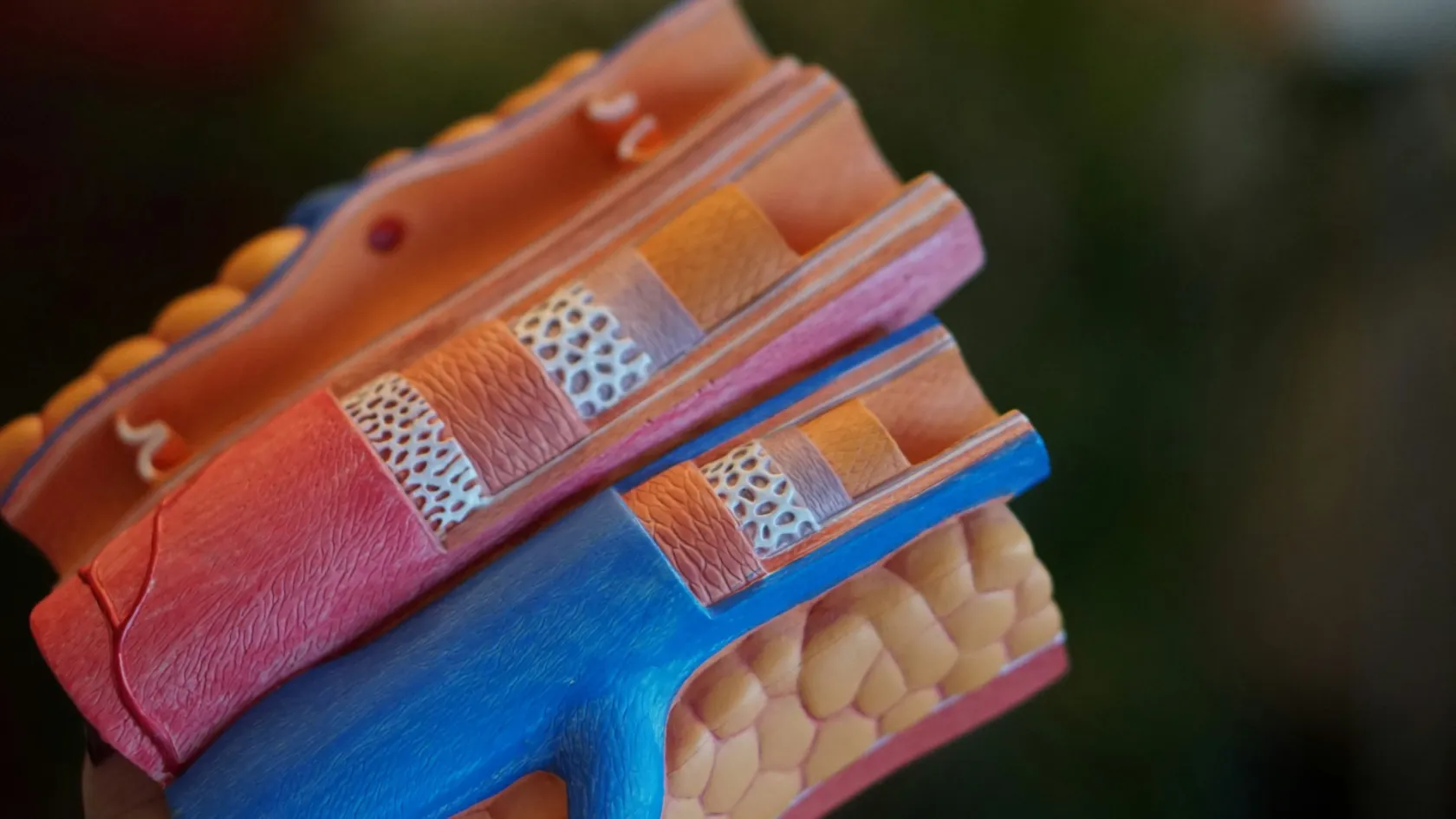
TEVAR - Thoracic Endovascular Aortic Repair
Across 11,473 thoracic stent graft procedures, 3.7 percent of patients suffered spinal cord injury and 2.1 percent were left with permanent damage. That risk has no equivalent in the abdominal version, and it should shape the whole conversation.
About This Department
Across 11,473 thoracic stent graft procedures in a national registry, 3.7 percent of patients suffered injury to the spinal cord, and in 2.1 percent it was permanent. Those patients were alive a year later 65 percent of the time, against 87 percent of everyone else. No comparable risk exists when the same treatment is done lower down in the abdomen, and it is the single fact that separates this procedure from the one people have usually read about.
Free consultation
Ask which aortic problem you have, then ask what the evidence says about that one
An aneurysm, a dissection, an injury and an ulcer are four separate conditions treated with the same device, and the published results for each are nothing like the others. Send the CT with contrast covering your chest and abdomen, any earlier scan so growth can be measured, the report of any previous aortic operation, your kidney function and your medication list. A vascular surgeon reviews the file and tells you which condition you have, whether a stent graft is the right answer for it, and what open surgery would mean instead. No fee, no obligation, and a coordinator replies in your own language.
Four diseases, one device
Find out which of those four is written on your report before you read any further about outcomes. In the registry that opens this page, the chance of spinal cord injury ranged from 1.1 percent for a traumatic tear to 5.7 percent for an intramural hematoma, which is a fivefold spread inside one procedure name.
The spinal cord
None of that makes the procedure unreasonable. It makes the extent of coverage a subject worth discussing and not a technical detail, and it explains why a surgeon may propose covering less of the aorta than the disease appears to require.
Why the chest is different
Anybody who has read about the abdominal version of this treatment arrives with a set of expectations, and three of them need adjusting, beginning with the fact that the device is delivered the same way through the arteries in the groin. After that, almost everything diverges.
The abdominal aorta feeds the kidneys, the gut and the legs, and all of those tolerate a graft sitting inside them. The chest aorta feeds the spinal cord and gives off the artery to the left arm and brain. Lining it is a decision about what you are prepared to cover.
Disease is the second difference. Most abdominal repairs treat one condition, a bulge that might burst. In the chest the same device is used for a bulge, for a tear in the lining, for an injury and for an ulcer, and the reason for treating is different every time. Reading a general statistic about thoracic stent grafting and applying it to your own diagnosis is how patients end up frightened by numbers that were never about them. The third is what the graft is trying to achieve. In an aneurysm it excludes a sac. In a dissection it covers a tear so that a false channel in the wall clots and the true channel opens back up, which is a slower, more biological result and one measured on scans over months and not settled on the day.
What can go wrong
- Spinal cord injury. Covered above, at 3.7 percent overall and permanent in 2.1 percent. The complication that defines this procedure and the one your consent conversation should spend the most time on.
- Stroke. The graft is manipulated in the arch of the aorta, close to the arteries feeding the brain, and debris can be dislodged. Risk rises the further into the arch the graft has to land, which is one of the trade-offs in deciding how much coverage to accept.
- Loss of the left arm artery. Covering it is often necessary to get a seal, and the consequences and the fix are dealt with in their own section below.
- Blood leaking into the space around the graft. Blood still reaching the aneurysm sac or the false channel despite the graft. Leaks at the sealing edges are treated, and the scans described further down exist to find them before they cause harm.
- New tearing caused by the device. Specific to dissection. The metal frame can injure a wall that is already fragile, creating a fresh entry point at either end of the graft, which is why oversizing is kept modest in dissection and generous in aneurysm.
- Trouble in the groin arteries. Thoracic devices are larger than abdominal ones, so the vessels they pass through matter more, and tearing, blockage and wound problems all occur, which is why the CT of those vessels is part of deciding whether this route works for you.
- Kidney injury from contrast. Dye is needed for the procedure and for the follow-up scans. Poor kidney function is both a risk factor for cord injury and a reason to plan the follow-up imaging differently.
Type B dissection
Dissection is where the treatment decision gets genuinely difficult, and where timing matters more than technique. Three situations are distinguished and they carry different answers.
Complicated, meaning something is already failing
Blood supply cut off to the gut, a kidney or a leg, pain that will not settle, blood pressure that cannot be controlled, or an aorta that is leaking. Here the case for covering the tear is not disputed and the alternative is an operation carrying substantially higher risk. Nobody argues about this group, and no randomized trial has ever been thought necessary, which is worth saying because the absence of a trial is sometimes presented as an absence of evidence. It is the opposite. Nobody would now consider it ethical to randomize a patient whose gut or leg is losing its blood supply to medical treatment alone.
Uncomplicated and recent
One trial randomly assigned 61 patients with a recent uncomplicated dissection either to medical treatment alone or to medical treatment plus a stent graft. At one year the aorta had remodeled far better in the treated group, with incomplete clotting of the false channel in 43 percent against 97 percent, and the overall composite outcome favored treatment. There were no deaths at thirty days in either group, one death in the treated group by a year, and three patients in the medical arm crossed over within a week because their dissection progressed. The graft changed the shape of the aorta. It has not been shown to change survival in that first year, and the trial's own authors said long-term results were needed. That is the honest summary of a small trial with a surrogate endpoint, and it is also the reason the three crossovers matter more than they look, since each one was a patient whose dissection was progressing while being managed the conservative way.
Uncomplicated and settled
A larger trial followed 140 patients with a stable dissection for five years, with 72 receiving a stent graft alongside medical treatment and 68 receiving medical treatment alone. Death from any cause was 11.1 percent against 19.3 percent, which did not reach significance. Death specifically from the aorta was 6.9 percent against 19.3 percent, and disease progression 27.0 percent against 46.1 percent. Both of those did. What the authors then showed with a look at years two to five is the finding that changed practice, since in that window nobody in the treated group died at all against 16.9 percent of the untreated. The same pattern appeared for aortic death and for disease progression, and the mechanism the authors point to is straightforward, because the false channel had clotted in 90.6 percent of the patients who received a graft.
Read that last figure with care. It came from a retrospective analysis of a randomized group, it counts only patients who had already survived two years, and the trial had 140 people in it, which means a handful of events moves it a long way. It is a real signal and it is not the same grade of evidence as a headline trial result, which is exactly why guidelines describe stent grafting in this group as reasonable and not as required, and why a surgeon who presents it as settled is overselling it. Weigh it against the alternative rather than against certainty, because the comparison group in that trial was not doing nothing either, and 19.3 percent of them had died of their aorta by five years. It is a real signal and it is not the same grade of evidence as a headline trial result, which is why guidelines describe stent grafting in this group as reasonable rather than as required.
Two things follow for a patient with a dissection. Medical treatment is not the do-nothing option, since blood pressure and heart rate control is doing real work and the trials above tested a graft added to it and not instead of it. And the decision has a clock on it, because the aorta remodels far better when the graft goes in while the wall is still fresh and pliable, and far less well once months have passed and the tissue has stiffened. A patient told their dissection is stable should still ask when the window for treating it closes, and should get an answer with a date attached to it instead of a shrug.
The left subclavian artery
Ask three questions if the plan involves covering that artery. Whether it will be rerouted, whether that happens before or during the procedure, and what your left arm and hand should feel like afterwards.
Thoracic aneurysm
Here the argument runs along familiar lines and the numbers are worth setting out in full, because a page that quotes half of them is common and misleading. Pooled data from 29 studies covering 49,972 patients compared stent grafting with open surgery for aneurysms of the descending thoracic aorta. Death around the procedure was significantly lower with the graft, at an odds ratio of 0.57. Freedom from dying of an aortic cause also favored the graft, with a hazard ratio of 0.71. Then the picture turns. Freedom from a further aortic procedure was substantially worse after grafting, with a hazard ratio of 2.10, meaning roughly twice the rate of reintervention. And when the analysis looked past the early period, patients who had open surgery had better survival from all causes beyond sixteen months, with a hazard ratio of 1.64 against the graft. Three cautions belong with that. The studies pooled were observational rather than randomized, so the patients sent for open surgery were younger and fitter as a group and the patients sent for grafting were not. Statistical heterogeneity between the studies was high, which makes the pooled figures less stable than they look. And a separate meta-analysis of 14,580 patients reported early mortality going the other way, higher after grafting at 4.4 percent against 3.2 percent, which is a direct contradiction that has not been settled. The reasonable reading is the one the authors of both papers reach. A stent graft is easier to survive in the short term for most patients, it commits you to more procedures later, and no randomized trial exists to settle the long-term question the way it was settled for the abdominal aorta.
Injury from an accident
Trauma is where this treatment has changed practice most completely, and where a patient's family is asked to absorb a decision within hours. The facts are simpler here than anywhere else on this page.
- It has become the standard approach. In a national trauma database covering 3,628 patients treated for a torn thoracic aorta, 87.9 percent received a stent graft and 12.1 percent an operation. Open repair fell from 7.4 percent of such admissions in 2007 to 1.9 percent in 2015.
- Survival favors the graft. Death occurred in 8.8 percent of graft patients and 12.8 percent of open repair patients, with open repair carrying an odds ratio for death of 1.63. Kidney injury was less common too, at 5.6 percent against 9.0 percent.
- The older pooled evidence points the same way. A review of 7,768 patients from 139 studies found mortality of 9 percent after a graft, 19 percent after open repair and 46 percent with no operation, though its authors graded the whole evidence base as very low quality and their recommendation as weak.
- Spinal cord risk is lowest here. At 1.1 percent in the large registry, well under a third of the rate seen with a dissection, because these patients are young, their arteries are otherwise healthy and the length of aorta covered is short.
- The comparison is not clean. Every one of those studies is observational, and the small number of patients still receiving open repair in recent years are the ones whose injuries or anatomy ruled out a graft. Some of the survival gap belongs to that selection and not to the treatment.
One practical note for families. Guidelines in this setting suggest repairing after other injuries have been stabilized rather than immediately, observing the most minor tears without treating them, and rerouting the left arm artery only where there is a reason to, so a plan that involves waiting a day is often the considered one.
The day itself
A planned thoracic graft takes one to three hours and is usually done under general anesthetic, since the blood pressure has to be controlled precisely at the moment the device opens and holding still matters. Preparation is where this procedure differs most from its abdominal cousin. A drain may be placed in the spinal fluid space beforehand in higher-risk cases, and lines are placed to measure blood pressure continuously so it can be dropped for a few seconds while the graft is released and then deliberately raised afterwards to protect the cord. If the left arm artery is being rerouted, that is often done first through a small incision in the neck. The graft itself goes in through a femoral artery in the groin, either by needle puncture or through a short cut, and is tracked up the aorta over a stiff wire under X-ray. Contrast shows exactly where the arch branches leave, the device is positioned against that landmark and released, and a completion picture confirms the seal and looks for any leak. Afterwards the first day is about the legs. Nurses check movement and sensation hourly, blood pressure is held high on purpose, and any weakness triggers an immediate response including raising the pressure further and draining spinal fluid if a drain is in place. Cord injury that appears late and is treated quickly recovers in a proportion of cases, which is the entire reason for that hourly routine.
Scans afterwards
Imaging after a thoracic graft is not the same task as after an abdominal one, because what is being watched depends on why the graft was put in. An aneurysm is followed for sac size and leaks. A dissection is followed for how the two channels are behaving along the whole length of the aorta, including well below where the graft ends.
This table scrolls sideways on a narrow screen. Swipe or drag to see every column.
| If you were treated for | The scans watch | Why it matters |
|---|---|---|
| An aneurysm | The diameter of the sac, any leak at either sealing edge, and whether the graft has moved. | A sac that keeps growing is the warning that the seal is not doing its job, and it produces no symptoms at all. |
| A dissection | Whether the false channel has clotted, whether the true channel has opened up, and the diameter along the whole aorta including the abdomen. | The dissection continues below the graft. That uncovered segment is where later enlargement usually shows up. |
| An injury | Graft position and shape in an otherwise healthy young aorta, at longer intervals once things are stable. | Reintervention in this group is uncommon, and the surrounding aorta is not diseased, so the burden is lighter. |
| An ulcer or wall bleeding | The treated segment and the rest of the aorta, since these conditions often signal a wall that is unwell along its length. | A second lesion appearing elsewhere is a recognized pattern and is found on a scan and not by symptoms. |
| Everybody | A scan before discharge, one at around a month, one at a year, then yearly for life. | Reintervention after a thoracic graft runs at roughly twice the open surgical rate, and it is found on imaging long before it is felt. |
Blood pressure control does as much work as any scan. A dissected or aneurysmal aorta is a diseased artery along its whole length, the graft treats one segment of it, and the medication keeping your pressure down is protecting everything the graft does not cover. Ask for your target as a number and for the reading you are actually achieving at home, because a target nobody has written down and nobody is measuring is not a treatment. Patients who buy a home monitor and keep a record are the ones whose doctors can adjust anything, and it costs less than a single scan.
When open surgery is better
- Connective tissue disorders. Marfan syndrome and its relatives leave the wall abnormal everywhere, so a graft has nothing dependable to seal against and tends to injure the tissue it presses on. Open replacement is generally preferred outside an emergency.
- Infection in the aorta. Placing foreign material into an infected field rarely ends well. The infected tissue usually has to come out.
- No landing zone. A healthy segment of aorta is needed above and below the disease. If the aneurysm reaches into the arch or down past the arteries feeding the gut and kidneys, a standard graft has nowhere to seal and the alternatives are an operation or a branched device in a specialist center.
- Arteries too narrow to deliver through. Thoracic devices are large, and the vessels in the pelvis and groin have to accommodate them. This is a common reason for turning a patient down.
- Youth, and a long life ahead. The reintervention rate and the absence of any randomized long-term comparison both weigh heavier the more decades you expect to live with the result.
Ask what your open operation would involve and what its risk would be for you specifically, and note that a unit performing both, which can say how many of each it did last year, is the one whose recommendation carries weight.
Recovery and flying home
Recovery after a planned thoracic graft is quick by the standards of aortic surgery and closely watched in a way that surprises people who expected a keyhole procedure to mean a quiet ward. The first day is the reason.
This table scrolls sideways on a narrow screen. Swipe or drag to see every column.
| Stage | What happens | What to watch for |
|---|---|---|
| First 24 hours | Monitored care with hourly checks of leg movement, blood pressure held deliberately high, any spinal drain still in place. | Weakness, heaviness or numbness in either leg. Say so immediately. Late cord injury treated fast can recover. |
| Days two to four | Drain removed, lines out, walking, eating normally, kidney function rechecked after the contrast dye. | Headache after a spinal drain, and the groin sites for swelling or a lump that pulses. |
| Leaving hospital | Usually three to five nights for a planned case, longer after a dissection or if the arm artery was rerouted. | A discharge scan and a written blood pressure target, both in your hand before you leave the building. |
| Weeks one to three | Ordinary activity returns steadily. No heavy lifting or straining for a week or so while the groin punctures seal. | A left arm that aches on use if that artery was covered. Mention it rather than assuming it is permanent. |
| Flying | Usually cleared around ten to fourteen days, once the groins are settled, kidney function has recovered and the scan has been reported. | A doctor examines you and writes that date. Take an aisle seat, walk hourly and wear compression stockings. |
Recovery from the wound is quick and the medical work is permanent. Blood pressure medication, stopping smoking and regular imaging carry on for life, because the graft has treated a segment and not the artery.
Reading a quote
No figure appears on this page, and here the reason is that the plan itself is not knowable until somebody has measured your aorta. How many graft components you need, whether an artery has to be rerouted and how many monitored nights your case requires all come out of the CT, and all three move the total substantially.
Six things to establish in writing before anybody books anything. Whether the planning CT and its specialist measurement sit inside the number or are billed separately, since that measurement is the assessment itself and it is skilled work. How many graft pieces the quote assumes, since covering a long segment can take two or three, and whether rerouting the left arm artery is included, given that it is a separate operation with its own theater time. How many nights of monitored care are budgeted and what an extra one costs, which matters more here than in most procedures because the first day is deliberately intensive. Whether a spinal drain and its management are covered. And what happens financially if the CT shows the anatomy is unsuitable and open surgery is recommended instead.
Your own file moves the total as much as the technique does. Kidney disease, an emergency instead of a planned one, previous aortic surgery and a long segment needing coverage each lengthen the expected stay.
Packages published by Turkish hospitals and medical travel agencies for aortic procedures generally include the airport transfer, pre-procedure testing, the operator and anesthesia fees, the angiography suite, the graft, the planned nights, an interpreter and the review before departure. They generally exclude flights, insurance, extra graft components, additional monitored nights, treatment of a complication and hotel stays past an agreed number. Compare what you receive against both lists.
Coming to Istanbul
Allow around three weeks for a planned case. Assessment and imaging fill the first days, the procedure occupies part of one day, three to five nights in hospital follow, and the balance is hotel recovery until somebody clears you to fly. The CT is the whole assessment, so send the images and not the report. Measurement of the landing zones, the position of the arch branches, the length of aorta that would be covered and the size of your pelvic arteries decides whether this is possible, which device is used and what your spinal cord risk is. If you have earlier scans, send those too, since the change between them tells a surgeon how fast the problem is moving. Say in your first message which condition you have been told you have, because a dissection and an aneurysm produce entirely different consultations from the first sentence onward. Your case is discussed by a vascular team, and you will be told what they concluded, how much of the aorta they plan to cover and why. One coordinator holds your file from the first message to discharge. The international patients team works in English, Arabic, French, Russian, Serbian, Romanian and Spanish, with other languages arranged on request, and a request for a female physician in your first message lets the department plan for it.
Someone can stay overnight with you on the ward, where the rooms carry a companion bed, while the monitored unit follows its own visiting rules. Hospital meals cover halal, vegetarian and diabetic diets and a prayer room is available. The international patients office books accommodation for both of you, arranges the airport transfer and daily transport, and prepares the appointment confirmation and the invitation letter naming the hospital and your treating doctor for a medical visa application, generally about ten days ahead of travel.
Take home a file instead of a discharge letter. It should name the graft system and every component with its size, state which segment of aorta was covered and whether any branch artery was covered or rerouted, carry the discharge scan images and report, record kidney function before and after, give the blood pressure target in numbers with the drugs prescribed to reach it, and set the date of the first scan at home. Address it to a vascular surgeon and book that appointment before you fly, because the years once you are back home are when a leak, a growing segment or a change below the graft would first appear. Your coordinator stays reachable on the same WhatsApp number, so a question about a groin bruise or a scan report reaches somebody holding your notes. Get in touch straight away about weakness or numbness in a leg, a lump that pulses or swelling in the groin, a fever lasting several days, a left arm that becomes painful or cold, and about sudden severe pain in the chest, back or abdomen, which needs an ambulance and not a message.
TEVAR FAQ
What is my actual risk of paralysis?
Is this the same as the abdominal procedure I have read about?
I have an uncomplicated dissection. Do I need a stent graft at all?
Will my left arm work afterwards?
Does a spinal drain prevent paralysis?
Will I need more procedures later?
My aorta was torn in an accident. Is a graft the right answer?
How long should I plan to be in Turkey?
References
- Scali ST, Giles KA, Wang GJ, Kubilis P, Neal D, Huber TS, Upchurch GR, Siracuse JJ, Shutze WP, Beck AW. National incidence, mortality outcomes, and predictors of spinal cord ischemia after thoracic endovascular aortic repair. Journal of Vascular Surgery. 2020;72(1):92-104.
- Wong CS, Healy D, Canning C, Coffey JC, Boyle JR, Walsh SR. A systematic review of spinal cord injury and cerebrospinal fluid drainage after thoracic aortic endografting. Journal of Vascular Surgery. 2012;56(5):1438-1447.
- Nienaber CA, Kische S, Rousseau H, Eggebrecht H, Rehders TC, Kundt G, Glass A, Scheinert D, Czerny M, Kleinfeldt T, Zipfel B, Labrousse L, Fattori R, Ince H. Endovascular repair of type B aortic dissection, long-term results of the randomized investigation of stent grafts in aortic dissection trial. Circulation Cardiovascular Interventions. 2013;6(4):407-416.
- Brunkwall J, Kasprzak P, Verhoeven E, Heijmen R, Taylor P, Alric P, Canaud L, Janotta M, Raithel D, Malina M, Resch T, Eckstein HH, Ockert S, Larzon T, Carlsson F, Schumacher H, Classen S, Schaub P, Lammer J, Lonn L, Clough RE, Rampoldi V, Trimarchi S, Fabiani JN, Bockler D, Kotelis D, von Tenng-Kobligk H, Mangialardi N, Ronchey S, Dialetto G, Matoussevitch V. Endovascular repair of acute uncomplicated aortic type B dissection promotes aortic remodeling, 1 year results of the ADSORB trial. European Journal of Vascular and Endovascular Surgery. 2014;48(3):285-291.
- Liu J, Gou D, Xu K, Lu Z, Li P, Lei Y, Wang Y, Yang Y, Liu S, Zhu G. Comparison of short-term and long-term outcomes between endovascular and open repair for descending thoracic aortic aneurysm, a systematic review and meta-analysis. International Journal of Surgery. 2025;111(3):2662-2674.
- Chen X, Wang J, Premaratne S, Zhao J, Zhang WW. Meta-analysis of the outcomes of revascularization after intentional coverage of the left subclavian artery for thoracic endovascular aortic repair. Journal of Vascular Surgery. 2019;70(4):1330-1340.
- Grigorian A, Spencer D, Donayre C, Nahmias J, Schubl S, Gabriel V, Barrios C. National trends of thoracic endovascular aortic repair versus open repair in blunt thoracic aortic injury. Annals of Vascular Surgery. 2018;52:72-78.
- Lee WA, Matsumura JS, Mitchell RS, Farber MA, Greenberg RK, Azizzadeh A, Murad MH, Fairman RM. Endovascular repair of traumatic thoracic aortic injury, clinical practice guidelines of the Society for Vascular Surgery. Journal of Vascular Surgery. 2011;53(1):187-192.
Editor's note
Written by the Biruni Hospital medical editorial team. Reviewed by Op. Dr. Mohammed N.I. SKAIK, Cardiovascular Surgery.
Medically reviewed by
Op. Dr. Mohammed N.I. SKAIK
Cardiovascular Surgery
Related Treatments
View All-
Aortic Aneurysm Repair - Aortic Aneurysm Surgery
Aortic aneurysm repair prevents rupture, which killed 37 percent of patients in emergency surgery against 6 percent in planned repair in a large screening trial. This guide explains when to watch and when to repair, size thresholds for men and women, EVAR against open surgery, long-term trial results, recovery and the stay in Istanbul.
-
Aortic Valve Repair
Being told the aortic valve leaks is usually followed by a conversation about which prosthesis to have. For a large group of patients there is a third answer, which is keeping the valve they were born with, and this page explains who qualifies.
-
Aortic Valve Replacement
The catheter valve wins the first year and the five-year data show the two routes ending up level. This page sets out what that means for a patient of 60 as opposed to a patient of 85, and which prosthesis belongs in each of them.
-
Coronary Angioplasty
In 5,179 patients with stable coronary disease, opening the narrowings did not reduce heart attacks or deaths. In somebody having a heart attack, the same procedure saves lives. Almost every argument about stents comes from confusing those two situations.
-
Endovascular Aneurysm Repair - EVAR
Three randomized trials compared keyhole aneurysm repair with open surgery and disagreed about long-term survival. They agreed on something else, and it is the part that decides how you will spend the next twenty years.