
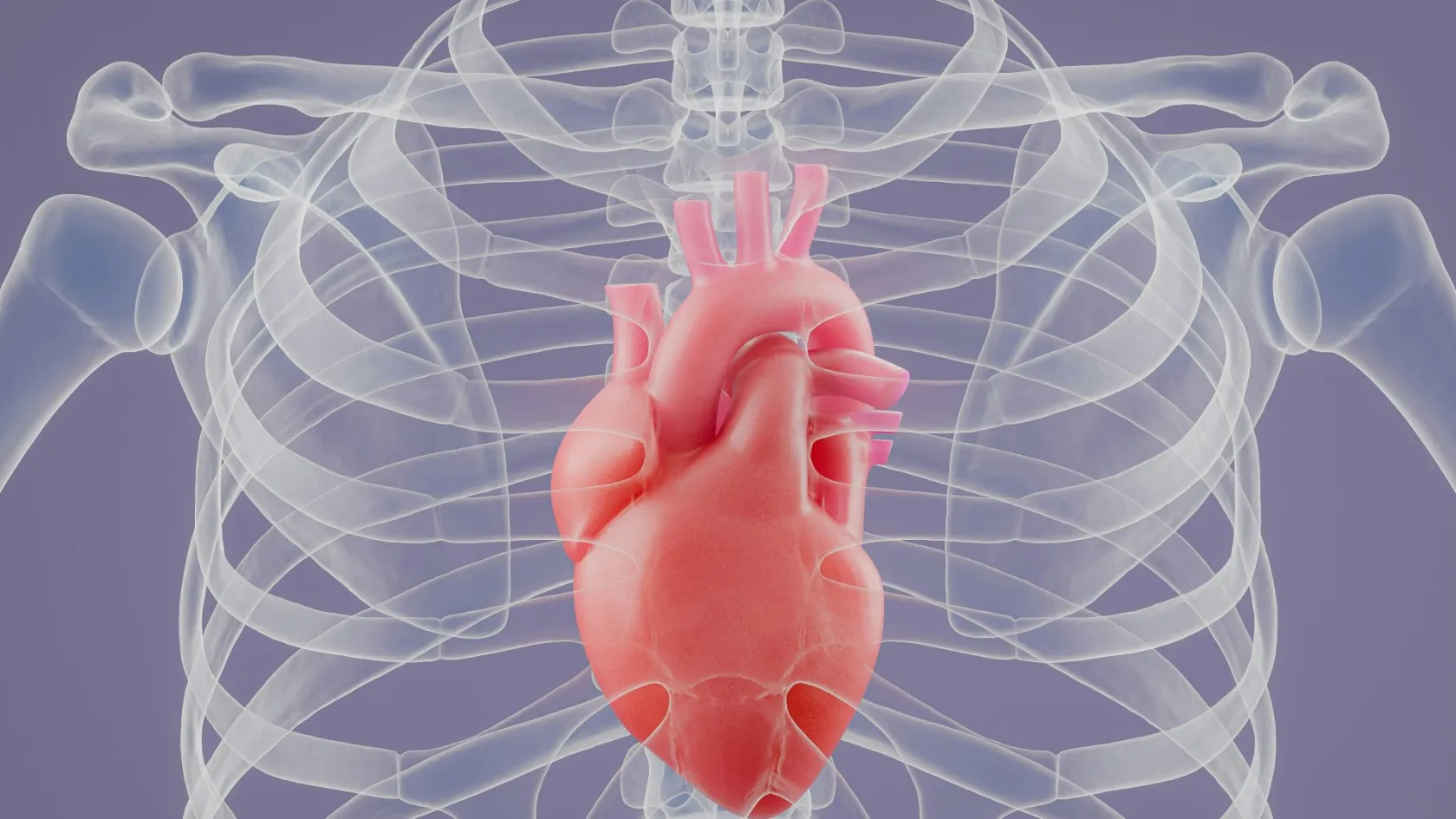
Heart Valve Surgery
Repair and replacement gave the same survival in a randomized trial. Only durability separated them, and the leak returned in 32.6 percent against 2.3 percent.
About This Department
Repair and replacement gave exactly the same survival. The leak came back in one repaired valve out of three.
A randomized trial gave 251 patients with a severe leaking mitral valve either a repair or a replacement. Survival came out the same. The leak, however, came back in 32.6 percent of the repaired valves within a year and in 2.3 percent of the replaced ones.
What the valves do, and how they fail
Four valves keep blood moving one way through the heart, and each is a set of thin leaflets that open under pressure and fall shut behind it. Two of them carry almost the entire burden of valve disease in adults. The aortic valve sits at the exit from the main pumping chamber, and the mitral valve sits between the upper and lower chambers on the left side. Between them they account for the overwhelming majority of valve operations performed anywhere.
Valves fail in exactly two ways, and the distinction runs through everything that follows.
A valve can narrow, which surgeons call stenosis, so the heart has to generate far more pressure to push blood through an opening that has stiffened and calcified. Or it can leak, which is called regurgitation, so a fraction of each beat travels backward and the heart moves the same blood twice. Narrowing makes the muscle thicken against the resistance. Leaking makes the chamber stretch to hold the extra volume. Both eventually exhaust the heart, but they do it on different timescales and they announce themselves differently. Breathlessness on the stairs, chest tightness on exertion and a blackout during exercise all point at a valve. The symptom that matters most is the one people dismiss. Fatigue that has crept up over two years, and that the person explains away as age, is the commonest way severe valve disease presents, and it is the reason an echocardiogram belongs in the workup of unexplained tiredness in anyone past middle age. That tiredness is the signal. Once symptoms appear in severe aortic stenosis the outlook without treatment changes sharply, which is the moment heart valve surgery stops being something to weigh up at leisure over the coming year and becomes a decision with a clock attached to it, and that shift catches people who have spent months being told their murmur was nothing to worry about.
Aortic and mitral are two different problems
Patients arrive having read about valve surgery in general. Surgeons think about the two valves as separate diseases with separate answers, and the gap between those two framings causes most of the confusion in a first consultation.
Narrow screens scroll this table sideways. Swipe or drag to reach every column.
| Aortic valve | Mitral valve | |
|---|---|---|
| Usual failure | Narrowing, from calcium laid down over decades or on a valve born with two leaflets instead of three | Leaking, from leaflet tissue that has stretched and prolapsed, or from a heart attack that pulled the valve out of shape |
| What the tissue looks like | Rigid, thickened, often bone-hard with calcium. There is nothing left to reshape | Frequently soft and intact, with a specific segment that has failed while the rest still works |
| Standard answer | Replacement, whether through the chest or by catheter | Repair wherever the anatomy allows it, replacement when it does not |
| Timing pressure | Symptoms change the outlook quickly, so waiting has a measurable cost | Chamber size and pump function on serial scans drive the timing, sometimes before symptoms arrive |
| Where the unit matters most | Choosing between surgery and a catheter valve for your particular age and anatomy | Whether the surgeon can actually repair it, which varies more between surgeons than any other question on this page |
The question that separates units
Ask any surgeon proposing to operate on your mitral valve what proportion of degenerative mitral valves they repair, and ask when that judgment is made. Before the operation is the answer you want. A team that repairs most of them is describing a different skill set from a team that replaces most of them, and the difference will not appear in any brochure. The aortic valve asks a gentler version of the same question, because replacement is the answer either way, and the real decision is which route and which prosthesis.
Repair, or replace
Repairing a valve means keeping the patient's own tissue and correcting what has gone wrong with it, usually by removing a redundant piece of leaflet, replacing broken cords with artificial ones, and fitting a ring around the valve opening to hold the new shape. Replacing it means cutting the valve out and sewing a prosthesis into its place. Repair keeps the architecture of the chamber intact, spares you a prosthesis, and, in degenerative mitral disease, spares you lifelong blood thinning as well, which together explain why a surgeon who can repair your valve will almost always argue for doing so. Where it works, it is the better operation. Nobody seriously disputes that.
Where it works. Those three words carry the whole section.
According to PubMed, a randomized trial assigned 251 patients with severe mitral regurgitation caused by previous heart attacks to either repair or chordal-sparing replacement. At twelve months the chambers had remodeled to the same degree, deaths ran at 14.3 percent after repair and 17.6 percent after replacement with no significant difference, and quality of life measured the same. What did differ was durability. Moderate or severe leak had returned in 32.6 percent of the repaired valves against 2.3 percent of the replaced ones (Acker et al, 2013).
Replacement provided a more durable correction of mitral regurgitation, but there was no significant between-group difference in clinical outcomes.The investigators' own conclusion, in a trial designed to prove repair superior and honest enough to publish that it had not.
Read that result narrowly, because it is narrow. These were valves damaged by heart attacks, where the leaflets themselves are healthy and the problem lies in a heart muscle that has changed shape around them. Repairing the valve does not repair the muscle, so the geometry pulls the repair apart again. In degenerative disease, where a segment of leaflet has prolapsed and the muscle is normal, repair is durable and remains the standard of care. The lesson is not that repair fails. It is that the cause of the leak, and not the word repair, is what predicts whether the correction lasts, and any surgeon offering you a repair should be able to say which of those two situations you are in and what their own recurrence rate looks like in it.
Mechanical or tissue, and what you are really choosing
Once replacement is decided, one question remains and patients are usually given it as a choice between a valve that lasts forever and a valve that does not. That framing is wrong. The error matters.
Columns run past the edge on a small screen. Slide the table across to read them.
| Mechanical | Tissue | |
|---|---|---|
| Made from | Carbon and metal, engineered to outlast the patient | Treated animal tissue, usually cow or pig, mounted on a frame |
| Blood thinning | Warfarin for life, with regular blood tests and a target range that food, illness and other medicines all disturb | Usually none beyond an early period, unless another condition demands it |
| Second operation | Unlikely for wear, though possible for clot or infection | Expected in a younger patient, since the tissue stiffens and calcifies over years |
| Sound | An audible click, which some people stop noticing within weeks and others never do | Silent, always |
| Pregnancy | Complicated, because warfarin carries risk to the fetus and the alternatives are harder to manage | Considerably simpler, which is why a tissue valve is often chosen for a woman planning a family |
The survival question has been studied directly, and the answer surprises people. According to PubMed, a single-center analysis matched 527 pairs of patients receiving mechanical and tissue aortic valves at an average age of about 54, and followed the whole cohort of 6143 patients. In-hospital complications and deaths were the same. By fourteen years, 32 percent of the tissue valve group had undergone a second operation against 8 percent of the mechanical group, and yet risk-adjusted survival at eighteen years came out at 60 percent for tissue valves and 58 percent for mechanical ones. Survival five years after a reoperation ran at 85 percent, so the second operation, when it came, did not shorten life (Attia et al, 2022). The authors concluded that the prospect of reoperation on its own should not rule out a tissue valve in a younger patient. So the real trade is not length of life. It is a choice between a daily anticoagulant and its restrictions on one side, and a likely second operation in your fifties or sixties on the other, and reasonable people choose differently once they understand that both roads lead to roughly the same place. Guidelines lean mechanical below about 50 and tissue above about 65, and the decade and a half in between stays a conversation in which your own circumstances carry more weight than any threshold, because two patients of the same age can reach opposite conclusions and both be right. Age, plans for pregnancy, how far you live from a clinic that can monitor warfarin reliably, and how you feel about the prospect of returning to an operating theater in fifteen years all belong in it.
TAVI, and who it is actually for
Transcatheter aortic valve implantation threads a collapsed valve up an artery in the groin and opens it inside the diseased one, crushing the old leaflets aside. No chest incision, no heart-lung machine, and for many patients no intensive care night. It began as a rescue for people too frail for surgery and has moved steadily into lower risk groups, and it is now the reason a great many patients arrive asking why they should have an operation at all.
Drag the table sideways where a phone cuts off the last column.
| Outcome | Which way it points |
|---|---|
| Death, any cause | No overall difference at 30 days, one year or five years |
| Stroke | No overall difference at any of those time points |
| Favors the catheter route | Less major bleeding, less kidney injury, less new atrial fibrillation, and about three days less in hospital |
| Favors surgery | Fewer permanent pacemakers, fewer major vascular complications at the access site |
| Still open | How the catheter valve behaves past ten years, since most long-term data come from older, higher risk patients |
That pooled picture comes from a systematic review of nine randomized trials, which found no overall change in the risk of death or stroke while several other outcomes moved in opposite directions, and which concluded that neither approach is dominant and that the decision belongs to the individual patient (Swift et al, 2021), according to PubMed. That is an unusually honest conclusion for a field this commercially active. Hold onto it when a clinic presents the catheter route as simply the modern answer.
The practical reading is this. Where life expectancy is measured in years rather than decades, the catheter route offers the same survival with a far gentler recovery and the durability question never becomes relevant. Where a patient is in their fifties or sixties with a normal life ahead, that question becomes the whole argument, and it is why surgical replacement remains the standard for younger patients in most guidelines. A hospital that recommends one route without discussing the other, or without a heart team that includes both a surgeon and an interventional cardiologist, is describing its own organization. Your valve has not entered into it.
Opening the chest, or opening less of it
Surgical valve operations reach the heart in one of three ways, and the choice affects the scar, the recovery and, according to the pooled evidence, the risk of the operation itself.
Three routes to the same valve
A full sternotomy divides the breastbone along its length, gives complete access to every structure in the chest, and remains the route a surgeon takes whenever the plan involves more than one valve, a bypass alongside the valve, or an aorta that has to be replaced at the same sitting. A mini-sternotomy divides only the upper portion of it, so the lower breastbone and every rib attached to that lower section stay whole. A right mini-thoracotomy skips the bone entirely and works between the ribs on the right side of the chest. All three use the heart-lung machine, all three stop the heart to open it, and all three fit the same prostheses. What changes is how much of the chest wall has to heal afterward.
What the pooled comparison found
According to PubMed, a network meta-analysis brought together 42 studies and 14925 patients, restricted to randomized trials and propensity matched series, to compare the three routes for aortic valve replacement. Operative mortality was significantly lower with mini-sternotomy than with full sternotomy, at a risk ratio of 0.60, and lower again than with right mini-thoracotomy, at 0.51. Right mini-thoracotomy carried a higher risk of returning to theater for bleeding than mini-sternotomy, at a risk ratio of 1.65. Hospital stay was shorter with mini-sternotomy by about 0.89 days. By one year, mortality was the same across all three (Ogami et al, 2022). Two things follow. The smallest incision is not automatically the safest one, and the middle option carried the best early numbers in this analysis. And by a year the differences have washed out, so the choice governs your first few weeks and nothing beyond them. Anatomy limits the choice anyway. A patient needing a valve and a bypass at the same time, or work on two valves, is having a full sternotomy, and a surgeon who says so is being straight with you.
Theater day and the first nights
An isolated valve operation runs about three to five hours from anesthetic to closing, and the parts that frighten people are the parts that are most routine.
What the first two nights are like
Waking happens in intensive care, and the breathing tube usually comes out within a few hours of arriving there. Drains in the chest, a urinary catheter, temporary pacing wires taped to the skin and several monitoring lines all count as ordinary, and most of them come out within two days. One night in critical care covers an uncomplicated case, with a further four to six on the ward. Sitting out of bed on the first day and walking the corridor on the second sound like small achievements and are the two things that most reduce chest infections and clots, so the physiotherapy team will be insistent about both. Breathing exercises hurt after any sternotomy and doing them anyway is the treatment.
What can go wrong
Valve surgery in an otherwise well patient is among the more predictable operations in cardiac surgery, and the risks worth understanding are specific ones. Bleeding needing a return to theater, an irregular heart rhythm in the first days, kidney injury, chest infection and a wound that heals slowly make up most of what happens. Stroke tops the list of complications patients ask about, and it arises when calcium or air escapes into the circulation, which is why surgeons clear the calcium carefully and empty the air from the heart deliberately before restarting it.
Three problems belong to valve surgery alone.
A permanent pacemaker becomes necessary in a minority of patients, because the heart's own electrical pathway runs directly beneath the aortic valve and swelling or stitches near it can interrupt conduction. That happens more often after the catheter route than after surgery, and more often again where the valve carried heavy calcium. Infection of the new valve, called prosthetic endocarditis, is uncommon and serious, and it is the reason dental and skin infections get treated promptly for the rest of your life. And a leak around the outside of the sewing ring, distinct from a leak through the valve itself, can appear where the tissue was too calcified to hold stitches evenly, which is exactly what the check on the operating table exists to catch.
Living with a new valve
The operation ends. A set of long habits begins. Most of them are undemanding, and the two that are not deserve explaining before you consent.
If you have a mechanical valve
Warfarin becomes permanent, and you manage it by keeping a blood test called the INR inside a target range that your surgeon sets according to which valve you carry and where it sits. Too low and the valve can clot, which is an emergency. Too high and bleeding becomes the risk instead. Green vegetables, alcohol, antibiotics, painkillers and a great many herbal preparations all shift that number, so the discipline is less about avoiding foods than about keeping intake steady and telling whoever monitors you when anything changes. Home testing devices exist and suit some people very well indeed. What matters most for an international patient is arranging the monitoring before you travel, because a mechanical valve fitted abroad and an INR service organized afterward is the wrong order.
Dental care, for either valve
Any prosthetic valve raises the risk that bacteria entering the bloodstream will settle on it. Dental treatment is the classic route, so tell every dentist you see that you have a prosthetic heart valve, and have outstanding dental work done before the operation wherever the timing allows. Skin infections, boils and infected wounds deserve the same seriousness. A fever lasting more than a few days in someone with a prosthetic valve is a reason to be examined that week, and saying so plainly is more useful than a list of antibiotic regimens that vary by country.
Recovery, week by week
A divided breastbone is a broken bone that has been wired back together, and it behaves like one, which is the single fact that explains almost every restriction you will be given.
Planning the trip
Two to three weeks in the country is the realistic plan for an isolated valve operation. Two or three days at the start cover echocardiography, bloods, dental review, anesthetic assessment and the consultation itself, and a coronary angiogram belongs in there for anyone past middle age, since finding coronary disease after the valve plan is fixed changes the operation. The admission runs five to seven nights in an uncomplicated case. The remainder covers wound review, a repeat echocardiogram, the start of warfarin dosing where a mechanical valve was fitted, and a fitness-to-fly assessment. Flight clearance is a separate decision from discharge and a more conservative one, because cabin pressure, immobility and a healing sternum combine badly, so the surgical team owns the departure date.
What is arranged for you, and what is not
A coordinator handles the case from your first message through to discharge, and stays reachable on the same WhatsApp number after you fly home. Reviewing your echocardiogram and reports remotely comes first and costs nothing. Once a plan is agreed, an invitation letter and an appointment confirmation are issued about ten days before travel for the countries that require a medical visa, and airport and hotel transfers are arranged directly. The international patients office covers English, Arabic, French, Russian, Serbian, Romanian and Spanish, with other languages arranged on request, and it also arranges accommodation on either side of the admission. A companion bed sits in the patient rooms, which matters more here than in most operations, since the first fortnight after a sternotomy is genuinely hard to manage alone. Halal, vegetarian and diabetic diets are catered for on the ward, there is a prayer room in the hospital, and a female physician can be requested and is accommodated wherever the rota allows. Two things sit at your end and both get forgotten. A cardiologist near home who will take over follow-up, identified before you travel. And, for a mechanical valve, an INR monitoring service with an appointment already booked for the week you land.
What moves the price
Two patients told they need heart valve surgery can receive quotes that look nothing alike, and no dishonesty is required for that to happen. Which valve is being operated on, whether it is repaired or replaced, and which prosthesis goes in are the three largest levers, and a mechanical valve, a stented tissue valve and a catheter valve occupy different price categories entirely. Operating on two valves in one sitting, or adding a coronary bypass to a valve operation, lengthens theater time and intensive care alike. The route into the chest carries its own consumables, since minimally invasive access uses specific instruments and cannulas that a full sternotomy does not. Time on the heart-lung machine, the number of intensive care nights, the blood products a case consumes and the histology and microbiology on anything removed all sit inside the total in ways that vary case by case. Patient factors move it too, among them kidney function, lung disease, previous cardiac surgery and how well the pump is working before anyone starts. Published packages in this market ordinarily cover surgeon and anesthesiologist fees, theater time, an agreed number of intensive care and ward nights, pre-operative imaging and blood work, the prosthesis itself, and transfers with interpreting. Flights, accommodation past the stated nights, treatment of a complication, a return to theater and any pacemaker implanted afterward sit outside them by default.
Back home, and the follow-up
Carry the operative note naming exactly which valve was treated and what was done to it, the make, model and size of any prosthesis with its implant card, the discharge summary listing every medication and its dose, your INR target where one applies, and a copy of the echocardiogram performed before discharge. That implant card is not a souvenir. A cardiologist reading a scan in ten years needs to know which valve sits in there and what its normal gradients look like. Without the card, that reading becomes guesswork, and a patient who cannot name their own prosthesis has been let down by the paperwork.
Who does what, once you are home
Follow-up belongs to a cardiologist near where you live, beginning with a review at four to six weeks and an echocardiogram that becomes the baseline against which every later scan gets measured, so it matters that the images themselves travel home with you and not merely a report describing them. After that, annual review is the usual pattern for a mechanical valve and for a tissue valve once it passes about five years, with earlier scans if a new murmur, breathlessness or reduced exercise tolerance appears. INR monitoring runs on its own schedule alongside all of it. Remote follow-up with the operating team earns its keep through the first months, particularly for a wound that is slow to settle or a sternum that clicks, and here the coordinator who handled your case stays on the same WhatsApp number after you fly home, with reports and imaging produced by your own doctors reviewed at no charge.
One instruction outlasts every other. A fever that persists for more than a few days, in anyone carrying a prosthetic valve, needs blood cultures taken before antibiotics are started, and telling that to a doctor who does not know your history could matter more than anything else in this article.
The review costs nothing and commits you to nothing. Send the echocardiogram images themselves rather than only the written report, since the mechanism of the leak or the narrowing is what decides between repair and replacement, along with any CT or coronary angiogram, and a note of your age, kidney function, previous chest surgery and current blood thinners. A cardiac surgeon will tell you what your valve actually needs.
Frequently asked questions
Can my valve be repaired instead of replaced?
For a mitral valve leaking because a segment of leaflet has prolapsed, usually yes, and repair is the preferred operation where the anatomy allows it. For a mitral valve leaking because a heart attack changed the shape of the muscle around it, repair is possible but the leak returned in 32.6 percent of cases within a year in a randomized trial, against 2.3 percent after replacement. For a calcified aortic valve, no. There is nothing left to reshape, and replacement is the answer whichever route is used to deliver it.
Should I choose a mechanical or a tissue valve?
Below about 50 the guidelines lean mechanical, above about 65 they lean tissue, and the years in between are a genuine conversation. The decision is not about how long you will live. In a matched analysis of patients averaging 54 years old, eighteen year survival came out at 60 percent with a tissue valve and 58 percent with a mechanical one, while second operations ran at 32 percent against 8 percent by fourteen years. You are choosing between lifelong warfarin and a likely second operation, and both roads reach a similar place.
Why would anyone have surgery when TAVI exists?
Because the durability question is unanswered for younger patients. Pooled randomized data show no difference in death or stroke between the two routes, with less bleeding and a shorter stay after the catheter route and fewer pacemakers after surgery. But at five years, mild or worse leak around the valve was present in 24.5 percent of catheter valves against 6.3 percent of surgical ones, and nobody yet knows what that looks like at twenty years. Where life expectancy is long, that uncertainty is the whole argument.
Is a smaller incision safer?
Not automatically. In a network meta-analysis of 42 studies and 14925 patients, mini-sternotomy had lower operative mortality than both full sternotomy and right mini-thoracotomy, and right mini-thoracotomy carried more returns to theater for bleeding. By one year, mortality was identical across all three routes. The smallest incision was not the best performer, and anatomy decides the question anyway, since a second valve or a bypass in the same operation means a full sternotomy.
When can I fly after heart valve surgery?
Two to three weeks in the country covers an isolated operation, with two or three days for assessment, five to seven nights as an inpatient, and the balance for wound review, a repeat echocardiogram and a fitness-to-fly check. Clearance depends on the wound, on how you are moving and, for a mechanical valve, on the INR sitting stably in range. It is decided at a review before departure rather than promised when the trip is booked.
Will I need a pacemaker?
A minority of patients do, because the electrical pathway of the heart runs immediately beneath the aortic valve and swelling or stitches near it can interrupt conduction. The risk is higher after the catheter route than after surgery, and higher again where the valve was heavily calcified. Temporary pacing wires are placed during every operation as a precaution and removed within days when they are not needed.
References
- Acker MA, Parides MK, Perrault LP, et al. Mitral-valve repair versus replacement for severe ischemic mitral regurgitation. N Engl J Med. 2014;370(1):23-32.
- Hahn RT, Ternacle J, Silva I, et al. 5-year echocardiographic results of transcatheter versus surgical aortic valve replacement in low-risk patients. JACC Cardiovasc Imaging. 2025;18(6):625-640.
- Attia T, Yang Y, Svensson LG, et al. Similar long-term survival after isolated bioprosthetic versus mechanical aortic valve replacement. A propensity-matched analysis. J Thorac Cardiovasc Surg. 2022;164(5):1444-1455.
- Ogami T, Yokoyama Y, Takagi H, et al. Minimally invasive versus conventional aortic valve replacement. The network meta-analysis. J Card Surg. 2022;37(12):4868-4874.
- Swift SL, Puehler T, Misso K, et al. Transcatheter aortic valve implantation versus surgical aortic valve replacement in patients with severe aortic stenosis. A systematic review and meta-analysis. BMJ Open. 2021;11(12):e054222.
Editor's note
Written by the Biruni Hospital medical editorial team. Reviewed by Assoc. Prof. Dr. Muhammed BAYRAM, Cardiovascular Surgery.
Medically reviewed by
Assoc. Prof. Dr. Muhammed BAYRAM
Cardiovascular Surgery
Related Treatments
View All-
Aortic Aneurysm Repair - Aortic Aneurysm Surgery
Aortic aneurysm repair prevents rupture, which killed 37 percent of patients in emergency surgery against 6 percent in planned repair in a large screening trial. This guide explains when to watch and when to repair, size thresholds for men and women, EVAR against open surgery, long-term trial results, recovery and the stay in Istanbul.
-

Aortic Valve Repair
Being told the aortic valve leaks is usually followed by a conversation about which prosthesis to have. For a large group of patients there is a third answer, which is keeping the valve they were born with, and this page explains who qualifies.
-
Aortic Valve Replacement
The catheter valve wins the first year and the five-year data show the two routes ending up level. This page sets out what that means for a patient of 60 as opposed to a patient of 85, and which prosthesis belongs in each of them.
-
Coronary Angioplasty
In 5,179 patients with stable coronary disease, opening the narrowings did not reduce heart attacks or deaths. In somebody having a heart attack, the same procedure saves lives. Almost every argument about stents comes from confusing those two situations.
-
Endovascular Aneurysm Repair - EVAR
Three randomized trials compared keyhole aneurysm repair with open surgery and disagreed about long-term survival. They agreed on something else, and it is the part that decides how you will spend the next twenty years.