
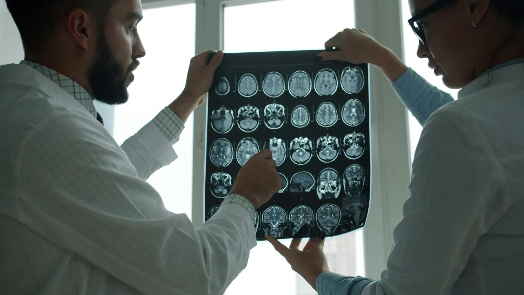
Spinal Cord Tumor Surgery
Three different operations hide behind one diagnosis, and where the tumor sits decides which one you are having. This page sets out the three compartments a spinal tumor can occupy, what the monitoring electrodes are listening for while the cord is open, why a surgeon who stops short is sometimes protecting your legs, and why the condition you arrive in predicts your result better than anything else.
About This Department
Where the tumor sits matters more than what it is called. Inside the cord, the monitor decides.
Three different operations hide behind one diagnosis, and the address of the tumor selects which one you are having. This page sets out how surgeons tell them apart, what the electrodes in the operating room are listening for, and why stopping short is sometimes the right call.
What a spinal cord tumor is
Your spinal cord is a cable roughly the thickness of a finger, running down a bony tunnel from the base of the skull to around the bottom of the ribs. It carries every instruction your brain sends to your legs and every signal your body sends back. A tumor anywhere near that cable causes trouble by pressing on it, by growing through it, or by both.
Space is the scarce thing here.
Around the cord sits a tough sleeve called the dura, and the whole assembly sits inside the spinal canal with almost no room to spare, so a growth of two centimeters in the spine does damage that the same growth would not do in the abdomen. Most of these tumors are benign in the sense that pathologists use the word. That word does less reassuring work than patients expect, because a benign tumor pressing on the cord still produces weakness, numbness and bladder trouble, and because the cord recovers from pressure slowly and incompletely once the damage has been done. So the clock matters more than the pathology in many of these cases, and the questions that decide an operation turn on location and timing instead of on how dangerous a cell looks under a microscope.
The address decides the operation
Patients arrive having read their pathology report and wanting to discuss the tumor type. Surgeons want to discuss where it sits. Those two conversations feel like the same conversation and are not, because the surgical difficulty, the risk of paralysis, the chance of a complete removal and the recovery timetable are all governed by the address first and the histology second. Address first.
A meningioma beside the cord and a meningioma inside it are different operations.
The second one barely exists, which is the point. Certain tumor types live at certain addresses, so the address and the likely diagnosis travel together, and a radiologist who says the lesion is intramedullary has already narrowed the list to three or four possibilities and set the risk level of the operation before anybody has seen a slide. That explains why the rest of this page runs by address, and why the first thing we look at in a remote review is the position of the lesion relative to the dura and the cord rather than the report that came with it.
Three addresses, three operations
Every spinal tumor falls into one of three compartments, and the names sound technical until you picture the anatomy, at which point they become obvious.
Roughly speaking, the deeper the address, the harder the operation and the more the outcome depends on things nobody controls, such as whether a clean boundary exists between the tumor and the tissue around it. Depth sets difficulty. Everything downstream follows from that single fact.
Symptoms, and why they get missed
Pain comes first in most of these patients. A study of 203 people operated on for tumors beside the cord found pain as the opening symptom in 63 percent, and pain in the back is the most common complaint in medicine, which is precisely the problem. That is the trap.
The features that separate this pain from ordinary back pain
Night pain that wakes somebody from sleep, or pain that is worse lying down than standing, runs against the pattern of mechanical back pain and deserves a scan, and pain in a band around the chest or abdomen at one level, often described as a tight belt, points at the cord instead of at a disc. Numbness that climbs from the feet upward over weeks, clumsy hands, a foot that catches on stairs, a change in how hot water feels on one side of the body, difficulty starting or stopping urination, all of these belong on the list. Any one of them in somebody with months of back pain changes the situation from a physiotherapy referral into an imaging request. These cases rarely run late because the signs were subtle. Back pain is simply so common that the signs sitting beside it get read as part of the same story.
What the MRI can settle
MRI with contrast answers the address question almost completely, which is a great deal more than most scans manage in most diseases, since it shows which compartment the lesion occupies, how many levels it spans, whether a cyst sits above or below it, and whether the cord looks swollen around it. The address is legible.
What it cannot do is tell you what will happen in the room. The one thing that governs an intramedullary operation, whether a surgeon can find and follow a boundary between tumor and cord, does not appear reliably on any scan taken beforehand, and surgeons who tell you otherwise are describing a probability as a promise. An ependymoma carries that boundary in most cases, and complete removal follows from it. An astrocytoma does not, and complete removal does not follow. Yet the two can look similar enough on an MRI that the honest position before surgery is a ranked list of possibilities and a plan for each, and the actual answer arrives twenty minutes after the cord has been opened.
Tumors beside the cord
Tumors beside the cord do well as a group, and they also form the largest group of primary spinal tumors, with meningiomas growing from the lining, schwannomas from the sheath of a nerve root, and both of them pushing the cord aside instead of growing into it.
What a large modern series reports
Across 203 consecutive patients followed for an average of thirty months, complete removal was achieved in 84 percent, with schwannoma the commonest diagnosis at 36 percent and meningioma next at 30 percent. Tumor size did not predict the final functional result. What predicted it came down to the neurological state the patient arrived in, the tumor type, how complete the removal was, and whether a complication such as a hematoma occurred afterward, and that first item carries the practical lesson for anybody reading this with a scan in hand and an appointment in three months. Size is negotiable. Nerve tissue already squeezed for a year is not, and the ceiling a patient can reach after surgery was largely set before they arrived at the hospital door. A patient who walks into the operating room walks out of it far more often than a patient who is carried in, and the single most effective thing you can do for your own outcome in this group is to be operated on before the cord has been squeezed for another year.
Tumors inside the cord, and the plane
Small group, large reputation. Intramedullary tumors make up a small share of the total and most of the difficulty. Surgeons open the cord along its midline, in a groove between the two halves where the fewest fibers cross, and reach the tumor from within.
Everything turns on whether a boundary exists
Surgeons call it the plane. An ependymoma, which is the commonest of these in adults, typically grows as a discrete mass that displaces cord tissue and leaves a boundary a careful surgeon can develop all the way around, and in a consecutive series of 100 such tumors operated on with full monitoring, complete removal was achieved in 89. An astrocytoma infiltrates, so the tumor and the cord blend into each other and the plane simply is not there, which turns the aim from cure into biopsy and debulking, while a hemangioblastoma sits in its own category, well demarcated but fed by vessels that have to be dealt with before the mass is touched. Three tumors, three different operations, one incision that looks identical from the outside. The plane therefore forms the real subject of these operations, and the surgeon's judgment about whether they still have one, minute by minute, is what the monitoring in the next section exists to inform.
What happens on the day
You are asleep throughout, lying face down, with electrodes placed at the scalp, along the limbs and, for tumors inside the cord, on a small strip slid into the spinal canal below the level being operated on. The anesthetic team picks agents that interfere with the recordings as little as possible, a genuine constraint and not a detail.
- Baseline recordings. Every signal is measured before the incision, because everything afterward is read as a change from that starting point rather than as an absolute value.
- Exposure. A midline incision, then removal of the bony roof over the levels involved, sometimes replaced at the end as a hinged flap in younger patients.
- Ultrasound. Before the dura is opened, a probe on the surface confirms the tumor lies where the MRI said and that the exposure covers it at both ends.
- Opening. The dura is opened under the microscope. For a tumor beside the cord the work starts here. For one inside it, the cord itself is opened next along the midline groove.
- Resection. The tumor is removed from the inside outward, staying on the boundary, with the monitoring running continuously and the surgeon pausing whenever a signal moves.
- Closure. A watertight dural closure, the bone replaced or left off according to the plan, and the wound closed in layers over a drain.
Those recordings repay a little attention, because they are what a family is really buying when it chooses a center for this operation. Three signals run at once, and they do not say the same thing.
This table scrolls sideways on a narrow screen. Swipe or drag to see every column.
| Signal | What it watches | What it predicts |
|---|---|---|
| Sensory potentials | The columns at the back of the cord that carry position sense | Sensory change, and a decline alone is no reason to abandon an operation |
| Muscle motor potentials | The whole motor pathway down to the muscle, tested in bursts | Weakness in the first days after surgery, and short term recovery |
| The D-wave | The main motor tract directly, from a strip electrode in the canal | Long term walking, and it is the signal a surgeon protects above all |
| All three together | Read as a pattern, since each covers what the others miss | Postoperative deficits better than any single signal on its own |
Cancer that has spread to the spine
Most tumors in the spine are not primary tumors at all. Most of them arrive as deposits from a cancer that started in the breast, the lung, the prostate or the kidney, they sit in the bone outside the dura, and when one of them presses hard enough on the cord it becomes an emergency measured in hours.
The trial that put surgery back in the pathway
Radiotherapy alone was the standard for years. Then 101 patients with cord compression from metastatic cancer were randomized to radiotherapy alone or to surgery followed by the same radiotherapy, and the trial was stopped early because the difference was large enough to meet a predefined rule. The gap was not small.
This table scrolls sideways on a narrow screen. Swipe or drag to see every column.
| Measure | Surgery then radiotherapy | Radiotherapy alone |
|---|---|---|
| Able to walk after treatment | 42 of 50, or 84 percent | 29 of 51, or 57 percent |
| Median time still walking | 122 days | 13 days |
| Regained walking, having arrived unable | 10 of 16, or 62 percent | 3 of 16, or 19 percent |
| Steroids and strong painkillers | Significantly lower requirement | Higher requirement |
Worse before better
One part of intramedullary surgery belongs in the conversation before the operation and not after it.
Day one is not the answer
A great many patients wake from these operations weaker than they went in. One series recorded persistent attenuation of the motor recordings during surgery in 71 percent of cases, and a large share of those patients had a new deficit in the first days, and most of them then improved over the following months. The monitoring alarms readily because the cord tolerates handling badly in the short term and recovers from it well in the long term, which is exactly why the signal that predicts the first week is a different signal from the one that predicts the first year. Studies that followed the same patients over time found the short term predictive value of the motor recordings falling away while their ability to identify the people who would be fine kept rising. In plain terms, a patient who is weaker on day one can be walking normally at twelve months, and a surgeon who is unbothered by a new deficit at the bedside on the first morning is reading the recordings and not being glib.
What surgery does not fix
Removing the tumor removes the cause. Two accounts run here. Damage already done to the cord is the second one, and it settles slowly and rarely in full.
The four things that tend to persist
Numbness and altered sensation outlast the weakness, since the midline opening in an intramedullary operation passes between the very columns that carry position sense, and bladder and bowel function that was already impaired before surgery improves less reliably than leg strength, which is the single hardest fact to hear when deciding whether to wait another six months. Bladder trouble is the warning shot. Neuropathic pain, the burning or electric kind that comes from the cord itself and not from the back, can persist or even begin after a technically perfect removal, and it answers to medication and time, never to more surgery. And the spine that was opened needs watching, because removing bone over several levels in a young patient can lead to a curve developing years later. None of this argues against operating. It argues for naming the target before the operation, so that a good result is recognized as a good result when it arrives.
Risks and complications
Risk in this operation is dominated by the address, so a single figure covering all spinal cord tumors would be meaningless. What follows separates them.
The complications specific to opening the spineCerebrospinal fluid leaking through the wound, which shows as clear fluid or a soft swelling under the skin and sometimes needs an extra stitch or a temporary lumbar drain for a few days. A collection of blood in the operative bed, which is rare and needs the operating room the same hour, since pressure on a cord that has just been handled is poorly tolerated. Wound infection, managed with antibiotics and occasionally with a return to theater. Instability or a developing curve of the spine after bone has been removed over several levels, which is why the bony roof is replaced as a hinged flap in children and younger adults wherever that is practical.
The risk that belongs to the decision rather than the operation
Operating too late is the commonest way these cases go wrong, and it rarely appears in a complication table because nothing went wrong in the room, since a patient watched for eighteen months while a meningioma quietly flattened the cord arrives for a technically excellent operation and then recovers to a worse ceiling than the one they would have reached a year earlier. The prognostic studies say the same thing every time, in the same words. The strongest predictor of the final result is the condition the patient was in beforehand.
Questions worth asking any surgeon
Ask all five. Each has a short factual answer, and anybody who does this work regularly can give all of them without leaving the room.
- Which compartment is my tumor in, and what does that make the realistic aim, complete removal or debulking?
- Which monitoring signals will run during my operation, will a D-wave electrode be placed in the canal, and who makes that decision for my case?
- What will you do if the signals change, and at what point would you stop rather than continue?
- Who reads the monitoring, are they in the room for the whole case, and have they worked with you before?
- Will the bone be replaced at the end, and what is the plan for watching my spine over the years that follow?
The third question is the one that separates departments. An answer describing a threshold and a plan is the answer you want, and an answer promising that the signals never change is not an answer at all.
Recovery and rehabilitation
Operating time runs from a few hours for a small tumor beside the cord to well past ten for a long intramedullary tumor with difficult anatomy. Pace varies. Direction does not. What follows runs broadly similar across the group, with the pace set by how much neurological ground has to be recovered.
A usual course after spinal cord tumor surgeryThe first night is spent under close neurological observation, with limb strength checked hourly. Sitting and standing begin on day one or two for most patients, with a longer spell lying flat where the dural closure gave trouble and the surgeon wants the repair left alone. Hospital stay runs several days for a tumor beside the cord and longer where a new deficit needs rehabilitation started before discharge. Physiotherapy begins in hospital and continues for weeks to months. Many people feel close to themselves again by six weeks, and those recovering from a genuine deficit measure progress in months instead. The wound heals long before the cord finishes its work.
Rehabilitation deserves more weight than it gets in a surgical consultation. A cord that has been decompressed or opened recovers through use, and the difference between a patient who works with a physiotherapist five times a week for three months and one who is discharged with a leaflet is visible at a year and permanent thereafter. Where a patient travels for this surgery, the rehabilitation plan has to be arranged at home before the flight out, because the weeks immediately after discharge are the ones that count most and they are the weeks a patient will be spending in their own country. Book the physiotherapist before the surgeon. A family that lands back home with a discharge letter and no appointment will lose three or four weeks to paperwork at precisely the point in the recovery when the cord is most responsive to being asked to work, and nothing later in the year buys those weeks back.
Follow up and the scans
Surgeons take the first scan within a day or two of the operation, while the operative field still looks the way they left it, and that image becomes the reference for everything afterward, so waiting a month to take it wastes its value, because by then blood and swelling have begun to change what the images show.
How often, and for how long
A completely removed benign tumor gets scanned at three months, at a year, and then at widening intervals, with the surveillance running for years and not months, because recurrences in this group arrive slowly. Ten years counts as an ordinary interval for a spinal meningioma to come back in, which is why a patient discharged from follow up at three years and told the matter is closed has been given a slightly rounder answer than the evidence supports. A tumor that was deliberately left behind is watched more closely and reviewed with a radiation oncologist, since some of these are treated with radiotherapy and some are simply followed. For a patient who has traveled, the practical arrangement is that once you are back home the scans happen there and the images come to us for comparison, which works only if the original postoperative scan traveled home with you in its raw form. We send a disc and not a report, along with the operation note describing exactly which levels were opened and what was found at the boundary, because those two documents are what any future surgeon will actually want.
Cost and having surgery in Istanbul
No price appears on this page, because the figure depends on the compartment, the number of levels and whether the spine needs fixing afterward, and a number written today would mislead you by the time you read it. Compartment first. What we can set out is how the remote assessment works, and for spinal cord tumors that assessment has to do more than usual, since the address of the lesion determines everything that follows and a good scan shows it plainly.
Send the MRI itself, with contrast, covering the whole spine and not one region, on a disc or a transfer link in its original form. Add a description of what you can and cannot do now, with dates, because the trajectory over the last six months tells a surgeon more than any single examination. Back comes a written answer naming the compartment, the realistic aim, which monitoring would be used, the expected length of stay and whether we think the operation should happen sooner than your current appointment.
Staff in the international patients office work in English, Arabic, French, Russian, Serbian, Romanian and Spanish, and interpreting for other languages is arranged before arrival. One coordinator handles your case from the first message until discharge and stays reachable on WhatsApp after you go home, which matters here because the questions that follow this operation concern rehabilitation and arrive over months. Rooms have a companion bed, hotel and transfers are arranged around the surgical dates, a female physician is available on request, and the invitation letter for a visa goes out ten days before travel. Halal, vegetarian and diabetic meals are routine, and a prayer room is open on site. Neurology, radiation oncology and physiotherapy all sit on the same campus as the operating rooms, which matters for this group in particular, since the pathology result after an intramedullary resection sometimes changes the plan and a patient who has to travel to a second institution for that conversation frequently never has it at all.
Plan on two to three weeks in the country for a straightforward case and longer where rehabilitation has to begin before you can fly. We will say which of those two you are in before you book anything, and we prefer to give the longer number early instead of discovering it with you afterward.
Send the scan of the whole spine. One region is rarely enough.
Spinal cord tumor FAQ
The questions below arrive in most first messages from abroad.
Will I be paralyzed by this operation?
Can this be done with a small incision?
My tumor is benign. Can I wait?
What is the D-wave and why does it keep coming up?
Why would a surgeon stop before removing everything?
Will I need radiotherapy afterward?
How soon can I fly home?
References
- Skrap B, Tramontano V, Faccioli F, Meglio M, Pinna G, Sala F. Surgery for intramedullary spinal cord ependymomas in the neuromonitoring era. Results from a consecutive series of 100 patients. Journal of Neurosurgery Spine. 2022;36(5):858-868.
- Tropeano MP, Rossini Z, Franzini A, Capo G, Olei S, De Robertis M, et al. Multimodal intraoperative neurophysiological monitoring in intramedullary spinal cord tumors. A 10-year single center experience. Cancers. 2023;16(1):111.
- Sala F, Skrap B, Kothbauer KF, Deletis V. Intraoperative neurophysiology in intramedullary spinal cord tumor surgery. Handbook of Clinical Neurology. 2022;186:229-244.
- Kimchi G, Knoller N, Korn A, Eyal-Mazuz Y, Sapir Y, Peled A, et al. Delayed variations in the diagnostic accuracy of intraoperative neuromonitoring in the resection of intramedullary spinal cord tumors. Neurosurgical Focus. 2021;50(5):E21.
- Patchell RA, Tibbs PA, Regine WF, Payne R, Saris S, Kryscio RJ, et al. Direct decompressive surgical resection in the treatment of spinal cord compression caused by metastatic cancer. A randomised trial. Lancet. 2005;366(9486):643-648.
- Ruella M, Caffaratti G, Saenz A, Villamil F, Mormandi R, Cervio A. Intradural extramedullary tumors. Retrospective cohort study assessing prognostic factors for functional outcome in adult patients. Neurocirugia. 2023;34(5):256-267.
- Raygor KP, Than KD, Chou D, Mummaneni PV. Comparison of minimally invasive transspinous and open approaches for thoracolumbar intradural-extramedullary spinal tumors. Neurosurgical Focus. 2015;39(2):E12.
Editor's note
Written by the Biruni Hospital medical editorial team. Reviewed by Assoc. Prof. Dr. Melih ÜÇER, Neurosurgery.
Medically reviewed by

Assoc. Prof. Dr. Melih ÜÇER
Neurosurgery
Related Treatments
View All
ALIF - Anterior Lumbar Interbody Fusion Surgery
Coming in from the front restores the curve of the lower back better than any posterior route and leaves the back muscles untouched. It also carries one risk most pages will not name, and this page names it.

Brain AVM Surgery - Arteriovenous Malformation Surgery
An unruptured brain AVM bleeds at roughly 2.2 percent a year, and surgery spends its whole risk on a single morning. This page sets those two shapes of risk side by side, explains what the ARUBA trial does and does not say about your case, and shows how a Spetzler-Martin grade changes the answer.
Brain Tumor Surgery - Brain Tumor Removal
Brain surgery is one trade repeated all day, how much tumor comes out against what the brain around it still does. This page puts the published numbers on both sides of that trade.
Cervical Microdiscectomy - Minimally Invasive Neck Disc Surgery
Neck disc surgery is done from the front, through the space between your windpipe and your carotid artery, and almost everything that goes wrong that is not the disc happens in that corridor. What has been measured in it.
Chiari Malformation Surgery
A Chiari malformation is measured in millimeters and treated according to symptoms, and those two things come apart more often than patients are told. Most people with the finding never need an operation. This page sets out who does, what posterior fossa decompression removes, why surgeons still argue over opening the dura, and what the pooled studies say about both sides of that argument.