
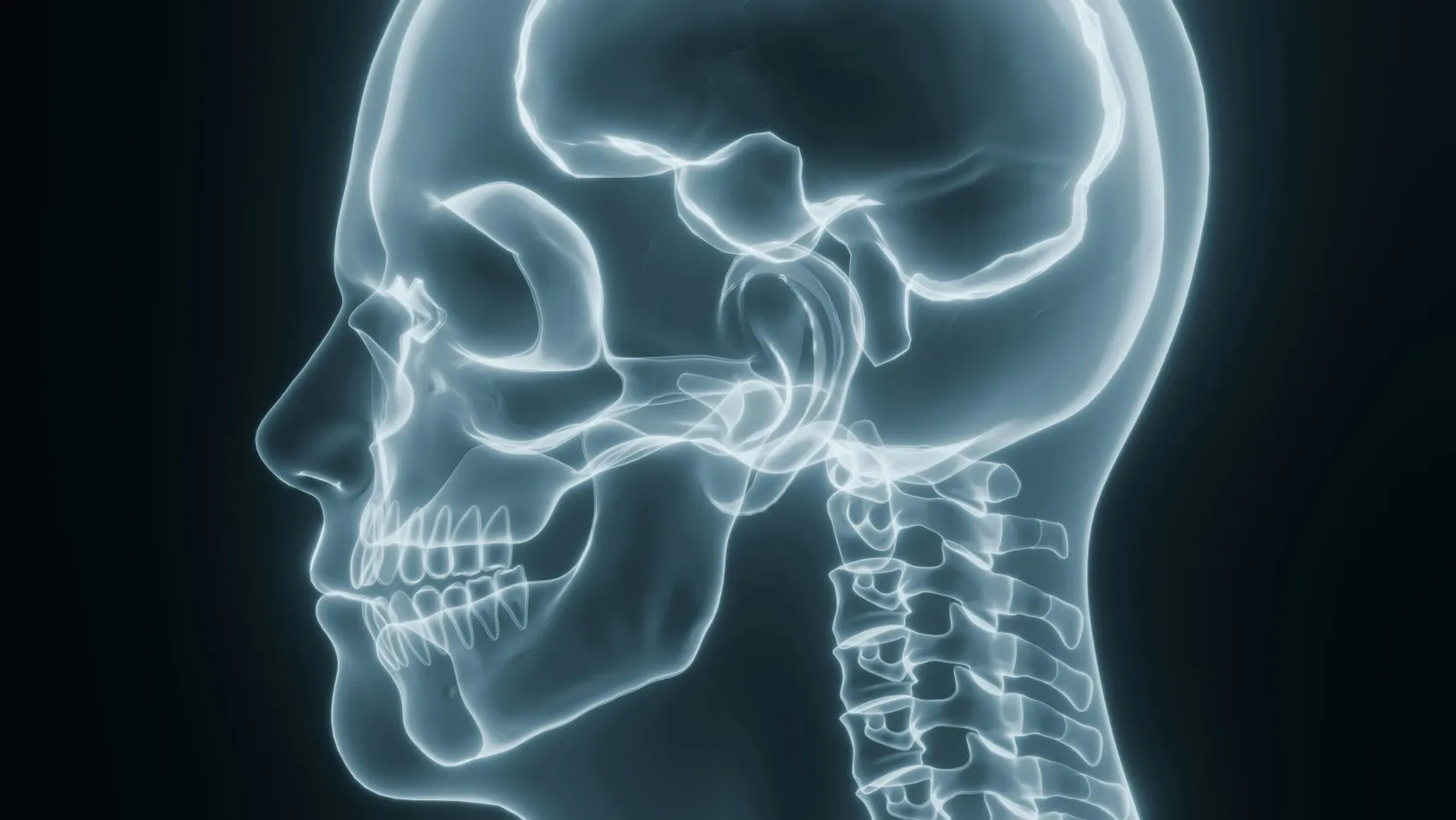
Chiari Malformation Surgery
A Chiari malformation is measured in millimeters and treated according to symptoms, and those two things come apart more often than patients are told. Most people with the finding never need an operation. This page sets out who does, what posterior fossa decompression removes, why surgeons still argue over opening the dura, and what the pooled studies say about both sides of that argument.
About This Department
A scan measures millimeters. Surgery answers symptoms.
Most people who are told they have a Chiari malformation will never need an operation. This page sets out who does, what the surgery removes, where the surgeons still disagree, and how to tell a careful recommendation from a reflex one.
What a Chiari malformation is
Two rounded lobes hang from the underside of the cerebellum, and neurosurgeons call them the cerebellar tonsils. In a Chiari I malformation they sit lower than they should, pushing down through the foramen magnum, which is the large opening where the skull ends and the spinal canal begins. Nothing has gone wrong with the brain itself. The space built to hold it is simply smaller than the contents.
Crowding is the whole problem.
Cerebrospinal fluid, the clear fluid that cushions the brain and cord, has to pass freely through that opening with every heartbeat and every cough, and when the tonsils plug it the fluid gets pushed around instead of flowing, which produces the pattern of symptoms this condition is known for. The classic one is a headache at the back of the head that arrives within seconds of coughing, laughing, straining or lifting, and that fades again within minutes. Some people also develop a cavity of fluid inside the spinal cord itself, called a syrinx, which slowly damages the nerve fibers crossing through it and produces numbness, weakness and loss of temperature sensation in a pattern that does not follow any single nerve. Others have swallowing trouble, hoarseness, unsteadiness, or sleep apnea. And a great many people have the anatomy and none of the consequences. That gap is where most of the confusion in this field begins.
The millimeters do not decide
A radiologist calls it a Chiari I malformation when the tonsils descend more than five millimeters below the foramen magnum. That number came from a convention, and it has spread far beyond what it can carry, because patients now arrive with a printed report, a figure in bold, and the belief that the figure determines whether they go to surgery.
It does not.
A prospective study followed 52 children with a Chiari finding and no syrinx for up to seven years, with annual imaging and annual examination, and it found the tonsil position to be a moving target instead of a fixed one. In half the children the position stayed where it started, in 38 percent it came back up, and in 12 percent it descended further. Twelve percent resolved altogether. Scans moved. Children did not. None of the radiological movement lined up with how the children were doing, and no child had surgery because of a change on a scan. Three eventually had surgery, and all three were operated on because of what they described in clinic. The authors concluded that routine surveillance imaging of an untroubled child changes nothing and can stop.
Our position follows from that. We read the scan carefully, we measure what has to be measured, and then we put the number to one side and listen to the history, because the history is where the decision actually lives.
Who the operation is actually for
Two bodies of evidence answer this, and they point the same way. One tracked what happens to people who are left alone, and the other tracked how often a quiet Chiari turns into an operated one.
So what tips the balance toward operating
Three things, broadly. A cough headache that is severe enough and frequent enough to shape somebody's week is the commonest reason, and it is also the symptom that answers surgery most reliably. An enlarging syrinx, or one that has already produced numbness or weakness, comes second, because the damage a syrinx does to the cord accumulates and does not come back once it has happened. Brainstem signs come third, meaning difficulty swallowing, a hoarse voice, choking on liquids, or sleep apnea traced to the same crowding, and outside those three, watchful waiting is a real and frequently better plan, so the person who tells you otherwise while pointing at a millimeter figure has skipped a step.
Symptoms that point toward surgery
Neurosurgeons listen for the five items below. Symptoms outside that list can still be real and still deserve investigation, and they carry much less weight in a decompression decision, and treating them with an operation disappoints everybody involved.
- Headache at the back of the head brought on within seconds by coughing, sneezing, laughing, straining or bending, easing within minutes afterward.
- Numbness, burning, weakness or loss of temperature sensation in the arms or across the shoulders, particularly when it does not match the territory of one nerve.
- Swallowing difficulty, a hoarse or weak voice, choking on thin liquids, or repeated chest infections from aspiration.
- Unsteadiness on the feet, clumsy hands, dropping things, or a change in handwriting that has crept in over the past few months.
- Sleep apnea, especially central apnea, in somebody who does not fit the usual picture for it.
Migraine sits outside the list. So does dizziness on its own, and so does neck pain on its own, and a scan showing tonsillar descent in somebody whose real problem is migraine is one of the most common traps in this whole area. That trap leaves a scar behind.
A syrinx changes the answer
A syrinx is a fluid cavity that forms inside the spinal cord when cerebrospinal fluid can no longer move freely across the craniocervical junction, and on an MRI it shows up as a dark channel running through the middle of the cord, sometimes over two or three vertebral levels and sometimes over ten. Around a third of people with a Chiari malformation have one.
What makes it different from the headache is the arithmetic of recovery. A cough headache that has troubled somebody for six years can disappear in the first two weeks after a successful decompression, because the mechanism producing it was pressure and the pressure has been taken away. A syrinx that has already destroyed fibers in the middle of the cord is a different case, since the cavity itself usually shrinks once the fluid is moving again, and yet the numbness and the weakness it produced may only partly recover or may not recover at all. That single asymmetry is the reason a syrinx moves a case up the queue instead of leaving it under observation, because waiting costs nothing at all when the only symptom is a headache and waiting costs spinal cord when the cavity is growing. The difference is permanent.
What the operation removes
The formal name is posterior fossa decompression, and the aim is to make the doorway bigger. Nothing is removed from the brain and nothing is put inside it.
The three layers a surgeon can open
Bone, band, dura. The first layer is bone. A small window of the occipital bone at the back of the skull comes out, around three centimeters across, and with it the back arch of the first cervical vertebra in patients whose tonsils sit low enough to warrant that. The second layer is a tough fibrous band on the inner surface of the bone, which is often thickened in this condition and can be split without opening anything underneath. The third layer is the dura itself, the leathery membrane wrapped around the brain and cord, and opening the dura is the step that turns a straightforward operation into a more powerful and more hazardous one. Which of those three layers a surgeon stops at is the real decision in Chiari surgery, and it belongs to the consultation and not to the operating room. Ask about it beforehand. A surgeon who cannot say in advance which layers they intend to open, and what would make them change their mind once they are inside, has left the most consequential judgment in the whole operation to whatever seems reasonable at two in the afternoon with your head already in a clamp.
Why stopping at the bone is a serious optionLeaving the dura closed keeps the operation outside the space where cerebrospinal fluid lives, and almost every serious complication of this surgery comes from having opened that space. A bone only decompression takes less time, the hospital stay is shorter, and the rate of fluid leaks, pseudomeningocele and meningitis falls sharply. The cost lands on the patients in whom bone alone fails to free the fluid, and they come back for a second operation, which is the trade this approach openly accepts. Balancing a lower risk today against a higher chance of returning is the entire argument, and it has been running for thirty years.
The dura question
When the dura is opened, it cannot simply be sewn shut again, because closing it tightly would undo the decompression that was just performed. A patch of material is sewn in to enlarge it, and that step is called duraplasty. So the two operations you will see named in every paper on this subject are decompression with the dura left alone and decompression with a patch sewn in.
Neither one settles every case, and any surgeon who says otherwise has described their own habits.
What makes this argument unusual is that it has never been settled by a randomized trial in adults, so the entire literature is built from cohorts that compared what different surgeons happened to do, which means the patients in the two groups were never quite alike to begin with. Several large pooled analyses have squeezed what they can out of that material, and although they disagree at the edges, they agree on the shape of the trade. The shape holds up. Opening the dura buys a better chance that symptoms settle and that a syrinx shrinks, at the price of a markedly higher chance of a fluid leak, an inflamed lining, a swelling under the wound or a second admission, and every study that has looked at it from a different angle has come back with a version of the same sentence.
What the pooled studies found
Most of the weight here rests on two meta-analyses, one pooling 3,618 patients across 17 studies and the other pooling 3,666 across 14. Read together they say four things.
Four findings a surgeon should be able to quote
- Symptom relief favors the patch. Clinical improvement was more likely with duraplasty, at a relative risk of 1.24 with a confidence interval of 1.07 to 1.44.
- Complications favor leaving the dura closed. Overall complications ran at a relative risk of 4.51 with duraplasty, and the interval stretched from 2.01 to 10.11.
- A syrinx does better with the patch. Syrinx shrinkage was more likely after duraplasty at a relative risk of 1.57, and patients with a syrinx improved more overall.
- Adults come back more often after bone only surgery. Reoperation in adults was less frequent with duraplasty, at a relative risk of 0.17, while in children the two approaches came out level.
Those specific complications deserve naming, since they are the ones a patient feels. Cerebrospinal fluid leaking through the wound ran at a relative risk of 5.23 after duraplasty, and aseptic meningitis, meaning inflammation of the linings without infection, at 4.02. So the practical translation runs like this. An adult with a syrinx has the strongest case for a patch, a child without one has the strongest case for leaving the dura alone, and everybody else sits in a middle ground where the surgeon should explain out loud which way they are leaning and why.
The patch, and why it matters
If the dura is going to be opened, something has to be sewn into it, and the choice of material turns out to move the numbers more than most patients are ever told.
What we use, and why
Our default remains the patient's own tissue, harvested as a sheet of pericranium from under the same incision, since it carries the lowest infection rate and the lowest return rate in that analysis and it costs nothing to obtain. The trade costs a slightly longer incision and a few extra minutes of operating time. Where the pericranium has grown too thin, or where a previous operation already used it, human donor tissue becomes the next choice, and any patient may ask what will be sewn into them before they sign anything, since a department that answers that question with a shrug has told you exactly how closely they follow their own results. Ask it anyway.
What happens on the day
You go to sleep under a general anesthetic and are turned face down, with the head held in a three pin clamp so that nothing moves. The incision runs down the midline at the back of the head and upper neck, three inches or so, and it sits inside the hairline where it is hidden once the hair grows back. Bone comes out, the thickened band beneath it is released, and the surgeon then follows the plan agreed with you beforehand. Nothing enters the brain. Most decompressions take two to three hours from first incision to last stitch.
The first night, and the days after it
Waking up, almost everybody reports neck stiffness and a sore, tight feeling across the back of the head, which comes from muscle and not from brain and which answers well to ordinary painkillers. Headache in the first few days is common and tells you nothing at all. You get up and walk the same evening or the next morning, you eat normally within a day, and you leave hospital in two to four nights depending on whether the dura was opened and how the wound behaves. Stitches or clips come out between day ten and day fourteen. The single most useful thing you can do in that first two weeks is avoid anything that raises pressure inside the head, which means no lifting beyond two kilograms, no straining, and telling somebody promptly if you develop a cough. That last point sounds small. A hard cough in week one drives fluid against a fresh dural repair harder than any weight you could pick up, which is the mechanism behind a good share of the leaks that bring people back to the ward, so a cough in the first two weeks gets treated rather than tolerated.
Risks and complications
Almost every complication on this list belongs to the group whose dura was opened. That single fact shapes the risk, and it explains why the dura conversation happens before the operation and not afterward.
This table scrolls sideways on a narrow screen. Swipe or drag to see every column.
| Problem | When it appears | What it usually needs |
|---|---|---|
| Fluid leak through the wound | First two weeks | Extra stitches, a lumbar drain, occasionally a return to theater |
| Pseudomeningocele | Two to six weeks | A soft swelling at the wound, watched, pressure dressing, sometimes drained |
| Aseptic meningitis | Days three to fourteen | Steroids, and a lumbar puncture first to rule out true infection |
| Bacterial meningitis or wound infection | First month | Intravenous antibiotics, sometimes removal of the patch |
| Symptoms that do not settle | Three to twelve months | Repeat imaging, and an honest review of whether the Chiari was the cause |
The complication nobody lists
Operating on the wrong problem belongs on this page too. A patient whose headaches were migraine all along, who had an incidental Chiari finding and a surgeon willing to act on it, wakes up with a scar, a month of recovery behind them and the same headaches in front of them. No published complication table counts that one. The consultation prevents it entirely, when somebody takes a proper history and proves willing to say that the anatomy on the scan is real and probably beside the point.
Recovery, week by week
Recovery from this operation turns mostly on the neck muscles that were moved aside, and the timetable below is the ordinary one. People who have been unwell for years sometimes expect to feel transformed within days, and a more realistic picture is a slow return over six weeks with the cough headache going early and the stiffness going last. Six weeks is the usual arc.
This table scrolls sideways on a narrow screen. Swipe or drag to see every column.
| Period | What you can do | What to hold back on |
|---|---|---|
| In hospital, two to four nights | Walking the same evening, eating normally within a day | Lying flat all day, which stiffens the neck faster than anything |
| Weeks one and two | Short walks several times a day, showering once the wound is sealed | Lifting beyond two kilograms, bending, straining, alcohol, nicotine |
| Weeks three and four | Longer walks, light desk work from home, gentle neck movement | Driving until your surgeon clears it, and any overhead work |
| Weeks four to six | Most people return to work, physiotherapy for the neck if it is tight | Contact sport, heavy lifting, anything jarring the head |
| Three to twelve months | Full activity, and the window in which a syrinx shrinks on imaging | Judging the result too early, since sensory change recovers slowly |
One part of the recovery deserves saying plainly, because it surprises people who were braced for something worse. The cough headache, if it was the reason you came, tends to be the first thing to go, and a substantial number of patients notice within the first week or two that coughing no longer produces the sensation they had learned to dread. Neck stiffness behaves in the opposite way and stays around for a month or two, easing gradually as the muscles that were retracted settle back down, and a short course of physiotherapy helps that more than rest does. Numbness and weakness that came from a syrinx follow their own timetable entirely, improving over six to twelve months if they improve at all, and that is the reason the scan at three months and the scan at a year get read together instead of separately. Judging the whole result at week six is the commonest mistake patients make. At six weeks the muscles still hurt, the numbness has not moved, and somebody who was promised a transformation concludes that the operation failed, when in fact the part that was ever going to change quickly has already changed and the rest is still in progress.
Follow up and what the scans show
Follow up after this operation is lighter than most brain surgery, and the scans that matter are few. The list is short. The usual pattern runs as follows, and your surgeon will adjust it if a syrinx was present or if the wound gave trouble.
Reading those scans without panic
Postoperative scans alarm patients with two findings that mean very little. The tonsils frequently stay exactly where they were, since the operation made the room bigger and never pulled them back up, so a report noting persistent tonsillar descent is describing the expected state of affairs. And a syrinx that has narrowed without vanishing is a success, because a thin residual channel that is no longer under pressure does no further damage. What matters on these scans is whether fluid is moving across the junction and whether a syrinx is smaller than it was, and both of those get answered by comparing images instead of by reading one in isolation.
What decompression does not fix
Surgery treats crowding at one small doorway. Any symptom generated somewhere else carries on exactly as before, and saying so in advance prevents a great deal of disappointment later.
Migraine continues, and often becomes more obvious once the cough headache has gone. Chronic daily headache without the cough trigger rarely answers this operation at all. Nerve damage that a syrinx already did to the cord may recover partly or not at all, since the operation stops the process and cannot rebuild what the process destroyed, and fatigue, brain fog and widespread pain, which many people carrying this diagnosis live with and which are genuinely disabling, have no established relationship with tonsillar descent and no good evidence that decompression touches them. Scoliosis in a child with a syrinx sometimes stabilizes after the syrinx shrinks and sometimes progresses regardless, so an orthopedic team follows it in its own right, and none of this argues against the operation for the people who need it, while all of it argues for knowing which of your symptoms is being treated before you agree to anything. Write your symptoms down in order of how much they cost you. Take that list to the consultation and ask, item by item, which ones the surgeon expects to change, because a good answer will sort your list into three groups and a poor one will treat the whole list as a single thing called Chiari.
Cost, coordination and having surgery in Istanbul
We print no price on this page, because a figure written today would be out of date by the time you read it and because the true answer depends on whether the dura is opened. What we can describe is how the assessment works, and for this condition the assessment carries more weight than usual, since a fair proportion of the people who write to us should not be having an operation at all.
What to send, and what comes backSend the MRI images themselves on a disc or a transfer link, covering the brain and the whole spine, along with a written account of your symptoms including when the headache comes, how long it lasts, and what sets it off. A neurosurgeon reviews both and answers three questions in writing. Whether the anatomy fits the symptoms. Whether an operation is likely to help those particular symptoms. Whether the dura would be opened in your case, and on what grounds. If the answer to the second question is no, that is what you will be told, and it is told for free.
Staff in the international patients office work in English, Arabic, French, Russian, Serbian, Romanian and Spanish, with interpreting for other languages arranged before you arrive. A single coordinator handles your case from the first message through to discharge and stays reachable on WhatsApp once you are home, which matters here because the questions that come up at week three tend to be wound questions and not brain questions. Your room has a bed for a companion, hotel and transfers are arranged around the surgical dates, a female physician can be requested, and the invitation letter for a visa application goes out ten days before travel. Halal, vegetarian and diabetic meals are routine, and there is a prayer room on site. None of that is decoration. A patient who has flown in from four time zones away, who cannot read the signage, whose companion is sleeping in a chair and whose only route back to the surgeon runs through a switchboard in a language they do not speak, will experience an ordinary recovery as a frightening one, and the arrangements above exist so that the recovery stays ordinary.
Plan on ten to fourteen nights in the country. That covers the preoperative assessment, the operation, two to four nights on the ward, and enough time afterward for the wound review before you fly, since airline pressure changes are best met with a wound that has already been checked. Back home, your local doctor manages the stitches and the early follow up, and we send them the operative note, the details of what was removed, and whether a patch was used and what it was made of, so that any future surgeon reading a scan knows exactly what was done.
Send the scan and the story. Both, or neither is much use.
Chiari surgery FAQ
Most first messages from abroad ask the questions below.
My tonsils descend 12 mm. Does that mean I need surgery?
Is the dura opened in every operation?
Will my headaches go away?
Will the syrinx disappear?
How long before I can fly home?
Can the operation be done again if it fails?
What will the scar look like?
References
- Langridge B, Phillips E, Choi D. Chiari malformation type 1. A systematic review of natural history and conservative management. World Neurosurgery. 2017;104:213-219.
- Whitson WJ, Lane JR, Bauer DF, Durham SR. A prospective natural history study of nonoperatively managed Chiari I malformation, and whether follow-up MRI surveillance alters surgical decision making. Journal of Neurosurgery Pediatrics. 2015;16(2):159-166.
- Leon TJ, Kuhn EN, Arynchyna AA, Smith BP, Tubbs RS, Johnston JM, et al. Patients with benign Chiari I malformations require surgical decompression at a low rate. Journal of Neurosurgery Pediatrics. 2019;23(4):498-506.
- Tam SKP, Brodbelt A, Bolognese PA, Foroughi M. Posterior fossa decompression with duraplasty in Chiari malformation type 1. A systematic review and meta-analysis. Acta Neurochirurgica. 2021;163(1):229-238.
- Chai Z, Xue X, Fan H, Sun L, Cai H, Ma Y, et al. Efficacy of posterior fossa decompression with duraplasty for patients with Chiari malformation type I. A systematic review and meta-analysis. World Neurosurgery. 2018;113:357-365.
- Jbarah OF, Aburayya BI, Shatnawi AR, Alkhasoneh MA, Toubasi AA, Alharahsheh SM, et al. Risk of meningitis after posterior fossa decompression with duraplasty using different graft types in patients with Chiari malformation type I and syringomyelia. A systematic review and meta-analysis. Neurosurgical Review. 2022;45(6):3537-3550.
Written by the Biruni Hospital medical editorial team. Reviewed by Dr Yunus Emre Yavuz, Neurosurgery.
Editor's note
Written by the Biruni Hospital medical editorial team. Reviewed by Assistant Professor Fikret BAŞKAN, Neurosurgery.
Medically reviewed by
Assistant Professor Fikret BAŞKAN
Neurosurgery
Related Treatments
View All
ALIF - Anterior Lumbar Interbody Fusion Surgery
Coming in from the front restores the curve of the lower back better than any posterior route and leaves the back muscles untouched. It also carries one risk most pages will not name, and this page names it.

Brain AVM Surgery - Arteriovenous Malformation Surgery
An unruptured brain AVM bleeds at roughly 2.2 percent a year, and surgery spends its whole risk on a single morning. This page sets those two shapes of risk side by side, explains what the ARUBA trial does and does not say about your case, and shows how a Spetzler-Martin grade changes the answer.
Brain Tumor Surgery - Brain Tumor Removal
Brain surgery is one trade repeated all day, how much tumor comes out against what the brain around it still does. This page puts the published numbers on both sides of that trade.
Cervical Microdiscectomy - Minimally Invasive Neck Disc Surgery
Neck disc surgery is done from the front, through the space between your windpipe and your carotid artery, and almost everything that goes wrong that is not the disc happens in that corridor. What has been measured in it.
Deep Brain Stimulation Surgery - DBS Surgery
Stimulation does not slow Parkinson's disease down, and what it gives back is hours of the day when the medication is working without the writhing that used to come with them. This page sets out who qualifies, what the trials actually measured, how the two targets trade against each other, and what the hardware asks of you for the rest of your life.