
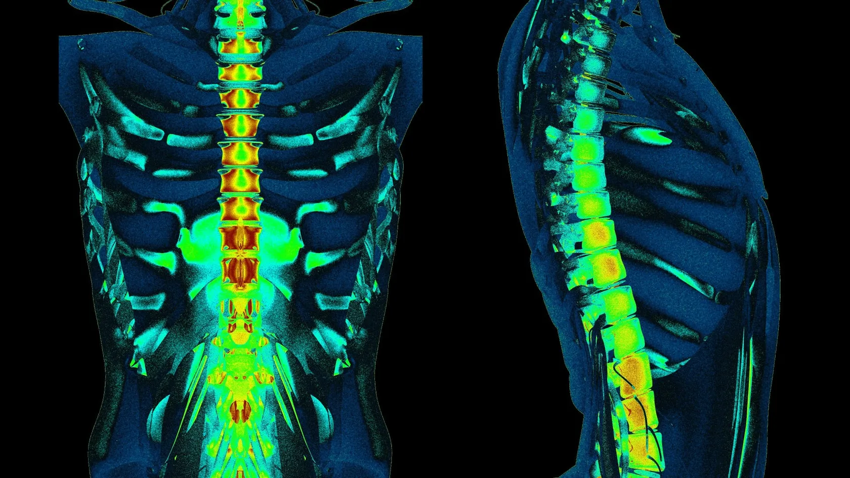
Cervical Microdiscectomy - Minimally Invasive Neck Disc Surgery
Neck disc surgery is done from the front, through the space between your windpipe and your carotid artery, and almost everything that goes wrong that is not the disc happens in that corridor. What has been measured in it.
About This Department
Reaching a disc in your neck does not involve going in from the back of it. The incision is made in the front of the throat, and the windpipe and gullet are held to one side while the carotid artery is held to the other. In a study that mounted a pressure sensor on the retractor blade, the pressure applied to the wall of the gullet averaged 92.7 millimeters of mercury, higher than the pressure in the patient's own arteries at the time. Almost everything that can go wrong in this operation that is not about the disc happens in that corridor, and almost nothing patients are told beforehand is about it.
Free consultation
Send the images and have a spine surgeon read them against your arm symptoms
Send the image files themselves along with the radiology report, a description of exactly where the pain, numbness or weakness runs in the arm and hand, how long it has been there and whether it is changing, and a note of anything already tried. Neck disc surgery has more than one route and the choice between them is decided by which nerve is compressed, from which direction, at which level, and whether the spinal cord is involved. A spine surgeon reads the images against your symptoms and tells you which routes are open and whether an operation is the right question yet. No fee, no obligation, and a coordinator replies in your own language, usually within the same working day.
Where the surgeon comes in
Cervical microdiscectomy means removing a disc fragment that is pressing on a nerve in the neck. The disc sits in front of the spinal cord, so the shortest and safest line to it runs from the front of the throat instead of from behind, and that is why the scar ends up in a skin crease under the jaw rather than at the back of the neck. A transverse incision two or three centimeters long is made on one side, and the surgeon then works down through a natural plane that already exists in the tissue. None of that is controversial and none of it is where the interesting decisions live, because the choice that shapes your recovery was made before the first incision, when somebody decided which side of your throat to come in on, how many levels to open, and whether anything would be implanted. That decision is worth asking about.
On the inner side of that plane sit the windpipe and the gullet, held gently across the midline, while on the outer side sit the carotid artery, the jugular vein and the vagus nerve, held firmly the other way. Running in the groove between the windpipe and the gullet on each side is the recurrent laryngeal nerve, which supplies the vocal cord. Behind all of it is the front of the spine, and the surgeon reaches it without cutting a single muscle that moves the neck. That is the genuine and considerable advantage of this route, and it explains why people walk out of hospital the next morning.
The price is that everything just listed has to be held out of the way throughout. Swallowing trouble, voice change, a rare injury to the gullet, swelling that narrows the airway and, very rarely, an injury to the artery that runs up through the bones of the neck are all complications of the corridor and not of the disc. They are also the ones patients hear least about, partly because they are usually temporary and partly because a consent conversation tends to be organized around the nerve instead of around the route to it. Naming them beforehand is not scaremongering, since a complication you were warned about is an inconvenience while the same complication unmentioned is a betrayal, and the difference between those two experiences is entirely a matter of what was said in the clinic before anybody signed anything.
This page is organized the other way round. The disc part of a cervical microdiscectomy is well described everywhere and is not in dispute, so what follows is about the corridor, what has actually been measured in it, and which of the alternatives avoid it altogether.
The one you will probably meet
Half of patients at one month
Swallowing trouble after anterior neck surgery is not a rare complication. In the prospective study that produced the grading scale everybody now uses, 249 consecutive patients were contacted at set intervals, and 50.2 percent reported difficulty swallowing at one month, 32.2 percent at two months, 17.8 percent at six months and 12.5 percent at twelve. Reassuringly, only 4.8 percent had moderate or severe difficulty at the six-month mark, so most of what that first figure describes is a nuisance and not a disability. It is still half of everybody, and it is the single most likely thing you will notice after this operation that has nothing to do with your arm. Half of everybody is not a footnote, and the reason it reads like one in most patient material is that the difficulty is usually mild, usually temporary and usually not what the operation was about, which makes it easy to leave out of a conversation already crowded with the nerve.
The two-year number nobody quotes
Researchers following 454 patients across 23 sites looked much further out and found something that consent conversations almost never mention. Thirty percent reported difficulty at three months, which is broadly in line with the study above. What is different is that the problem persisted in 21.5 percent of patients at six months and in 21.3 percent at twenty-four months. Roughly one patient in five was still describing swallowing difficulty two years after their operation, and the same cohort found that people reporting difficulty at three months went on to report higher disability and lower physical health at every later assessment. That figure has no confidence interval attached to it and comes from telephone follow-up, so read it as a signal about a fifth of patients and not a precise proportion, and read it anyway.
What makes it more likely
Number of levels is the factor that comes up in every study. In that same multicenter cohort the three-month rate rose from 19.8 percent after one level, to 33.3 percent after two, to 39.1 percent after three or more. Being female raised the risk at six months in the prospective grading study, and operating on multiple levels raised it at one and two months. A three-year prospective series of 133 patients added three more, finding that myelopathy, pre-existing swallowing difficulty and operating specifically at the C4 to C5 level all predicted trouble, with reflux disease predicting greater severity. That last group matters because two of those factors are known before anybody books an operating room. Reflux and existing swallowing difficulty can be looked up in your own notes today, which means the conversation about your particular risk of this particular complication is available well before anybody schedules anything and does not have to wait until afterward.
Why the number varies tenfold
Fifty percent, or nine percent, or eighty-three
Ask three surgeons how often swallowing trouble follows this operation and you can be told fifty percent, ten percent or eighty percent without anybody lying. A meta-analysis pooling 222 observational studies and 50,584 patients put dysphagia at 9.5 percent. The prospective grading study above found 50.2 percent at one month. A small prospective study that took 47 patients and actually filmed them swallowing under X-ray found difficulty in 83.0 percent at one week and 59.6 percent at one month, with material entering the airway short of the lungs in 36.2 percent, and yet not a single case of pneumonia. Those three numbers are not in conflict. They are answers to three different questions. Anybody quoting one of them without saying which question it answers is not being dishonest so much as imprecise, and imprecision of exactly that kind is what makes a patient feel misled later when their own experience matches the highest figure rather than the lowest.
Where the spread comes from
One scoping review screened 2,396 abstracts and extracted data from 280 studies, finding 317 different swallowing outcome measures in use. Forty-six percent of them were unvalidated patient-reported measures, another 16 percent gave insufficient detail, 13 percent came from chart review or a database and only 7 percent involved any instrumental assessment. Sixty-three percent of the studies were retrospective. Its conclusion is that our understanding of the mechanism, the timeline and the presentation of this complication is limited by the way it has been measured, which is an unusually blunt thing for a review to say about its own field.
Feeling it and having it are different
Twenty-one patients had what they said about their swallowing compared against what a videofluoroscopic swallow study showed, before surgery and six weeks afterward. Everything got worse on every measure, and the increases were larger on the patient-reported measures than on the instrumented one. More importantly, the two did not agree case by case. Its conclusion is that the presence or absence of symptoms may not correspond with the observed physical impairment, which is worth knowing in both directions, because the patient who feels fine may still be aspirating slightly and the patient who feels dreadful may look normal on film. Two things follow from that mismatch, since somebody who feels fine may be letting a little liquid past the vocal cords without noticing, and somebody who feels dreadful may look entirely normal on film and then be told, unhelpfully, that nothing is wrong. Both deserve a proper look.
The retractor, measured
One German group did something unusual, which was to stop theorizing about why swallowing goes wrong and put instruments on the retractor to find out. In 31 patients they placed a pressure transducer on the back of the blade holding the gullet aside and a second inflatable sensor inside the gullet itself. Pressure on the outside of the gullet averaged 58.3 millimeters of mercury before the retractor was fully opened and 92.7 millimeters afterward, against a mean arterial pressure in those same patients of 76. The pressure needed to perfuse the lining of the gullet is roughly 25. In other words, the retractor squeezes the food pipe harder than the heart is pushing blood into it. Measuring something everybody had previously been guessing about is worth doing even when the answer is uncomfortable, and the answer here is that a standard retractor presses on the wall of the gullet harder than the circulation is pushing back at it.
The same group then measured blood flow directly under the blade in fifteen patients using a laser probe. Perfusion averaged 107 units before the retractor was opened, fell to 30 while it was open and returned to 117 after it was closed, and every single one of the fifteen patients showed reduced blood flow while the retractor was in, ranging from a 21 percent drop to a 93 percent drop. Seven of them showed a surge of blood flow at the end of the procedure, four regained their starting level, and in four the flow was still below baseline after the retractor came out. The tissue is briefly starved and mostly recovers, which is a satisfying mechanism for a complication that mostly settles.
Then the same group tested the mechanism against the outcome in 92 patients, with 32 lumbar surgery patients as a comparison group, and found no correlation at all between how hard the retractor pressed and who could not swallow afterward. Almost half of the neck patients had swallowing disturbance and none of the lumbar patients did, so something local is clearly responsible. It is simply not the pressure, or not only the pressure. Pressure did fall to roughly three quarters of its starting value within the first hour, meaning the tissue accommodates, and the honest position is that the mechanism has been measured beautifully and still does not predict who suffers. Resisting a tidy mechanism is harder than it sounds, and the group that produced the tidy mechanism is the same one that went looking for the link to symptoms and then reported plainly that they could not find it, which is a good reason to trust the rest of their measurements.
Worth carrying away from all of that is a practical point. Surgeons who release the retractor periodically, use a wider blade, or keep the total retraction time short are acting on a mechanism that is real even if the link to symptoms is unproven, and none of those measures cost anything. A surgeon who can describe what they do about retraction has thought about the corridor.
The voice
Between two percent and a quarter of patients
The nerve that moves the vocal cord runs in the groove beside the windpipe, directly through the field of the retractor. A qualitative systematic review of 34 studies reports the incidence of vocal cord palsy after anterior cervical surgery as ranging from 2.3 percent to 24.2 percent, and that tenfold spread has the same explanation as the swallowing spread, since a study that looks at every larynx afterward finds far more than a study that counts referrals for persistent hoarseness. Retrospective series in this pack report 1.9 percent and 2.7 percent, both of which counted only patients who complained long enough to be sent to a laryngologist. Which of those two figures applies to you depends less on your own anatomy than on whether anybody bothers to look, and that is an uncomfortable thing to say about a complication rate while remaining the plainest reading of the evidence. Ask which kind of number you are being given.
Most of it comes back
One series identified 21 vocal fold paralyses among 411 operations, at 5 percent, and followed them. Of the eighteen with complete records, fifteen had resolved completely within twelve months and one more recovered at fifteen months. Symptoms in that group were hoarseness in eighteen, persistent cough in seven, difficulty swallowing in seven and aspiration in thirteen. One patient with paralysis on both sides presented with noisy breathing and respiratory distress and needed a tracheotomy, which is the rare and serious end of this complication and the reason it is worth naming at all. Reoperating through a previous anterior scar raises the risk, with one series reporting two events among 21 revision cases, and that is a small numerator carrying a large percentage. Recovery in that series was measured in months instead of weeks, which is a longer horizon than most people expect for something that sounds as minor as hoarseness, and saying so in advance turns a frightening symptom into a merely tedious one.
Left or right, and the cuff
Many surgeons approach from the left on the grounds that the nerve on that side runs a longer and more protected course. The evidence is thinner than the tradition. One review found a single moderate-strength study suggesting the right side raises risk, while two retrospective series of 328 and 418 patients found no association with the side of approach whatsoever. A historical review argues the rule descends from anatomical reasoning plus the early practice of one influential surgeon and notes that modern series have not shown a clear difference. What does have supporting data is the breathing tube cuff, since one prospective study of 242 patients found palsy in 1.3 percent when cuff pressure was reduced while the retractors were in against 6.5 percent when it was not, and the review above puts that finding at 6 percent falling to 2 percent while noting it has never been confirmed in a randomized trial.
The gullet
Rare, serious, and often late
A hole in the gullet is the complication of this corridor that nobody wants to discuss and everybody should hear named once. A systematic review gathered 65 articles describing 153 patients over 35 years, with an average age of 44.7, and the levels most often involved were C5 to C6 and C6 to C7. Mortality among those reported cases was 3.92 percent, and what that review cannot give is an incidence, because its denominator is the published literature rather than a population, so nobody should be quoted a percentage risk from it. What it can give is the shape of the problem. Naming a complication that produces a handful of published cases a decade may look disproportionate on a page like this, and it earns its place because the delayed form of it is the one thing on this list capable of presenting years later to a doctor who has no idea you ever had neck surgery.
The commonest cause is the hardware itself
Of those 153 cases, 31 followed hardware failure and 23 followed hardware erosion, against 14 caused by injury during the operation itself. The review divides presentation into three windows, meaning during the operation, within thirty days, and delayed, and the delayed group is the one that catches people out. A plate or a screw slowly wearing through the back wall of the gullet does not announce itself for months or years, and presenting symptoms across the series were difficulty swallowing in 63 patients, fever in 24, neck swelling in 23 and leakage from the wound in 18. A second review of 173 patients across 76 series found that the longer the injury went unrecognized, the more complex the reconstruction needed to fix it.
How rare is rare
Injury to the pharynx, the part above the gullet proper, is rarer still, and a systematic review published in 2024 could find only twelve patients in the entire world literature, eleven of them from published reports and one of them the authors' own. All but one were repaired surgically and all did well, and the authors state plainly that the long-term outlook remains unknown because there are not enough cases to know it. Rare enough to be describable patient by patient is a useful thing for a patient to hear, and it is a fair summary of where this complication sits. A literature containing twelve cases in total is a literature about something genuinely uncommon, and what to take from it is a reassurance instead of a probability, because the alternative to a countable literature is one large enough to have produced a rate. That absence is the point.
What the corridor risks
Numbers with proper confidence intervals are scarce in this literature, and the ones that exist are collected here. Read the right-hand column, because several of these come from designs that cannot support a precise figure.
Wide table. Swipe or drag it sideways on a narrow screen, because it scrolls instead of shrinking.
| Complication | Reported rate | Where it comes from and what limits it |
|---|---|---|
| Blood collection needing a second operation | 0.40 percent, with an interval of 0.33 to 0.46 | 37,261 patients in a national database. Roughly one in 250, and 37 percent of them happened after discharge. |
| Needing the breathing tube put back | About 0.20 percent within one day | Same figure across three separate datasets covering more than 280,000 operations, which is unusually consistent. |
| Weakness of the shoulder afterward | 5.3 percent overall, with an interval of 4.6 to 6.0 | 704 cases among 13,621 patients across 79 studies. Lowest after anterior discectomy at 3.3 percent. |
| Vocal cord palsy | Between 2.3 and 24.2 percent across studies | 34 studies with no agreed definition. The high figures come from examining every larynx, the low ones from counting complaints. |
| Injury to the artery in the neck bones | 0.07 percent across 163,324 operations | Surgeons reporting on their own careers in a survey. Treat as an order of magnitude and not a rate. |
| Failure of the bones to knit | 10.0 percent pooled | 222 studies and 50,584 patients, with no interval given. Rises steeply with the number of levels. |
| Death within the study window | 0.1 percent pooled | Same pooled analysis. Observational studies only, so under-reporting is likely rather than possible. |
Two things about that table deserve saying out loud. Every rate in it rises with the number of levels operated on, which is why a single-level microdiscectomy is a meaningfully different proposition from a three-level fusion even though both are described with the same words. And the rates that carry proper intervals are the ones drawn from large databases, which capture what got coded rather than what happened, so they are precise about something slightly narrower than what you want to know.
The first thirty-six hours
Swelling comes before bleeding
Reviewing 311 anterior cervical procedures, investigators found an airway complication in 19 patients, at 6.1 percent, with six needing the breathing tube replaced and one death. Symptoms appeared on average 36 hours after surgery. All but two of those complications were caused by swelling of the throat tissues instead of by a collection of blood, which matters because the two are managed differently and because swelling builds gradually instead of announcing itself. Risk rose with exposure of more than three vertebral bodies, blood loss over 300 milliliters, operations involving the upper neck and operating time beyond five hours, all of which describe far bigger procedures than a single-level microdiscectomy. Having a drain in place was not protective. Thirty-six hours is the number worth carrying out of that paragraph, since it falls squarely after the point at which a modern single-level patient would ordinarily have gone home, and it is the reason the next two paragraphs exist at all.
Bleeding is rarer and more sudden
Among 37,261 patients in a national surgical database, a collection of blood in the neck needing a return to the operating room within thirty days occurred in 0.40 percent, with a confidence interval of 0.33 to 0.46 percent. Risk was tripled by operating on three or more levels, roughly doubled by a raised clotting time before surgery, doubled by a higher anesthetic risk grade, and raised by male sex, anemia and a low body mass index. Patients who had one went on to have more pneumonia, more wound infection and more time in hospital. The number that matters most for anybody being sent home the day of surgery is that 37 percent of these events happened after discharge.
Going home the same day
Same-day discharge after single-level anterior neck surgery is now ordinary. A statewide registry compared 806 patients discharged the same day after one- or two-level anterior cervical surgery, split between hospital outpatient departments and free-standing surgery centers, and found no difference in complications, patient-reported outcomes at ninety days or one year, or return to work. Two database studies found the same for outpatient disc replacement and outpatient discectomy. Read those results alongside the sentence in the paragraph above rather than instead of it, because a null result in 162 patients at one type of center is weak evidence of safety, and because being at home is exactly where a third of the serious bleeds happen. Somebody should be with you for the first night, and you should know what to do at three in the morning. Selected patients doing well is a different claim from the operation being safe to send everybody home after, and the studies above establish the first of those without establishing the second.
Questions before you consent
Ten questions, all answerable in a sentence. The first four are about the route, the next three about what gets left inside, and the last three about afterward.
- Confirm how many levels you are operating on, because almost every risk on this page scales with that number.
- Say which side you are approaching from, and why that side.
- Say what you do about retraction during the operation, meaning blade width, release intervals and total time.
- Explain whether the route from behind is possible for my particular fragment, and if not, why not.
- Tell me whether a plate is going in, and what you gain and give up by using one.
- Say what the chance is that the bones do not knit at my number of levels, and what happens then.
- Confirm whether I have reflux or existing swallowing trouble, and what that changes about my risk.
- Give me your own rates for swallowing difficulty and hoarseness, and tell me how you count them.
- State exactly which symptoms in the first two days should make me call rather than wait.
- Confirm I leave with the operative note naming the level, the side and the implant, and my images.
The vertebral artery
Plate, cage or neither
Once the disc is out there is a gap, and what fills it is a separate decision with its own consequences for the corridor. A metal plate screwed to the front of the vertebrae is the traditional answer and it sits directly against the gullet. A low-profile cage with its own anchors avoids that, and putting nothing at all avoids both.
Another wide one. Drag it sideways on a narrow screen, because it scrolls instead of shrinking.
| Question | What was found | The qualifier |
|---|---|---|
| Does a low-profile cage reduce swallowing trouble | Yes at two, three and six months, across 7 studies and 409 patients | No difference immediately after surgery and no difference beyond a year. The advantage is a middle-months one. |
| Does every analysis agree | No. A pooled analysis of 687 two-level cases found swallowing rates comparable | Nine observational studies with no intervals published. The implant literature does not speak with one voice. |
| What does the cage cost | More sinking into the bone, at 15.1 against 8.8 percent, odds of 1.97 with an interval of 1.34 to 2.89 | Driven entirely by multilevel cases. At a single level the odds were 1.43 with an interval of 0.61 to 3.37. |
| What happens to the shape of the neck | Across 14 trials and 1,067 participants, the cage group lost more of the normal forward curve | The gap widened with longer follow-up. A smaller three-year series found no difference, so this is unsettled. |
| What if nothing is put in at all | In 170 single-level cases with graft alone, no plate and no collar, 94 percent fused | Every single patient lost curve at the operated level, averaging 7.4 degrees, though overall alignment held in 78 percent. |
| Does the choice matter at one level | Much less than at two or three. Most of the measured penalties appear only in multilevel work | Which is the most useful single sentence here for anybody having one disc removed. |
What happens on the day
When to call the same day
Five situations warrant contacting somebody immediately, roughly in this order. The first three are about the corridor and they cluster in the first two days.
- Difficulty breathing, noisy breathing, or a neck that feels tight and is visibly swelling. This is an emergency and it needs assessment within minutes. Airway problems appeared on average 36 hours after surgery in one series, and 37 percent of the bleeds that needed a second operation happened after patients had gone home.
- Choking on liquids, or coughing every time you drink. Ordinary swallowing soreness is expected. Liquid going the wrong way is a different symptom and it deserves a proper assessment rather than reassurance.
- A voice that has gone weak or breathy and is not improving. Most vocal cord palsies recover, with fifteen of eighteen resolving within a year in one series, and getting it looked at early changes how it is managed.
- New weakness in the arm, hand or shoulder. Shoulder weakness in particular can appear days after everything seemed fine, since roughly half of these palsies present at three days or later.
- Fever with worsening neck pain or anything leaking from the wound. Late fever and wound leakage were among the presenting features in the reported cases of injury to the gullet, and that is a diagnosis nobody wants made slowly.
Fusion that does not happen
Where a fusion is part of the operation, the two vertebrae are expected to knit into one over the following months, and sometimes they do not, which is called pseudarthrosis and is common enough to deserve a number rather than a shrug.
Pooling 222 observational studies and 50,584 patients, the overall complication rate after anterior cervical discectomy and fusion was 16 percent. The individual figures were excessive neck swelling 11.3 percent, failure of the bones to knit 10.0 percent, swallowing difficulty 9.5 percent, sinking of the cage or graft into the bone 9.4 percent, worsening of spinal cord symptoms 7.7 percent, hoarseness 2.3 percent and death 0.1 percent. More levels and older age were both associated with a higher overall complication rate.
Two of those figures should be treated with suspicion and it is worth saying which. The swallowing figure of 9.5 percent and the hoarseness figure of 2.3 percent are far below what the prospective studies in this pack report, because pooling observational studies pools their under-ascertainment as well as their patients. The pseudarthrosis, swelling and subsidence figures do not have that problem, since a fusion that has not united shows up on a scan whether or not anybody asked the patient a question.
Level count drives it. One series of 168 patients and 264 levels reported non-union in 5.4 and 3.4 percent after single-level surgery depending on the graft material used, rising to 7.1 and 8.1 percent at two levels and to 10 and 11.1 percent at three or more, with no significant difference between the two materials at any level count. Another series of 352 patients found non-union in 7.6 percent of those with a workers' compensation claim against 0.9 percent of everybody else, alongside a revision rate of 12.9 against 2.7 percent. That cohort also contained more smokers and more physically demanding jobs, so it does not isolate smoking, and it is the closest thing here to evidence on a question every surgeon will raise with you. Reading those three sets of figures together gives you a gradient instead of a number, and a gradient is the more useful object because it tells you what changes when the plan moves from one level to two.
Routes that avoid the front
Everything above is a reason to ask whether the corridor can be avoided. For some herniations it can, and the alternatives divide into going in from behind and going in from the front but through the bone rather than past the throat.
Wide again. Swipe the table sideways on a narrow screen, because it scrolls instead of shrinking.
| Route | What the evidence shows | Who it suits and what limits it |
|---|---|---|
| Open foraminotomy from behind | Across 3 randomized trials and 7 comparative studies, complications averaged 4 percent against 7 percent from the front | Reoperation within two years ran the other way, at 6 percent against 4 percent. The review graded the overall difference as none. |
| The same operation through a tube | Shorter hospital stays and less blood loss than the open version, with no difference in complications or reoperation | Two studies found more neck pain after the open version early on, and in both the difference disappeared with longer follow-up. |
| Endoscopic foraminotomy from behind | Pooling 9 studies and 486 patients, complications were lower than from the front and reoperation was similar | Stated indication is one-sided arm symptoms without spinal cord involvement. Commonest problem was temporary altered sensation. |
| Through the vertebral body from the front | Series of 36 and 26 patients report good results, with the drilled channel about 7.5 millimeters across and no collapse | Small, retrospective, from the groups that invented it, with no control arm. Read as promising rather than established. |
| Behind against through the bone | Across 40 studies and 1,661 patients, complications 7 percent with an interval of 5 to 10, reoperation 5 percent with 3 to 7 | Neither differed. The two arms were pooled separately instead of compared head to head, which limits what can be concluded. |
| All of them against each other | A network analysis of 23 randomized trials and 1,844 patients found no statistical difference between any of the treatments | The strongest evidence base on this page, and its answer is that the choice of operation matters less than people assume. |
Read down that table and a pattern appears. Nobody has shown one of these operations to be better than another for arm pain from a trapped nerve, and the strongest evidence available, drawn from randomized trials only, says the differences are not statistically distinguishable. What differs is which corridor is used and therefore which complications are on the table. Going in from behind puts nothing near the gullet or the voice box and does not require a fusion, and it is limited to fragments sitting out to one side without spinal cord compression. Going in from the front reaches everything and costs the corridor. Deciding between them on anatomy is sensible, and deciding between them on which one sounds more advanced is not.
The order decisions get made
Six decisions sit behind the phrase minimally invasive neck disc surgery, and they get made in roughly this sequence. Knowing the order lets you ask about each one at the point it is still open.
- Whether to operate now at all. Arm pain from a recently trapped nerve improves without treatment at a measurable rate, and the section below sets out by how much.
- Whether the spinal cord is involved. Cord compression changes the calculus entirely and rules out the smaller operations from behind.
- Which direction the fragment sits in. A fragment out to the side compressing one nerve can be reached from behind. One in the midline generally cannot.
- How many levels are involved. This is the number that drives swallowing trouble, bleeding, non-union and overall complications, all in the same direction.
- Whether a fusion is needed and what fills the gap. A plate, a low-profile cage or graft alone, each with the trade described in the table above.
- Where you spend the first night. Same-day discharge is reasonable and common, and a third of the serious bleeds happen after people have gone home.
Waiting instead
Before agreeing to any of this it is worth knowing what happens without it, and for recent arm pain from a trapped neck nerve there is a proper randomized answer. Two hundred and five patients at three Dutch hospitals, all with symptoms for less than a month, were assigned either to a semi-hard collar with rest, to twelve physiotherapy sessions over six weeks, or to carrying on with daily life and no specific treatment at all.
In the group that did nothing, arm pain fell by 3.1 millimeters a week on a hundred-millimeter scale, with a confidence interval of 4.0 to 2.2, which came to 19 millimeters over six weeks. A collar and physiotherapy each added a further 1.9 millimeters a week, giving about 12 millimeters more relief than doing nothing at the six-week mark. Neck pain in the untreated group did not improve significantly at all.
Notice which pain moved, because arm pain from the compressed nerve improved steadily on its own while neck pain did not budge at all, which is a useful warning for anybody being offered an operation mainly for a sore neck. Notice also the size of what treatment added, because 12 millimeters on a hundred-millimeter scale after six weeks of collars or physiotherapy is a real effect and a modest one. Anybody offered an operation primarily to relieve neck pain rather than arm pain should ask what evidence supports it, given that the untreated group in this trial saw their neck pain barely move across six weeks while their arm pain improved steadily on its own. The trial only enrolled people whose symptoms had lasted under a month, so it says nothing about somebody who has had arm pain for a year, and that limit matters as much as the result.
Discs in the neck can also disappear on their own, and the case literature includes a 76-year-old man with a large fragment behind a vertebral body and severe canal narrowing whose scan showed almost complete resorption at seven months after he declined surgery. A single case proves nothing about probability and the review accompanying it notes that larger extruded fragments have a greater chance of resorbing, which is the same counterintuitive pattern seen in the lower back. Nobody should plan around it, and everybody should know it happens.
The first six weeks
Coming to Istanbul
Cervical microdiscectomy FAQ
Why is neck disc surgery done from the front?
How long will swallowing be difficult?
Why do different surgeons quote such different risks?
Will my voice change?
Can the operation be done from the back of the neck instead?
Do I need a plate?
How often does the fusion fail to knit?
Should I try waiting first?
References
- Bazaz R, Lee MJ, Yoo JU. Incidence of dysphagia after anterior cervical spine surgery, a prospective study. Spine. 2002;27(22):2453-2458.
- Riley LH, Skolasky RL, Albert TJ, Vaccaro AR, Heller JG. Dysphagia after anterior cervical decompression and fusion, prevalence and risk factors from a longitudinal cohort study. Spine. 2005;30(22):2564-2569.
- Min Y, Kim WS, Kang SS, Choi JM, Yeom JS, Paik NJ. Incidence of dysphagia and serial videofluoroscopic swallow study findings after anterior cervical discectomy and fusion, a prospective study. Clinical Spine Surgery. 2016;29(4):E177-E181.
- Molfenter SM, Amin MR, Balou M, Herzberg EG, Frempong-Boadu A. A scoping review of the methods used to capture dysphagia after anterior cervical discectomy and fusion, the need for a paradigm shift. European Spine Journal. 2023;32(3):969-976.
- Jones-Rastelli RB, Amin MR, Anandhakrishnan M, Balou M, Crossman C, Herzberg EG, Johnson AM, Molfenter SM. Comparing videofluoroscopic and patient reported outcome measures of swallowing after ACDF surgery. Laryngoscope. 2025;135(10):3804-3814.
- Opsenak R, Snopko P, Sutovsky J, Micurova G, Matuska T, Richterova R, Kolarovszki B. Incidence and severity of dysphagia after anterior cervical discectomy and fusion with zero-profile spacer, prospective study with 3-years follow-up. Bratislava Medical Journal. 2023;124(12):879-885.
- Deng Y, Huang K, Liu H, Chen H, Rong X, Gong Q, Hong Y, Yang Y. An absorbable collagen biomembrane help improve swallowing function after anterior cervical spine surgery. Dysphagia. 2019;35(5):780-786.
- Ko JH, Han KS, Yoon SJ. Efficacy of laryngeal rehabilitation therapy on dysphagia after anterior cervical surgery, prospective, randomized control trial. Journal of Clinical Medicine. 2022;11(9):2470.
- Albanese V, Certo F, Visocchi M, Barbagallo GMV. Multilevel anterior cervical diskectomy and fusion with zero-profile devices, analysis of safety and feasibility, with focus on sagittal alignment and impact on clinical outcome. World Neurosurgery. 2017;106:724-735.
- Heese O, Schroder F, Westphal M, Papavero L. Intraoperative measurement of pharynx and esophagus retraction during anterior cervical surgery, part I, pressure. European Spine Journal. 2006;15(12):1833-1837.
- Heese O, Fritzsche E, Heiland M, Westphal M, Papavero L. Intraoperative measurement of pharynx and esophagus retraction during anterior cervical surgery, part II, perfusion. European Spine Journal. 2006;15(12):1839-1843.
- Papavero L, Heese O, Klotz-Regener V, Buchalla R, Schroder F, Westphal M. The impact of esophagus retraction on early dysphagia after anterior cervical surgery, does a correlation exist. Spine. 2007;32(10):1089-1093.
- Tan TP, Govindarajulu AP, Massicotte EM, Venkatraghavan L. Vocal cord palsy after anterior cervical spine surgery, a qualitative systematic review. The Spine Journal. 2014;14(7):1332-1342.
- Jung A, Schramm J. How to reduce recurrent laryngeal nerve palsy in anterior cervical spine surgery, a prospective observational study. Neurosurgery. 2010;67(1):10-15.
- Beutler WJ, Sweeney CA, Connolly PJ. Recurrent laryngeal nerve injury with anterior cervical spine surgery risk with laterality of surgical approach. Spine. 2001;26(12):1337-1342.
- Kilburg C, Sullivan HG, Mathiason MA. Effect of approach side during anterior cervical discectomy and fusion on the incidence of recurrent laryngeal nerve injury. Journal of Neurosurgery Spine. 2006;4(4):273-277.
- Morpeth JF, Williams MF. Vocal fold paralysis after anterior cervical diskectomy and fusion. Laryngoscope. 2000;110(1):43-46.
- Ba Z, Pan F, Liu X, Zhu J, Wu D. Do the complications increased in the anterolateral right-side approach to treat the cervical degenerative disorders, a retrospective cohort study. International Journal of Surgery. 2017;39:52-56.
- Johnson MD, Matur AV, Asghar F, Nasser R, Cheng JS, Prestigiacomo CJ. Right versus left approach to anterior cervical discectomy and fusion, an anatomic versus historic debate. World Neurosurgery. 2020;135:135-140.
- Halani SH, Baum GR, Riley JP, Pradilla G, Refai D, Rodts GE, Ahmad FU. Esophageal perforation after anterior cervical spine surgery, a systematic review of the literature. Journal of Neurosurgery Spine. 2016;25(3):285-291.
- Sharma A, Shabani S, Khan M, Padhya T, Mifsud M. Algorithmic approach to reconstruction of esophageal and hypopharyngeal injuries after anterior cervical spinal fusion. World Neurosurgery. 2021;155:e655-e664.
- Adam BE, Kilic O, Bozcan S, Kahraman MA, Simsek AT, Calis F, Mutlu A, Balak N. Pharyngeal perforations after anterior cervical spinal procedures, a systematic review. Heliyon. 2024;11(1):e41466.
- Bovonratwet P, Fu MC, Tyagi V, Bohl DD, Ondeck NT, Albert TJ, Grauer JN. Incidence, risk factors, and clinical implications of postoperative hematoma requiring reoperation following anterior cervical discectomy and fusion. Spine. 2019;44(8):543-549.
- Sagi HC, Beutler W, Carroll E, Connolly PJ. Airway complications associated with surgery on the anterior cervical spine. Spine. 2002;27(9):949-953.
- Wilson LA, Zubizarreta N, Bekeris J, Poeran J, Liu J, Fiasconaro M, Girardi F, Elkassabany N, Memtsoudis SG. Risk factors for reintubation after anterior cervical discectomy and fusion surgery, evaluation of three observational data sets. Canadian Journal of Anesthesia. 2020;67(1):42-56.
- Strony JT, Ahn J, Du JY, Ahn UM, Haase L, Ahn NU. The effect of high-normal preoperative international normalized ratios on outcomes and complications after anterior cervical spine surgery. Orthopedics. 2024;47(1):e26-e32.
- Stockman IG, Soliman MAR, Quiceno E, Aguirre AO, Baig M, Ansari A, Tanbour Y, Aslam A, Levy HW, Ghannam MM, Kuo CC, Im J, Burns E, Sood EM, Masood U, Francois H, Levy LC, Baig R, Khan A, Pollina J, Mullin JP. Incidence of C5 palsy and recovery rate after cervical spine surgery, a systematic review and meta-analysis. Neurosurgery. 2025;98(3):520-542.
- Shou F, Li Z, Wang H, Yan C, Liu Q, Xiao C. Prevalence of C5 nerve root palsy after cervical decompressive surgery, a meta-analysis. European Spine Journal. 2015;24(12):2724-2734.
- Takenaka S, Nagamoto Y, Aono H, Kaito T, Hosono N. Differences in the time of onset of postoperative upper limb palsy among surgical procedures, a meta-analysis. The Spine Journal. 2016;16(12):1486-1499.
- Lunardini DJ, Eskander MS, Even JL, Dunlap JT, Chen AF, Lee JY, Ward TW, Kang JD, Donaldson WF. Vertebral artery injuries in cervical spine surgery. The Spine Journal. 2013;14(8):1520-1525.
- Tong MJ, Xiang GH, He ZL, Chen DH, Tang Q, Xu HZ, Tian NF. Zero-profile spacer versus cage-plate construct in anterior cervical diskectomy and fusion for multilevel cervical spondylotic myelopathy, systematic review and meta-analysis. World Neurosurgery. 2017;104:545-553.
- Liu Z, Yang Y, Lan J, Xu H, Zhang Z, Miao J. Changes in cervical alignment of zero-profile device versus conventional cage-plate construct after anterior cervical discectomy and fusion, a meta-analysis. Journal of Orthopaedic Surgery and Research. 2022;17(1):510.
- Lu Y, Fang Y, Shen X, Lu D, Zhou L, Gan M, Zhu X. Does zero-profile anchored cage accompanied by a higher postoperative subsidence compared with cage-plate construct, a meta-analysis. Journal of Orthopaedic Surgery and Research. 2020;15(1):189.
- Chen L, Liu D, Wang M, Huang Y, Chen Z. Anterior cervical discectomy and fusion with zero-profile anchored spacer versus plate and cage for 3-level contiguous cervical degenerative disease, a systematic review and meta-analysis. World Neurosurgery. 2024;190:228-239.
- Lu VM, Mobbs RJ, Fang B, Phan K. Clinical outcomes of locking stand-alone cage versus anterior plate construct in two-level anterior cervical discectomy and fusion, a systematic review and meta-analysis. European Spine Journal. 2019;28(1):199-208.
- Chen Y, Chen H, Cao P, Yuan W. Anterior cervical interbody fusion with the Zero-P spacer, mid-term results of two-level fusion. European Spine Journal. 2015;24(8):1666-1672.
- Jagannathan J, Shaffrey CI, Oskouian RJ, Dumont AS, Herrold C, Sansur CA, Jane JA. Radiographic and clinical outcomes following single-level anterior cervical discectomy and allograft fusion without plate placement or cervical collar. Journal of Neurosurgery Spine. 2008;8(5):420-428.
- Tavanaei R, Ansari A, Hatami A, Heidari MJ, Dehghani M, Hajiloo A, Khorasanizadeh M, Margetis K. Postoperative complications of anterior cervical discectomy and fusion, a comprehensive systematic review and meta-analysis. North American Spine Society Journal. 2025;21:100596.
- Wang M, Chou D, Chang CC, Hirpara A, Liu Y, Chan AK, Pennicooke B, Mummaneni PV. Anterior cervical discectomy and fusion performed using structural allograft or polyetheretherketone, pseudarthrosis and revision surgery rates with minimum 2-year follow-up. Journal of Neurosurgery Spine. 2020;32(4):562-569.
- Tabaraee E, Ahn J, Bohl DD, Elboghdady IM, Aboushaala K, Singh K. The impact of worker's compensation claims on outcomes and costs following an anterior cervical discectomy and fusion. Spine. 2015;40(12):948-953.
- Wang T, Guo J, Long Y, Hou Z. Comparison of two anterior reconstructive techniques in the treatment of 3-level and 4-level cervical spondylotic myelopathy, a meta-analysis of last decade. Geriatric Orthopaedic Surgery and Rehabilitation. 2022;13:21514593221124415.
- Liu WJ, Hu L, Chou PH, Wang JW, Kan WS. Comparison of anterior cervical discectomy and fusion versus posterior cervical foraminotomy in the treatment of cervical radiculopathy, a systematic review. Orthopaedic Surgery. 2016;8(4):425-431.
- Zhang Y, Ouyang Z, Wang W. Percutaneous endoscopic cervical foraminotomy as a new treatment for cervical radiculopathy, a systematic review and meta-analysis. Medicine. 2020;99(45):e22744.
- Platt A, Gerard CS, O'Toole JE. Comparison of outcomes following minimally invasive and open posterior cervical foraminotomy, description of minimally invasive technique and review of literature. Journal of Spine Surgery. 2020;6(1):243-251.
- Rajjoub R, Nguyen R, Ghaith AK, El-Hajj VG, De Biase G, Onyedimma C, Yolcu YU, Jarrah R, Elmi-Terander A, Akinduro OO, Abode-Iyamah K, Bydon M. Minimally invasive posterior cervical foraminotomy versus the anterior transcorporeal approach for cervical radiculopathy, a systematic review and meta-analysis. Journal of Neurosurgery Spine. 2024;41(4):508-518.
- Gao QY, Wei FL, Zhu KL, Zhou CP, Zhang H, Cui WX, Li T, Qian JX, Hao DJ. Clinical efficacy and safety of surgical treatments in patients with pure cervical radiculopathy. Frontiers in Public Health. 2022;10:892042.
- Du Q, Wang X, Qin JP, Friis T, Kong WJ, Cai YQ, Ao J, Xu H, Liao WB. Percutaneous full-endoscopic anterior transcorporeal procedure for cervical disc herniation, a novel procedure and early follow-up study. World Neurosurgery. 2018;112:e23-e30.
- Chen X, Gao JA, Du Q, Qiao Y, Kong WJ, Liao WB. Percutaneous full-endoscopic anterior transcorporeal cervical discectomy for the treatment of cervical disc herniation, surgical design and results. Pain Physician. 2021;24(6):E811-E819.
- Gandhi SD, Park DK, Hu J, Zakko P, Tong D, Schultz L, Chang V, Nerenz DR, Aleem I, Kazemi N, Taliaferro K, Abdulhak M, Easton R, Perez-Cruet M, Khalil JG. Same-day spine surgery at an ambulatory surgical center versus hospital outpatient department, a propensity-matched analysis of complications and patient-reported outcomes. Journal of Neurosurgery Spine. 2026;44(6):835-846.
- Bovonratwet P, Fu MC, Tyagi V, Ondeck NT, Albert TJ, Grauer JN. Safety of outpatient single-level cervical total disc replacement, a propensity-matched multi-institutional study. Spine. 2019;44(9):E530-E538.
- Safaee MM, Chang D, Hillman JM, Shah SS, Wadhwa H, Ames CP, Clark AJ. Cost analysis of outpatient anterior cervical discectomy and fusion at an academic medical center without dedicated ambulatory surgery centers. World Neurosurgery. 2021;146:e940-e946.
- Kuijper B, Tans JTJ, Beelen A, Nollet F, de Visser M. Cervical collar or physiotherapy versus wait and see policy for recent onset cervical radiculopathy, randomised trial. BMJ. 2009;339:b3883.
- Cvetanovich GL, Hsu AR, Frank RM, An HS, Andersson GB. Spontaneous resorption of a large cervical herniated nucleus pulposus. American Journal of Orthopedics. 2014;43(7):E140-E145.
Editor's note
Written by the Biruni Hospital medical editorial team. Reviewed by Assistant Professor Özcan ÇIKLATEKERLİO, Neurosurgery.
Medically reviewed by

Assistant Professor Özcan ÇIKLATEKERLİO
Neurosurgery
Related Treatments
View All
ALIF - Anterior Lumbar Interbody Fusion Surgery
Coming in from the front restores the curve of the lower back better than any posterior route and leaves the back muscles untouched. It also carries one risk most pages will not name, and this page names it.

Brain AVM Surgery - Arteriovenous Malformation Surgery
An unruptured brain AVM bleeds at roughly 2.2 percent a year, and surgery spends its whole risk on a single morning. This page sets those two shapes of risk side by side, explains what the ARUBA trial does and does not say about your case, and shows how a Spetzler-Martin grade changes the answer.
Brain Tumor Surgery - Brain Tumor Removal
Brain surgery is one trade repeated all day, how much tumor comes out against what the brain around it still does. This page puts the published numbers on both sides of that trade.
Chiari Malformation Surgery
A Chiari malformation is measured in millimeters and treated according to symptoms, and those two things come apart more often than patients are told. Most people with the finding never need an operation. This page sets out who does, what posterior fossa decompression removes, why surgeons still argue over opening the dura, and what the pooled studies say about both sides of that argument.

Deep Brain Stimulation Surgery - DBS Surgery
Stimulation does not slow Parkinson's disease down, and what it gives back is hours of the day when the medication is working without the writhing that used to come with them. This page sets out who qualifies, what the trials actually measured, how the two targets trade against each other, and what the hardware asks of you for the rest of your life.