
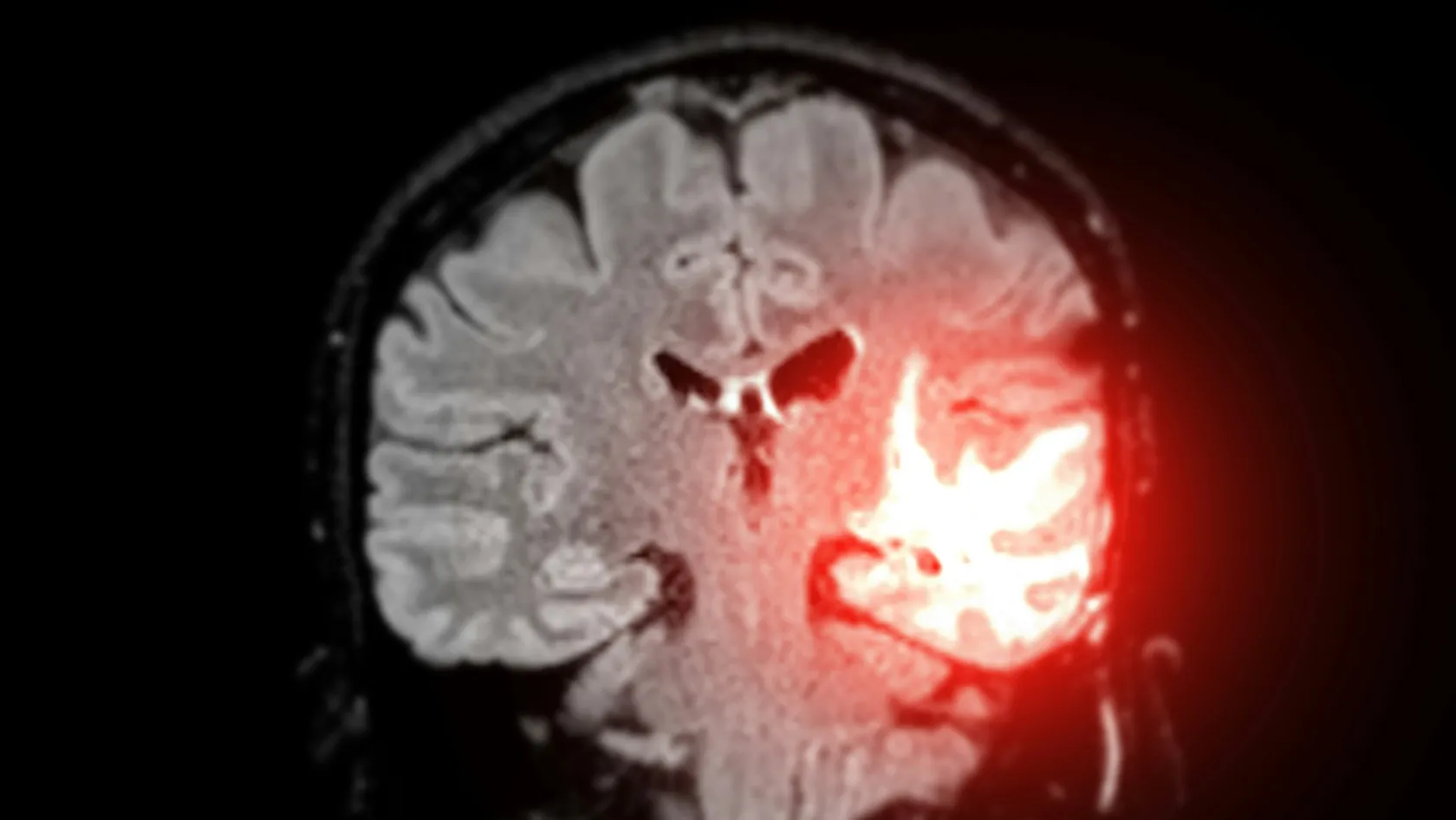
Brain Tumor Surgery - Brain Tumor Removal
Brain surgery is one trade repeated all day, how much tumor comes out against what the brain around it still does. This page puts the published numbers on both sides of that trade.
About This Department
Every brain operation is the same trade. How much comes out, against what still works.
Radiotherapy and chemotherapy are given on this same site, so a pathology report that changes the plan does not send you to another hospital to start again.
A scan has found something in your head and somebody has used the word tumor. Two facts rarely surface in the first hour of searching, and both of them belong at the front of your reading. The commonest primary brain tumor of all is meningioma, which grows from the covering of the brain, outside the brain tissue itself, is usually slow and benign, and can often be cured outright by taking it out completely. And of the tumors that do arise inside brain tissue, a large share are slow growing types where people live many years.
The scan alone cannot tell you which of these you have. Only the tissue can.
Surgeons offer an operation even when a tumor looks incurable on the pictures partly for that reason, because it gives diagnosis and treatment in one, and the molecular tests run on what comes out decide everything that follows. This page covers what brain tumor surgery involves, how surgeons decide how much to remove, the honest risk figures, and the parts a patient traveling from another country has to plan around.
What kind of tumor it is decides everything else
Brain tumor names a category. The diagnosis sits a level below it, and two people with the same size lesion on the same scan can face completely different operations, completely different follow up treatment and completely different outlooks, and reading survival figures for the wrong type is the fastest way to frighten yourself with information that has nothing to do with you.
This table scrolls sideways on a narrow screen. Swipe or drag to see every column.
| Type | What the operation aims at | What usually follows |
|---|---|---|
| Meningioma | Complete removal along with its attachment to the covering of the brain | Scans at intervals, and nothing else in most low grade cases |
| Low grade glioma | The largest safe removal, since more removed buys more time | Molecular testing, then watching or radiotherapy and chemotherapy |
| Glioblastoma and other high grade gliomas | Removing all the tissue that lights up on contrast, safely | Radiotherapy with chemotherapy, starting a few weeks after surgery |
| A secondary tumor from a cancer elsewhere | Taking out one accessible deposit that is causing symptoms | Focused radiotherapy, and treatment aimed at the original cancer |
| Pituitary tumor | Removal through the nose, with no opening in the skull at all | Hormone tests and endocrine follow up, sometimes lifelong |
| Lymphoma of the brain | A biopsy only, because removal helps nothing here | Chemotherapy, which this tumor responds to well |
Read the last row again, because it makes a point the rest of the page depends on. For one type of brain tumor the correct operation is to take a fragment and close, and a surgeon who removed it aggressively would have harmed the patient for nothing, so surgery does not automatically follow, and a team that says so about your scan is telling you something valuable.
Why surgeons push for more, and where they stop
For most tumors that grow inside brain tissue, how much comes out is the single surgical variable that changes the outcome, and the effect is larger than the word surgery suggests. A systematic review and meta-analysis in JAMA Oncology pooled 37 studies covering 41,117 patients with newly diagnosed glioblastoma. Compared with a partial removal, taking out all of the tissue that lights up with contrast reduced the risk of dying within a year by 38 percent, with a relative risk of 0.62, and it reduced two year mortality as well. Even a partial removal beat a biopsy alone.
Which raises the obvious question of why any surgeon stops early.
Because tumor and brain look almost identical under the microscope of the operating room, and the brain in question is running your right hand, or your ability to find the word for a spoon. A glioma has no capsule and no edge. It grows through healthy tissue like ink spreading in blotting paper, so the surgeon is not lifting an object out of a cavity, they are deciding, millimeter by millimeter, where tissue stops being expendable. Push a centimeter too far in the wrong place and the patient wakes with a weak arm that never recovers, loses their driving licence, loses their job, and gains nothing in survival because the tumor cells beyond that margin were never going to be reachable anyway. Two technologies moved that boundary outward, and both of them are ordinary practice now. Every serious neurosurgical department therefore uses the phrase maximal safe resection, with the second word doing as much work as the first.
Start with fluorescence. A patient drinks a dose of 5-aminolevulinic acid a few hours before surgery, malignant glioma cells convert it into a compound that glows pink under a particular blue light, and healthy brain does not. A randomized controlled trial across multiple centers, published in Lancet Oncology, tested this against ordinary white light in 322 patients and found complete removal of the contrast enhancing tumor in 65 percent of the fluorescence group against 36 percent with white light, with six month progression free survival nearly doubling, from 21.1 percent to 41.0 percent. Severe adverse events did not differ between the groups.
Mapping where function lives in this particular brain is the other, and the next section covers it in full.
Staying awake so your speech survives
Textbook diagrams of which part of the brain does what are averages. Your own language areas sit a centimeter or two away from where the diagram puts them, a tumor growing slowly for years may have pushed them somewhere else entirely, and no scan reads out that map with enough certainty to cut by. So for tumors near speech, movement or vision, the surgeon asks the brain directly, while you are awake and talking.
It sounds far worse than it turns out to be. You sleep through the painful parts, meaning the incision and the opening of the skull, then wake once the brain is exposed, which itself has no pain receptors. A neuropsychologist sits by your face for the next hour or so showing pictures to name, sentences to complete, or asking you to move a hand, while the surgeon touches a small electrical probe to the surface, and if you stop mid-word or your hand stops obeying, that patch of tissue stays. The map gets built point by point, on your brain, on that day. Then you go back to sleep for the closing.
People generally describe it as strange and tiring, and seldom as frightening or painful.
Awake surgery suits a tumor sitting in or beside a functional area, in a patient able to cooperate for an hour, so young children cannot do it, nor can anyone too confused or too unwell to follow instructions, nor a patient unable to lie still, nor someone whose anxiety no amount of preparation settles. Say plainly if you think you could not manage it, because the alternative is asleep surgery with electrical monitoring, which remains a good operation.
The operation, and the night that follows it
A tumor craniotomy commonly runs three to five hours, and longer for an awake case or a difficult location.
Only a strip of hair around the planned incision is shaved, since whole head shaving stopped being routine years ago, and a clamp then fixes your head so it cannot move by even a millimeter, while a navigation system links your pre-operative MRI to instruments in the surgeon's hand, so a pointer touched to the surface shows on screen exactly where it sits inside your scan. The surgeon opens the skin, drills a small number of holes, joins them with a fine saw, and lifts out a disc of bone that is kept for the end. Under the microscope the tumor is taken out piece by piece, with fluorescence or stimulation guiding the edges where the plan calls for them, then the bone goes back and is fixed with tiny plates, so nothing is missing from your skull afterward, and the skin is closed with stitches or clips. You wake in intensive care, and that is planned rather than a sign that something went wrong. Nurses check your pupils, your grip and your speech every fifteen to thirty minutes at first, because the point of that unit is to catch a problem in the window when it is still fixable. Most patients move to a normal ward the next morning. A scan within 72 hours records how much tumor was removed, which is the number that goes into every decision made afterward, and it is done early on purpose, before healing changes make the pictures harder to read.
Published patient information from large centers puts the whole hospital stay at around a week, and a straightforward meningioma or a single accessible deposit runs shorter than that.
What can go wrong, and how often
Risk in brain tumor surgery is dominated by location. Compare a tumor at the front of the right frontal lobe with one beside the speech area. Both carry the same anesthetic risk and completely different neurological risk, so any figure quoted for brain surgery in general is a starting point that your own surgeon should narrow for your own scan.
Ask your surgeon for their figure for your tumor, in your location, with your imaging in front of them. A neurosurgeon who answers with a range and a reason has thought about your case.
The first ten days, and what your family will see
Relatives arrive at the bedside expecting the person they know and meet somebody quieter, slower and stranger, then spend a night convinced the operation has gone wrong. This section exists to prevent that night.
Physiotherapy, speech therapy and occupational therapy start on the ward wherever they are needed, because the brain reorganizes fastest in the early weeks and waiting until you are home wastes that window. Steroids come down on a written schedule instead of stopping at once.
Stitches or clips come out around day seven to ten, and hair regrows over the scar and hides it within a few months.
When the pathology report changes the plan
Nobody knows exactly what the tumor is until the laboratory has finished with it, and while a frozen section during the operation gives the surgeon a rough answer within minutes, the full report takes several days and the molecular tests that now define these tumors take longer still. Those results routinely change what happens next, and for a patient who flew in for an operation and planned to fly out again, that is the part of the process most likely to go wrong logistically.
Understand three realistic changes before you travel. A tumor everyone expected to be benign returns as a higher grade, which turns a single operation into a course of radiotherapy. A glioma comes back carrying molecular markers that make chemotherapy clearly worthwhile, or clearly not. And a lesion assumed to be a primary brain tumor turns out to be a deposit from a cancer somewhere else, which changes the whole investigation and often the treating specialty. Here the arrangement at Biruni Hospital helps, and it should be said plainly. Radiotherapy and chemotherapy are both delivered on this site, on the same campus as the surgery. If your report calls for treatment to start a few weeks after the operation, you continue with the team that already knows your case, with no referral and no transfer of records between institutions. The alternative stays open too, and for many patients it is the better one, since going home to have radiotherapy near your family is easier to live through than six weeks in a hotel abroad. That route comes with a written handover to your own oncologist, including the operation note, the imaging and the full pathology.
Deciding that with the report in front of you beats deciding it in a panic at an airport, so say at the outset that you want the pathology discussed before you book your return flight, and build a flexible ticket into your budget.
Getting back to ordinary life
Six to eight weeks is the figure large centers quote for recovery from a craniotomy, and it describes the physical side reasonably well. Fatigue outlasts it. Brain fatigue after this operation is its own condition, distinct from ordinary tiredness, and it arrives without warning after concentrating for an hour, in a crowded room, or in a conversation with three people talking, and it improves over months and responds to planning your day in short blocks with real rest between them, which sounds trivial and turns out to be the single most useful piece of advice most patients are given. Walking daily from the first week. Light housework and short outings in the first month. Desk work part time from around six to eight weeks if the job allows it, with full time later and physical work later still, and no contact sport or anything that risks a blow to the head until your surgeon clears it, which can mean several months and permanently for certain sports.
People underestimate the driving restriction, which has nothing to do with how well you feel. Every country sets its own period off the road after a craniotomy or a seizure, often measured in months, and it is set by law rather than by your surgeon. Look up the rule that applies where you live before you make plans that depend on a car, and ask for the wording of your diagnosis in the discharge letter, because that is what your own licensing authority will want.
Alcohol, flying, swimming, hair dye and sexual activity all have sensible answers that depend on your wound, your medication and your seizure history, so write the questions down and ask them at the discharge conversation, where there is time, because searching for them at two in the morning three weeks later goes badly.
Flying home after a craniotomy
Opening the skull leaves some air inside the head. Surgeons call that air pneumocephalus, it shows on the scan afterward, and the body absorbs it over days to weeks. A cabin at cruising altitude is pressurized to roughly the equivalent of two thousand meters, so any trapped gas expands, and a pocket of air inside the skull that expands has nowhere to go. That physics, rather than the wound or the bone, is what governs when you can fly.
Practically, that means three things for you. Plan on being in the country for two to six weeks after the operation rather than a few days, and treat any package built around a shorter stay with suspicion. Ask for a scan before departure and a dated written fitness to fly opinion that names the pneumocephalus finding, because that is the document an airline or an insurer will ask for. And build the flight around seizure risk as well as air, meaning medication taken on schedule across time zones, sleep protected the night before, someone traveling with you, and no alcohol or sedatives that could disguise a change in your neurological state.
Flexible tickets cost less than changed ones.
Compression stockings and walking the cabin apply here as they do after any long operation.
What moves the cost of treatment
Brain tumor treatment makes the clearest case in medicine for publishing no price at all. Surgery forms one item on a bill whose other items stay unknown until the pathology comes back, so a number quoted before the diagnosis exists prices the wrong treatment.
Six things move the total. Where the tumor sits and how long the operation therefore takes, which separates a straightforward convexity meningioma from a deep tumor beside the brainstem, and whether the case is done awake, which adds a neuropsychologist and a longer list. Whether fluorescence, navigation and continuous nerve monitoring are used, each of which carries its own consumables. How many nights in intensive care and how many on the ward. The pathology itself, including the molecular tests that now define these tumors and that are not cheap. Rehabilitation during the stay. And any radiotherapy or chemotherapy that the report calls for afterward, which is a course of treatment rather than a line item.
Your own condition moves it too, through age, how well you are before surgery, steroid and anti-seizure medication, blood thinners, and other illnesses that lengthen the monitoring. Published packages in this market usually cover the transfers, the pre-operative tests, the surgeon and anesthesia fees, a stated number of nights including intensive care, the post-operative scan, an interpreter and a set number of hotel nights. They usually leave out flights, insurance, extra intensive care beyond the plan, the treatment of a complication, and, importantly here, the whole of any oncology treatment that follows.
The oncology bill is a separate conversation.
Five questions that make a quote comparable
Does this figure include the full pathology report and the molecular tests, or only the operation? How many intensive care nights are assumed, and what is the cost of one more? Are navigation, fluorescence and intraoperative monitoring inside the figure or billed separately? Is the scan within 72 hours of surgery included? If radiotherapy or chemotherapy is needed, when will that quote be given, and will treatment at home be supported if I prefer that?
Send the MRI and any biopsy result you already have. A neurosurgeon reading them will tell you whether the operation is worth traveling for, and that review costs nothing and commits you to nothing.
Traveling with someone who has a brain tumor
Most people reading this paragraph are not the patient. The daughter, the wife or the son organizes everything while the patient sleeps, and the practical detail below is written for them.
A companion counts as essential here in a way that other operations do not demand. Someone has to hold the medication schedule, notice a change in behavior that the patient cannot notice in themselves, and sit through the pathology conversation with a notebook, so patient rooms carry a second bed, one person stays overnight for the whole admission, and the international patients office arranges hotel accommodation for both of you on the nights either side, the airport transfers and every journey between hotel and hospital. Seizure risk alone makes independent taxi travel a poor plan in the first weeks.
Language matters more in neurosurgery than almost anywhere else in a hospital, for a reason that only becomes obvious at the bedside. Half the assessments performed on you are language tasks, and testing word finding through an improvised translation gives an answer nobody can rely on, and that is the reason the international patients team works in English, Arabic, French, Russian, Serbian, Romanian and Spanish, other languages are arranged on request, and one coordinator carries your case from the first message through to discharge. Raise your language at the very first contact if awake mapping is a possibility, because the mapping session has to be run in a language you are fluent in. A request for a woman surgeon or physician goes to the department and is met wherever the rota allows. The visa paperwork, meaning an appointment confirmation and an invitation letter naming the hospital and the treating doctor, is issued roughly ten days before travel and is what most consulates want with a medical visa application. Halal, vegetarian and diabetic meals come from the hospital kitchen, and there is a prayer room on the site.
Bring the original scan files on a disc or a drive, along with the report and a list of every medication with doses. Printed pictures will not do.
Scans, follow up, and who to call
Follow up after a brain tumor is scan driven and it lasts for years, so the file you leave with is what makes it work from another country, and it should contain the operation note, the discharge summary, the post-operative imaging, the full pathology including the molecular results, the steroid and anti-seizure schedules with their end dates, and the plan for the next scan. Any neurosurgeon or oncologist anywhere can pick that up.
Your coordinator stays reachable on the same WhatsApp number once you are home, so a photograph of a wound or a question about a dose is answered quickly, and scans done locally can be sent for review by the team that operated. A named doctor at home, arranged before you travel, handles the wound, the medication and the referrals.
Some symptoms cannot wait for the next scheduled contact, and after brain surgery that list is specific.
New weakness, new numbness, new trouble speaking or new loss of vision, or a first seizure, or a seizure different from your usual pattern, or headache that gets steadily worse over days, particularly with vomiting or drowsiness. Fever with a stiff neck, or a wound that is red, swollen or leaking, or clear fluid running from the wound or from the nose. Confusion that the people around you notice before you do. Swelling and pain in one calf, or sudden breathlessness. For any of these, the nearest emergency department the same day, with your operation note in hand, and a message to your coordinator once you have been seen.
Brain tumor surgery FAQ
How long do I need to stay before flying home?
Will I be awake during brain tumor surgery?
Can my family stay with me in the hospital?
Will my whole head be shaved?
What happens if the pathology shows I need radiotherapy?
Will someone speak my language on the ward?
When can I drive again?
Written by the Biruni Hospital medical editorial team. Reviewed by Dr Yunus Emre Yavuz, Neurosurgery.
References
- Brown TJ, Brennan MC, Li M, Church EW, Brandmeir NJ, Rakszawski KL, et al. Association of the extent of resection with survival in glioblastoma. A systematic review and meta-analysis. JAMA Oncology. 2016;2(11):1460-1469.
- Stummer W, Pichlmeier U, Meinel T, Wiestler OD, Zanella F, Reulen HJ. Fluorescence-guided surgery with 5-aminolevulinic acid for resection of malignant glioma. A randomised controlled multicentre phase III trial. The Lancet Oncology. 2006;7(5):392-401.
- Nowacka A, Sniegocki M, Atkins W, Ziolkowska E. Awake craniotomy versus general anesthesia for resection of high-grade gliomas. A systematic review and meta-analysis. Journal of Clinical Medicine. 2026;15(4):1431.
- Di Carlo DT, Cagnazzo F, Anania Y, Duffau H, Benedetto N, Morganti R, Perrini P. Post-operative morbidity ensuing surgery for insular gliomas. A systematic review and meta-analysis. Neurosurgical Review. 2020;43(3):987-997.
- Mampre D, Rupp C, Wilson J, Kelbert J, Riha G, Richardson M, et al. Postoperative recommendations for commercial air travel after neurosurgical intervention. Aerospace Medicine and Human Performance. 2026;97(6):411-418.
Editor's note
Written by the Biruni Hospital medical editorial team. Reviewed by Assistant Professor Fikret BAŞKAN, Neurosurgery.
Medically reviewed by
Assistant Professor Fikret BAŞKAN
Neurosurgery
Related Treatments
View All
ALIF - Anterior Lumbar Interbody Fusion Surgery
Coming in from the front restores the curve of the lower back better than any posterior route and leaves the back muscles untouched. It also carries one risk most pages will not name, and this page names it.
Brain AVM Surgery - Arteriovenous Malformation Surgery
An unruptured brain AVM bleeds at roughly 2.2 percent a year, and surgery spends its whole risk on a single morning. This page sets those two shapes of risk side by side, explains what the ARUBA trial does and does not say about your case, and shows how a Spetzler-Martin grade changes the answer.
Cervical Microdiscectomy - Minimally Invasive Neck Disc Surgery
Neck disc surgery is done from the front, through the space between your windpipe and your carotid artery, and almost everything that goes wrong that is not the disc happens in that corridor. What has been measured in it.
Chiari Malformation Surgery
A Chiari malformation is measured in millimeters and treated according to symptoms, and those two things come apart more often than patients are told. Most people with the finding never need an operation. This page sets out who does, what posterior fossa decompression removes, why surgeons still argue over opening the dura, and what the pooled studies say about both sides of that argument.
Deep Brain Stimulation Surgery - DBS Surgery
Stimulation does not slow Parkinson's disease down, and what it gives back is hours of the day when the medication is working without the writhing that used to come with them. This page sets out who qualifies, what the trials actually measured, how the two targets trade against each other, and what the hardware asks of you for the rest of your life.