
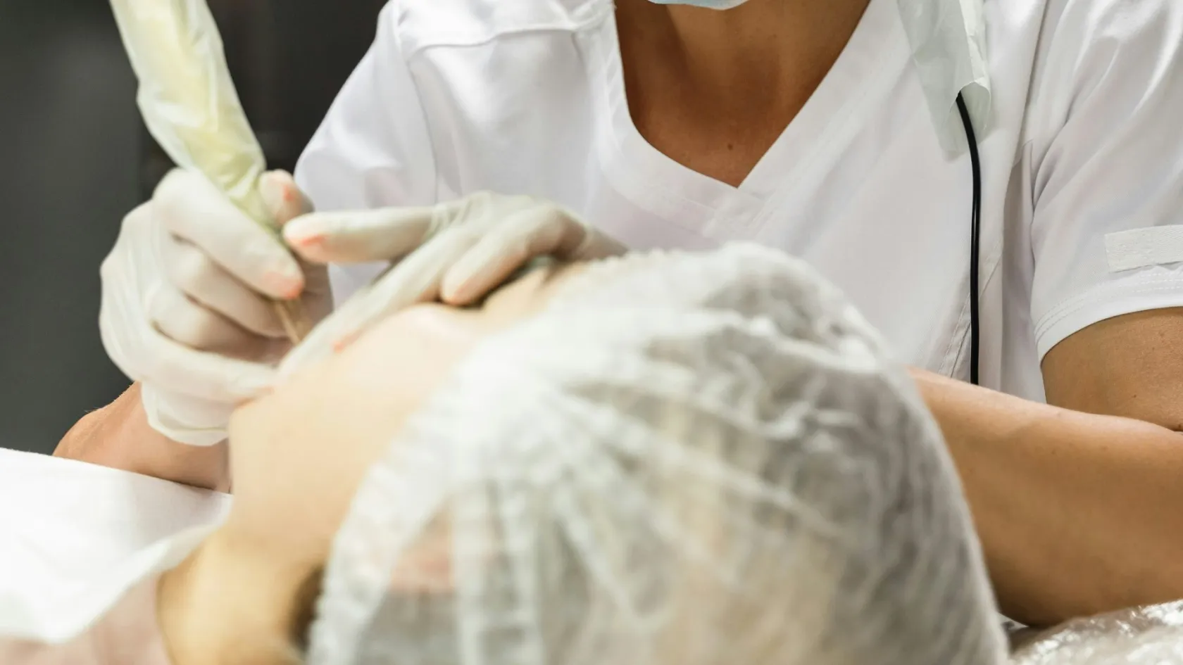
Soft Tissue Tumor Excision
The hard part is not the operation. It is deciding whether the lump should be scanned first, because that order cannot be corrected afterwards.
About This Department
Somebody has probably told you the lump is nothing to worry about and can come out whenever suits you. For most lumps that advice is correct. The difficulty is that the small number of lumps where it is wrong look almost identical from the outside, and the order in which things are done decides how much that matters. This article sets out which lumps need imaging before anyone operates, what a soft tissue tumor excision involves once that is settled, and how the answer changes between a lipoma in the back and a sarcoma in the thigh.
Free consultation
Send a description of the lump before you book anything
This review costs nothing and does not commit you to anything. Send its size, how long it has been there, whether it has changed, and any ultrasound or MRI already done. A surgeon will tell you whether the lump can simply be removed, whether it needs imaging first, and whether the operation belongs in a day unit or in a theater with a cancer team behind it.
Which lumps need removing
Soft tissue means everything between the skin and the bone, so fat, muscle, tendon, nerve, blood vessel and the fibrous tissue that binds them. Lumps arise in all of it, and in adults the overwhelming majority are harmless. Lipomas alone account for around half of all benign soft tissue masses, with ganglion cysts, epidermal inclusion cysts and schwannomas making up much of the remainder.
Reasons a benign lump comes out
Plenty of those never need touching at all. A small, soft, unchanged lipoma that causes no symptoms can be left indefinitely, and removing it is a decision about comfort or appearance alone. Excision earns its place when a lump hurts, catches on clothing or a waistband, presses on something, keeps coming back, or when the diagnosis is uncertain enough that leaving it would mean not knowing.
That last reason is where the sequence starts to matter.
The features that change the order of events
Nothing about the following list predicts cancer on its own. What it does is separate the lumps that can go straight to a minor operating list from the ones where a scan should come first, and the distinction exists because the two situations are almost impossible to tell apart by touch. A sarcoma sitting deep in a thigh is typically painless, firm, and has been quietly there for months, which is exactly how a harmless deep lipoma presents as well. Pain is a poor guide in either direction. Growth over weeks or months is the single feature most worth taking seriously, and a lump that a patient says has definitely got bigger deserves imaging even when everything else about it looks reassuring.
Get imaging before anyone operates if the lump
Measures 5 cm or more in any direction, which is roughly the size of a golf ball.
Sits deep to the fascia, meaning it feels fixed under the muscle layer instead of rolling freely under the skin.
Is growing, or appeared suddenly without any injury to explain it.
Feels tethered to the tissue around it instead of moving as a separate object.
Has come back after being removed once already.
Any one of these is reason enough for an ultrasound or an MRI with contrast before a decision about surgery. Roughly one malignant mass in ten measures under 5 cm at diagnosis, so a small lump with the other features still deserves a scan.
None of this means a lump with a red flag is cancer. Most turn out to be nothing. What it means is that the scan costs a few days and answers the question, while removing an unrecognized sarcoma piecemeal turns a single clean operation into two, and the second one takes far more tissue than the first would ever have needed, so the days spent waiting for imaging buy something real even in the great majority of cases where the answer comes back reassuring.
Is it a lipoma or something else?
Fatty lumps generate more anxious searching than every other kind combined, and the reason is that a lipoma and its awkward relative can look the same to a hand and nearly the same on a scan. That relative is the atypical lipomatous tumor, also called well differentiated liposarcoma. It does not spread to the lungs the way an aggressive sarcoma does, so it is not the disaster the word liposarcoma suggests. It does come back locally if it is shelled out like an ordinary lipoma, which is the whole reason for telling them apart first.
What the scan can settle, and where it cannot
MRI carries most of that decision, and the published detail is more useful to a worried reader than any reassurance. According to PubMed, a study of 79 patients with confirmed fatty tumors found that lesions showing an even fat signal throughout and measuring under 8 cm were lipomas in every case, while those with internal partitions of 2 mm or thicker, more than one non-fatty nodule inside them, or a maximum size of 12.8 cm and above were characteristically atypical lipomatous tumors (Moran et al, 2025). Between those two descriptions sits an uncertain group where the imaging cannot settle it and a biopsy is needed, and the same study put a specialist musculoskeletal radiologist's accuracy at 90 percent sensitivity against 66 percent specificity, which is a candid way of saying the scan rarely misses the dangerous ones and frequently over-calls the harmless ones.
A biopsy with a test for the MDM2 gene settles those remaining cases. That test is the reference standard.
For a patient this translates into something practical. A soft mobile fatty lump under 8 cm on your arm almost certainly needs nothing more than a straightforward excision if it bothers you. A firm 15 cm mass deep in your thigh needs an MRI and a specialist opinion before anyone reaches for a scalpel, however painless it has been.
Two operations wearing the same name
Soft tissue tumor excision covers two procedures with very little in common beyond the words.
A marginal excision follows the natural capsule around a benign lump and lifts it out along that plane. Local anesthetic often suffices, the incision is short, the whole thing takes half an hour or less for a modest lipoma, and the patient goes home the same day. That operation is entirely correct for a lump already known to be benign. It is not a compromise. The wide version is a different animal. Instead of following the capsule, the surgeon deliberately stays outside it and removes the tumor inside a continuous envelope of normal tissue, which means sacrificing healthy muscle and fascia around the lesion so that no tumor cell reaches the cut surface. General anesthesia is standard, the incision is long, muscle is divided, drains go in, and the defect sometimes needs a plastic surgical flap to close. That is the operation a sarcoma requires, and doing the marginal version instead leaves disease behind in tissue that looked completely normal.
This table scrolls sideways on a narrow screen. Swipe or drag to see every column.
| Marginal excision | Wide excision | |
|---|---|---|
| Suits | A lump already known to be benign | A confirmed or suspected sarcoma |
| Anesthetic | Often local, in a day unit | General, in a main theater |
| What comes out | The lump, along its own capsule | The lump plus a cuff of healthy muscle and fascia |
| Going home | The same day | After several days, once drains are out |
Everything in the earlier sections exists to make sure the right one of those two gets chosen the first time.
Lumps that sit on a nerve
A schwannoma grows from the sheath around a nerve and is benign. Patients typically notice a firm lump that sends an electric tingle down the limb when pressed, and surgeons describe the operation to remove it as simple, because the tumor can be shelled out while leaving the nerve fibers themselves intact. The published numbers are more sobering than that description suggests.
According to PubMed, a series of 30 schwannomas removed from major nerves in the lower limb found that 23 patients, meaning more than three quarters, woke up with some neurological deficit after surgery (Kim et al, 2012). Most of it resolved. At an average of just under five years, 19 of the 30 had no residual deficit at all, 9 had symptoms they found tolerable, and 2 were left with significant weakness and sensory loss. Larger tumors carried the higher risk, and one tumor recurred.
The honest framing
So the honest framing for a schwannoma on a major nerve is that most people end up fine, temporary numbness or weakness is the norm rather than the exception, and a small number keep a permanent deficit. Weigh that against how much the lump is troubling you now. A painless schwannoma that is not growing is a reasonable thing to leave alone and watch.
When leaving it alone is the treatment
Desmoid tumors, also called aggressive fibromatosis, occupy strange ground. They invade locally and recur stubbornly after surgery, yet they never spread to distant organs, and a substantial share of them simply stop growing or shrink without any treatment at all.
That behavior has changed the standard advice completely.
Active surveillance is a plan, not an absence of one, and it has parts.
- Scheduled MRI at set intervals, close together at first and stretching out as the tumor proves it is behaving.
- A named point of escalation agreed in advance, so that growth triggers a decision rather than a debate.
- Symptom control in the meantime, since these tumors can hurt while they are being watched and pain is not a reason to abandon the strategy on its own.
- A clear statement of what would change the plan, usually continued growth across two consecutive scans or a threat to a nerve, a vessel or a joint.
Watchful waiting, meaning scheduled scans and no operation, is now the preferred first approach for most desmoids, and surgery is reserved for tumors that keep progressing or that threaten something important nearby. According to PubMed, roughly 80 percent of desmoid tumors regress spontaneously or stabilize during first-line active surveillance, so treatment for the progressive minority is escalated in steps from the least toxic option upwards (Tsukamoto et al, 2022). A separate review of radiotherapy for these tumors reaches the same starting point, noting that up to half remain stable or regress and that watchful waiting is the preferred initial management (Looi et al, 2021). Both papers describe a disease where doing nothing, carefully and with scans, outperforms the instinct to cut, and that runs against everything patients expect to hear when a tumor is found.
Watchful waiting is the preferred initial management.
Two independent reviews reach the same starting point for desmoid tumors, in a disease where doing nothing, carefully and with scans, outperforms the instinct to cut.
If you have been told you have a desmoid and offered immediate surgery, that recommendation deserves a second opinion. It may well be right for your particular tumor. It is no longer the automatic answer, and asking why surgery now is a perfectly reasonable question to put to your team.
Radiotherapy before or after the excision
For a soft tissue sarcoma in a limb, radiotherapy accompanies the excision in most cases, and the choice of when to give it is a genuine trade-off with consequences the patient feels directly. This decision does not arise for benign lumps at all.
The evidence here comes from a randomized trial that has shaped practice for two decades. Per PubMed, 190 adults with limb soft tissue sarcoma were randomly assigned to radiotherapy before surgery at 50 Gy in 25 fractions or after surgery at 66 Gy in 33 fractions, and wound complications within 120 days occurred in 35 percent of the preoperative group against 17 percent of the postoperative group, a difference of 18 percentage points (O'Sullivan et al, 2002). Tumor size and site independently affected that risk, and overall survival was marginally better in the preoperative group.
This table scrolls sideways on a narrow screen. Swipe or drag to see every column.
| Timing | What it buys you | What it costs you |
|---|---|---|
| Before surgery | A lower total dose over a smaller treated area, which spares more tissue in the long run and can shrink the tumor before it is removed | Roughly double the rate of wound healing problems in the four months after the operation |
| After surgery | A markedly lower risk of the wound breaking down while it heals | A higher total dose across a wider field, which is linked to more long-term stiffness, scarring and swelling in the limb |
Neither column is the right answer on its own. A large tumor low down in the leg, where skin is thin and healing is slow, may argue for giving the radiotherapy afterward. A tumor in a thigh with generous soft tissue cover, in a patient who will live for decades with whatever stiffness the treatment leaves behind, more often argues for giving it first. Ask which way your own team is leaning and what specifically about your tumor points them there. Ask one logistical question alongside it, because the answer decides how the trip is shaped. Whether the radiotherapy is delivered on the same site as the surgery. Here it is, which means preoperative radiotherapy and the operation that follows it can be sequenced by one team instead of being fitted around two hospitals and a flight in between.
What goes wrong, and how often
Risk scales with the operation. One figure covering both versions of this surgery would mislead in both directions.
After a straightforward benign excision
Bleeding into the cavity the lump left behind, a wound infection, a collection of fluid called a seroma, and a scar the patient dislikes cover most of what happens. Numbness in a patch of skin beyond the scar is common, and it settles over months as small cutaneous nerves recover. Recurrence is the other one to raise beforehand, since an incompletely removed lipoma or ganglion cyst comes back often enough that hearing about it afterward feels like a surprise nobody warned you about.
After a wide excision for sarcoma
Wound healing dominates, and preoperative radiotherapy pushes that risk to around a third of patients within four months. A wound that breaks down over a radiotherapy-treated field heals slowly, sometimes needs a return to theater, and occasionally needs a flap of tissue moved in to cover it. Beyond the wound, removing muscle costs strength permanently, dividing lymphatic channels can leave a limb swollen, and a nerve running through the resection field may have to be sacrificed with a predictable and permanent deficit that should be described to you by name before you sign anything. Local recurrence remains possible even after a technically clean operation, which is what the surveillance scans afterward are looking for. Blood clots deserve their own mention too, in anyone having a long operation on a limb with a cancer diagnosis, since the disease raises clotting risk before surgery adds to it, and it is fair to ask what the unit does about that, because the answer should involve compression, early walking and, in many cases, injections that continue for a period after you go home.
From a day case to a long recovery
Weeks, after a small benign excision
Recovery from a small benign excision is measured in weeks and mostly consists of leaving the wound alone. Expect the dressing to stay dry for a couple of days, stitches or clips out at around ten to fourteen days, and a return to desk work within a few days once the anesthetic has worn off and the site is comfortable. Heavy lifting and sport wait until the wound has strength behind it, three to four weeks in most cases, and longer where the lump sat over a joint or under a strap of muscle that gets loaded every time you move. The scar itself goes on fading and softening for a full year after everything else has settled.
Months, after a sarcoma resection
Sarcoma surgery runs on a different clock entirely.
Drains stay in for days, until the daily output falls. The wound gets watched closely, and where radiotherapy came first it gets watched more closely still. Physiotherapy starts early and continues for months, aimed at restoring what the removed muscle used to do through the muscles that remain, and where a large volume of tissue came out, some loss of power or endurance persists no matter how diligently the exercises are done. Swelling in the limb can take months to settle and sometimes needs compression garments and specialist lymphedema therapy. Most people return to ordinary daily activity within two to three months, while the final ceiling on function is not clear for six to twelve, which is a long time to spend not knowing how much of your old strength is coming back and one of the reasons a physiotherapist who has handled sarcoma patients before is worth seeking out at home.
Stay, flying and what fits a trip
Travel plans are where the two operations diverge most sharply, and a general answer would serve nobody.
A benign lump, on a short trip
A benign excision suits a short trip well. Consultation and surgery can often fall on consecutive days, the procedure itself is a day case, and the wound itself is what constrains flying. Allow several days after the operation for a wound check before departure, since a wound reviewed once before you fly is far easier to manage than one first inspected at home. A short flight a few days after a small excision on the arm is a very different proposition from one after a groin or thigh wound that will be sat on for six hours.
A sarcoma, on a much longer one
Sarcoma surgery needs a completely different plan. Inpatient stay runs into days, drains have to come out before departure, the wound needs review over a longer window, and the histology confirming margin status arrives after a week or two, frequently once the patient is already home. Where radiotherapy forms part of the plan, a course before surgery adds several weeks in the country by itself, and coordinating that from abroad is precisely the arrangement that goes wrong when two teams in two countries each assume the other is holding the timeline.
Flight clearance for either version rests on the wound, on mobility and on clotting risk, and it is decided at a review before departure. No date can honestly be promised in advance. Ask for a fitness-to-fly letter before discharge, since airlines sometimes require one after surgery, and if the excision was anywhere you will be sitting on for the length of the flight, say so when the clearance is being discussed rather than discovering the problem at the gate.
What separates a small excision from a big one
Two quotes for a soft tissue tumor excision can differ by an order of magnitude, and nothing dishonest has to happen for that to be true.
The anesthetic sets the floor, since a lump removed under local anesthetic in a treatment room costs a fraction of the same lump removed under general anesthetic in a main theater. Size and depth then drive almost everything else, because a superficial lump needs a short procedure while a deep one near vessels and nerves needs a long one with more monitoring. A wide excision for sarcoma adds theater time, a frozen section service, drains and an inpatient bed. Reconstruction is the largest single step change, since a defect that will not close directly needs a skin graft or a plastic surgical flap and a second surgical team. Where imaging and pathology are involved, the MRI, the biopsy and the specialist analysis of the specimen all carry their own line items, and a test for the MDM2 gene on a fatty tumor is not the same cost as a routine report. Radiotherapy, when it forms part of the plan, is a separate course of treatment and gets priced on its own.
This table scrolls sideways on a narrow screen. Swipe or drag to see every column.
| Small benign excision | Wide excision for sarcoma | |
|---|---|---|
| Anesthetic | Local, in a treatment room | General, in a main operating theater |
| What sets the length | The lump sits superficially and comes out in minutes | Depth, and proximity to vessels and nerves that have to be dissected clear |
| Bed | None, you walk out | An inpatient bed, with drains and monitoring |
| Pathology | A routine report on the specimen | Frozen section during surgery, then specialist analysis, and a MDM2 test where the tumor is fatty |
| Closing the gap | Direct closure | A skin graft or a plastic surgical flap, and a second surgical team with it |
| Radiotherapy | Not part of it | A separate course of treatment, priced on its own |
Patient factors matter less here than in bigger cancer surgery. Age, diabetes, blood thinning medication and previous surgery at the same site still affect how the case runs and how the wound behaves afterward.
Packages published in this market for soft tissue lump surgery generally cover the surgeon and anesthesiologist fees, theater time, the histopathology on whatever was removed, dressings and the follow-up visits before departure. Flights, accommodation past the stated nights, treatment of a complication, a return to theater and any radiotherapy sit outside them.
Before accepting a figure, pin down which anesthetic it assumes, whether the histopathology on the specimen is included or billed afterward, what happens financially if the pathology comes back malignant and a second wider operation becomes necessary, and whether the pre-operative MRI sits inside the quote. That third question is the one most people forget to ask, and it is the one that turns a small planned expense into a large unplanned one. A surgeon who has seen your imaging is the only person who can answer it, and that review costs nothing.
After you get home
Every lump that comes out goes to a pathologist. That includes the ones everybody was confident about, and the report is the single most important thing to chase once you are home.
It sometimes contains a surprise. A lump excised as a straightforward lipoma occasionally returns as an atypical lipomatous tumor, and that changes the follow-up plan even though it rarely changes the immediate outcome. Make sure the report reaches you and your own doctor directly and in writing, so it never sits in a file you would have to ask for.
Take the operation note home too, with any imaging.
Practical follow-up after a benign excision is short. A wound check near home at around two weeks for stitch removal, and nothing further unless the lump returns. After a sarcoma excision it runs for years, with scheduled scans of the operated area and of the chest, arranged where you live because nobody flies back for routine imaging. Keep a line of contact to the operating surgeon through all of it, since a scan showing something ambiguous at the operated site is far easier to interpret with input from the person who made the excision and knows exactly which tissue they took and which they deliberately left, and agreeing that line of contact before you are discharged is much easier than establishing it from another country when something has already started to worry you.
Frequently asked questions
Can I fly home the day after a lump is removed?
Does every lump need a scan before it is removed?
What happens if the pathology comes back as a sarcoma?
Will removing a lump on a nerve leave me with numbness?
Should a desmoid tumor be removed straight away?
References
- O'Sullivan B, Davis AM, Turcotte R, et al. Preoperative versus postoperative radiotherapy in soft-tissue sarcoma of the limbs. A randomized trial. Lancet. 2002;359(9325):2235-2241.
- Moran LM, Li Cai CY, Ramirez A, Royuela A. Differentiation of Atypical Lipomatous Tumors from Lipomas. Our Experience with Visual Analysis of Conventional Magnetic Resonance Imaging. J Imaging. 2025;11(2):47.
- Kim SM, Seo SW, Lee JY, Sung KS. Surgical outcome of schwannomas arising from major peripheral nerves in the lower limb. Int Orthop. 2012;36(8):1721-1725.
- Tsukamoto S, Takahama T, Mavrogenis AF, et al. Clinical outcomes of medical treatments for progressive desmoid tumors following active surveillance. A systematic review. Musculoskelet Surg. 2022;107(1):7-18.
- Looi WS, Indelicato DJ, Rutenberg MS. The Role of Radiation Therapy for Symptomatic Desmoid Tumors. Curr Treat Options Oncol. 2021;22(4):34.
Editor's note
Written by the Biruni Hospital medical editorial team. Reviewed by Assistant Professor Şafak SAYAR, Orthopedics and Traumatology.
Medically reviewed by
Assistant Professor Şafak SAYAR
Orthopedics and Traumatology
Related Treatments
View All-
Achilles Tendon Repair
Achilles tendon repair stitches a torn tendon back together and cuts the risk of a second rupture, while a boot with early movement gives the same function at one year in trials. This guide covers the signs of a tear, surgery or a boot, open and minimally invasive repair, rehabilitation week by week and the stay.
-
Bone Tumor Resection
Most people arrive expecting to lose the limb. For the large majority of bone tumours today the limb is saved, the bone is rebuilt with an implant or a graft, and survival is no worse for it. What decides the answer is the margin, and the margin is planned from an MRI of the whole bone before anything is cut.
-
Chondrosarcoma Surgery and Bone Tumor Resection
Chemotherapy and radiation barely touch this tumor, which sounds like the worst news on the page and is the reason a single operation cures it outright. For a grade 1 chondrosarcoma in an arm or a leg the bone is scraped clean from the inside and the natural joint stays where it is.
-
Complex Knee Revision Surgery
Infection now causes more failed knee replacements than any other single reason, 21.6 percent across registries in seven countries, and it can turn a revision into two operations with weeks of antibiotics between them. This page explains how the cause is found before surgery, when cones or a hinge are needed, and how a two-stage revision fits around travel.
-
Complex Trauma Reconstruction
A fracture that has not healed after a year, healed crooked or became infected can still be repaired, and the treatment often runs for months in an external frame. This page explains nonunion, malunion and bone loss repair, the choice between bone transport and the Masquelet technique, and how a long treatment fits around travel to Istanbul.