
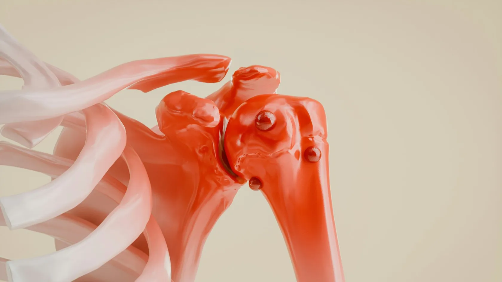
Chondrosarcoma Surgery and Bone Tumor Resection
Chemotherapy and radiation barely touch this tumor, which sounds like the worst news on the page and is the reason a single operation cures it outright. For a grade 1 chondrosarcoma in an arm or a leg the bone is scraped clean from the inside and the natural joint stays where it is.
About This Department
Cartilage makes this cancer, and chondrosarcoma behaves nothing like the bone cancers most people have already read about by the time they reach this page. Chemotherapy barely touches it. Radiation barely touches it. That sounds like the worst sentence on the page and it is actually the reason for optimism, because most chondrosarcomas are low grade, they grow slowly, they almost never spread, and one properly planned operation cures them outright.
Free consultation
Send the MRI, the biopsy slides and the CT, and get a grade, a margin plan and a reconstruction plan back
An orthopedic oncologist reviews the MRI of the whole bone, the biopsy report and the chest CT, then tells you what grade the tumor most likely is, whether the joint stays, how much bone would come out, and what would go in its place. Pathology slides are re-read here before anything is planned, because the grade on the first report changes more often than patients expect. The review costs nothing and carries no obligation, and a coordinator answers in your own language.
What the grade decides
Every meaningful decision in chondrosarcoma surgery follows from one number that a pathologist assigns under a microscope. Grade 1 tumors, now called atypical cartilaginous tumors when they sit in an arm or a leg, are locally troublesome and almost never lethal. Grade 2 tumors spread in a minority of patients. Grade 3 tumors behave like any other high grade sarcoma and send deposits to the lungs. The operation changes completely between the first of those and the last, which is why a surgeon who has not seen the slides cannot tell you what your operation will be. Nine in ten chondrosarcomas are conventional, meaning ordinary cartilage-forming tumors graded 1 to 3. The rest fall into rarer subtypes with their own rules. Dedifferentiated chondrosarcoma carries a high grade sarcoma growing out of a low grade cartilage tumor and it is the most dangerous of the group. Mesenchymal chondrosarcoma turns up in young adults and responds to chemotherapy, which the conventional tumors do not. Clear cell chondrosarcoma sits at the top of the femur or the humerus, grows very slowly, and is cured by removing it properly.
Age separates this disease from the sarcomas that affect teenagers. This cancer belongs to adult and middle life, with the peak between forty and seventy, and that changes the conversation over reconstruction, because a sixty-year-old who needs a hip replaced with a tumor prosthesis is asking a different question from a fifteen-year-old whose implant has to last another sixty years.
Grade by grade, at a glance
The table below scrolls sideways on a narrow screen. Swipe or drag to see every column.
| Grade or subtype | Usual operation | What it means for you |
|---|---|---|
| Grade 1, arm or leg | Curettage with a local adjuvant, cavity filled | Natural joint kept, short stay, cure expected |
| Grade 1, pelvis or spine | Wide resection, because recurrence here kills | Bigger operation than the same grade in a femur |
| Grade 2 | Wide resection and reconstruction | Chest CT surveillance for years afterwards |
| Grade 3 | Wide resection, wider than grade 2 | Real risk of lung spread, close follow-up |
| Dedifferentiated | Wide resection, chemotherapy considered | The one subtype where drugs enter the plan |
| Mesenchymal | Chemotherapy and surgery together | Treated much closer to Ewing sarcoma |
The cartilage lump that is probably not cancer
Many people reading this were told of a cartilage lesion after a scan done for something else entirely. A knee X-ray after a football injury. A shoulder MRI for a rotator cuff. The radiologist reports a cartilage lesion in the upper humerus or the lower femur, the word chondrosarcoma appears in the differential, and a frightening week follows.
Most of those lesions are enchondromas. Enchondromas behave themselves. They sit quietly in the marrow for decades, need no operation, and operating on one gains the patient nothing while costing them a scar, a hospital stay and a risk of infection. The difficult cases sit on the boundary between an enchondroma and a grade 1 tumor, and that boundary is genuinely hard, because the two look similar down a microscope and pathologists disagree over individual cases more than anyone would like.
So what moves a lesion from watch to operate. Pain at the site that is present at rest and at night, and that cannot be explained by the joint next to it. Growth between two scans taken months apart. Scalloping of the inner cortex deeper than two thirds of its thickness, or swelling of the bone with a soft tissue mass pushing out of it. Uptake on a bone scan that is clearly hotter than the surrounding skeleton. When several of those appear together, the lesion is treated as a tumor.
Send both scans if you have them and not the newest one alone, because the comparison between an image from two years ago and an image from last month answers the question faster than any single study can.
Why drugs and radiation do not cure this tumor
Conventional chondrosarcoma resists chemotherapy and radiotherapy, and every serious source on the disease says so plainly. The tumor consists mostly of cartilage matrix with few dividing cells scattered through it, and it has a poor blood supply, so drugs that kill fast-dividing cells in a well perfused tumor arrive in small amounts and find little to kill. Radiation runs into the same wall. Nobody enjoys writing that sentence, so here is what it actually means for you, starting with the comparison every patient makes, because in osteosarcoma the surgeon is one part of a treatment that also involves months of drugs, and there the drugs carry much of the cure. Here the surgeon carries all of it. That places an unusual amount of weight on a single day in an operating theatre, and it explains everything else on this page, from why the pathology slides get re-read before planning, to why the margin is measured in millimeters, to why the choice of unit matters more in this disease than in almost any other cancer.
What that means in practice
One practical consequence lands well with patients. No months of chemotherapy means no months of hair loss, mouth ulcers, low blood counts and repeated admissions. For a conventional chondrosarcoma removed with clear margins, treatment is an operation and then a follow-up schedule, and that is the whole of it.
Grade 1 in an arm or a leg, with the joint kept
Now the best news in this disease. A grade 1 chondrosarcoma in a long bone does not need the bone segment removed. Surgeons treat it from the inside.
The surgeon opens a window in the cortex over the tumor, scrapes the cavity out completely with curettes, then enlarges it with a high speed burr so that the walls are taken back into healthy bone well past the tumor edge. A local adjuvant is applied to the cavity to kill whatever cells the instruments missed. Bone cement then fills the hole, sets hard immediately, supports the bone at once and shows up clearly on later X-rays, so any recurrence stands out against it. The joint next to the tumor is never opened. Nothing is replaced.
Most patients walk on the limb within days and keep the knee or the shoulder they were born with, for life.
Oncological results support this. A Dutch series published in the Journal of Surgical Oncology in 2021 followed 55 enchondromas, 119 atypical cartilaginous tumors and 5 grade 2 tumors of the long bones treated by curettage with cryosurgery as the adjuvant, with a median follow-up of 53 months, and reported seven recurrences in the whole cohort, three of them in the atypical cartilaginous tumor group.
Patients ask what fills the hole. Bone cement usually does, for three reasons that have nothing to do with cost. It sets within minutes, so the bone carries weight the same week instead of waiting months for a graft to unite. The heat it gives off while it cures kills a further layer of cells at the cavity wall. And on every X-ray afterwards it stands out white against bone, so a recurrence growing at the edge of the cavity announces itself years before it would in a graft-filled defect. Bone graft is preferred where the cavity sits directly under a joint surface and cement would sit against cartilage.
Follow-up for these patients is light. An X-ray of the site and a clinic review, first at three months and then at widening intervals, with no chest CT unless the pathology was upgraded on the specimen, because a genuine grade 1 tumor in a limb does not spread to the lungs and scanning the chest of someone who has one achieves nothing except worry. Two conditions have to hold before this route is offered. The tumor must genuinely be grade 1, confirmed on slides that a bone tumor pathologist has read. And it must sit in a limb bone, since in the pelvis, the spine and the sacrum the same grade behaves far more dangerously because a recurrence there cannot be dealt with as easily.
Grade 2 and grade 3, and what a wide resection removes
Once the grade rises above 1, the tumor comes out inside a continuous envelope of normal tissue that is never breached during the operation. That envelope is the whole point. The surgeon does not see the tumor at any stage of a properly executed wide resection, because seeing it would mean cutting into it, and cutting into a chondrosarcoma spills cells that implant and grow wherever they land. Planning starts from an MRI of the entire bone from one joint to the next, which shows how far the tumor has travelled inside the marrow. From that, the surgeon marks the level of the bone cuts, decides which muscles come out attached to the specimen because they lie against the tumor, and identifies the nerves and vessels that have to be freed and preserved. The biopsy track is included in the specimen and removed with it, since tumor cells were dragged along it when the needle came out, and every decision of that kind happens before the first incision, so the operation executes a plan that already exists on paper.
Local recurrence is not a setback in this disease. It is the event that changes the outcome.
Evidence from 2023 makes the point. A study in the Iowa Orthopaedic Journal combining a single institution cohort with 516 cases from the SEER database found that patients who developed a local recurrence were 17.5 times more likely to die within a year after adjusting for age, and the same study found better five year survival after limb salvage than after amputation, with a hazard ratio of 1.49.
That last finding surprises people, so here is the explanation behind it. Amputation does not fail because it is a bad operation. Surgeons choose it for larger and more advanced tumors, and the size of those tumors drives the survival difference. What the finding does establish is that saving the limb costs nothing in survival when a clear margin is achievable, which is the question every patient facing this operation actually asks.
How much margin is enough
Guidelines say a wide margin without saying how wide, which is unhelpful when the tumor sits a few millimeters from the iliac vessels and the surgeon has to decide in theatre what is acceptable. A 2019 multicenter study in Clinical Orthopaedics and Related Research put a number on it. Fifty patients with peripheral chondrosarcoma of the pelvis were followed for a median of seven years, and local recurrence-free survival at ten years was 100 percent when the margin measured at least 1 mm, against 52 percent when it measured less than 1 mm. One millimeter. What that millimeter has to be is continuous, uninterrupted and microscopically clear, all the way around the tumor, and a centimeter of muscle taken carelessly is worth less than a millimeter taken deliberately. That number liberates more than it frightens, because it means limbs, nerves and organs that would be sacrificed under a rule of thumb are preserved instead, provided the surgeon knows exactly where the tumor edge is and works with that precision.
Achieving that millimeter is a matter of knowing where the tumor edge is, so the operation is planned from an MRI of the whole bone and never from the area the radiologist happened to report. Cartilage tumors push a rim of reactive tissue ahead of themselves, and the surgeon has to cut outside that rim without cutting through the artery, the nerve or the bladder that happens to lie on the other side of it, which is the whole difficulty of this operation expressed in one sentence. Where the plane is genuinely tight, the specimen is oriented and inked in theatre so the pathologist can report exactly which face was closest, and that report is what tells you whether anything further is needed.
The same study reported something else that patients deserve to know before they travel. The grade on the preoperative biopsy matched the grade on the resected specimen in only 15 of 41 patients, which is 37 percent. Chondrosarcoma grows unevenly, a needle samples a few millimeters of a tumor several centimeters across, and the most aggressive area is exactly the part a needle is most likely to miss. Practical consequences follow from that. Surgeons plan the operation for the grade the imaging and the whole clinical picture suggest, and never for the grade written on a biopsy report alone. And the definitive grade only exists after the whole specimen has been examined, which is why the conversation after surgery sometimes differs from the conversation before it.
The pelvis and the sacrum
After the femur, the pelvis holds more of these tumors than any other site, and it makes for the hardest operations in the specialty. A pelvic tumor grows large before it causes symptoms, because there is room in the pelvis for a mass to grow quietly, and by the time it is found it sits among the iliac vessels, the sciatic and femoral nerves, the bladder, the ureter and the bowel. Clearing it while keeping all of those working is a long operation, frequently with a vascular surgeon scrubbed and sometimes with a urological or colorectal colleague, Transfusion is planned in advance and not improvised.
Grade 1 does not mean a small operation here. In the pelvic series described above, patients died from local recurrence pressing on structures that could not be decompressed, including patients whose original tumors were grade 1. A recurrence in a limb can usually be cut out again. In the pelvis it frequently cannot, and that single difference explains why the same pathology is treated more aggressively in this location. Reconstruction depends entirely on which part of the pelvis came out. Removing the wing of the ilium sometimes needs nothing at all, and patients walk with a slight difference in leg length and remarkably good function. The socket coming out means the hip has to be rebuilt so that the femur has something to articulate with. Sacral tumors raise a separate and difficult question over the nerves that control the bladder and the bowel, and where those nerves have to be divided, the consequences are permanent and they are explained in detail before anyone signs a consent form.
Surgeons describe pelvic resections by zone. Zone one is the wing of the ilium, zone two is the socket of the hip, zone three is the pubis and the ischium at the front, and zone four is the sacrum. Which zones the tumor occupies decides almost everything that follows, from how long the operation runs to whether you will walk with a limp, an aid or neither, and the plan is written in those terms before you travel so that you know which conversation you are having.
Zone three resections surprise people. A tumor of the pubic bone can be taken out with no reconstruction at all, and function afterwards is close to normal, because the front of the pelvic ring carries far less load than patients assume, which is one of the few places in this operation where the anatomy is on your side.
Sacral tumors are the other end of the scale. The nerves that leave the sacrum control the bladder, the bowel and sexual function, and how much of that is preserved depends on the level of the cut, so a resection below the third sacral nerve roots leaves continence intact in most people, while a higher one may not. Those consequences are permanent, they are explained in detail with the specific level marked on your own scan, and no one is asked to sign a consent form until that conversation has happened in a language they are comfortable in.
Rehabilitation after a pelvic resection is slower than after any limb operation. Expect several weeks of supervised physiotherapy before the flight home and a further several months of strengthening after it.
A SEER analysis of 623 spinal and pelvic chondrosarcomas published in Spine in 2021 built a model of cancer-specific death and found radical resection strongly protective, with a subdistribution hazard ratio of 0.38, while high grade raised the risk with a ratio of 2.68 and distant metastasis raised it with a ratio of 5.18.
Removing the tumor completely, at the first attempt, is the single most powerful thing anyone can do for a patient with a tumor in this location.
Chest wall, scapula, spine and skull base
Ribs, sternum and shoulder blade
Chondrosarcoma of a rib or the sternum is removed with the ribs above and below it and with the pleura underneath, and the chest wall is then rebuilt with a mesh and a rigid layer so that the chest does not move paradoxically with each breath. Breathing dominates recovery here, and physiotherapy in the first week works on the chest. Scapular tumors come out with part or all of the shoulder blade while the arm and the hand are kept entirely, and although the shoulder loses overhead reach afterwards, the hand keeps its full function, which is what determines whether someone can work.
The mobile spine
Vertebral tumors come out in one piece where the anatomy allows, which means an operation in two stages through the front and the back, followed by instrumented fixation to hold the spine together where the vertebra used to be. Even in patients whose disease has already spread, taking out the primary tumor changes the outlook. A SEER analysis of 110 metastatic spinal chondrosarcomas published in Spine in 2020 reported a hazard ratio of 0.262 for overall survival in patients whose primary tumor was resected, with the benefit concentrated in conventional subtypes, grades 1 to 3 and patients under seventy.
Skull base and head and neck
Chondrosarcoma at the base of the skull grows near the carotid artery and the cranial nerves, and it is approached jointly with neurosurgery and with head and neck surgery, and while the goal stays the same, meaning removal of as much tumor as the anatomy safely allows, a truly wide margin is rarely achievable in that space. Residual disease at the skull base is where particle radiotherapy has its clearest role, and a plan that needs it is written into the discharge documents with a named referral, so the patient is not left to work it out at home.
When the pathology changes the plan
Because a needle biopsy gets the grade right in a third of cases, the report on the whole specimen occasionally says something the pre-operative plan did not anticipate. Two findings change what happens next.
Dedifferentiated chondrosarcoma is the first. A low grade cartilage tumor that has been sitting in a bone for years develops a high grade sarcoma inside it, and the specimen shows both parts side by side under the microscope. This subtype behaves aggressively and it is the one place in conventional practice where chemotherapy enters the discussion, since the dedifferentiated component is not made of cartilage and can respond to drugs the cartilage would ignore. Mesenchymal chondrosarcoma is the second. It appears in younger patients, it is sensitive to chemotherapy, and it is treated on a protocol much closer to the one used for Ewing sarcoma. Being in one hospital instead of three matters at exactly this moment. Radiotherapy and chemotherapy are both delivered at Biruni Hospital, on the same site as the surgery, so a patient whose specimen comes back dedifferentiated does not need a referral to another institution, a second set of appointments in a new city, or a transfer of records that takes weeks. The pathology goes to the same tumor board, the medical oncologist reads the same file, and the first cycle can start once the wound has healed.
Going home for that treatment stays open and it is a perfectly reasonable choice. Patients who prefer to have their chemotherapy near their own family leave with the full histopathology, the staging scans and a written summary addressed to their own oncologist, and the coordinator here stays reachable while that handover happens.
Rebuilding the bone
Removing a segment of femur or humerus leaves a gap. Four methods fill it, and which one suits you depends on your age, on whether a joint has to be replaced, on how much muscle is left to power the limb, and on how quickly you need to bear weight.
The four options
A tumor prosthesis is not a hip replacement and should not be described as one.
These implants sit in bone that has lost its muscle attachments, they carry loads that a standard joint replacement never sees, and published series report substantial rates of revision over the years, from infection, from loosening of the stem, from wear, and from soft tissues failing to hold the joint. None of that argues against the implant. It argues for hearing the numbers before the operation instead of after the first revision, and for choosing a unit that keeps the same patients under review for decades.
Risks, and what is done for each one
Complication rates after tumor resection run higher than after ordinary orthopedic surgery, because the operations are longer, the wounds are larger and the soft tissue cover is thinner once muscle has been removed with the specimen. Each of the following has a standard response, and they are set out here so that you recognize a problem early.
The late one
Mechanical failure of a reconstruction belongs on this list too, although it arrives years later. Stems loosen, bearings wear, allografts occasionally fracture, and a proportion of patients with a large implant will need a revision at some point in their lives, and knowing that from the beginning turns a later operation into an expected part of the plan instead of a sign that something went wrong.
Recovery, week by week
Physiotherapy starts the day after the operation and the reconstruction sets the pace from there. A cavity filled with cement after curettage allows weight through the limb almost at once, so those patients are walking within two or three days and go home with little more than a wound to look after. Megaprostheses also allow early weight bearing, with standing on the first or second day and walking with a frame inside the first week. An allograft or a fibula has to be protected until the bone unites, which means crutches and limited weight for two to four months, and that difference belongs in the conversation before you choose between them. The first month belongs to the wound, the swelling and persuading the muscles around the reconstruction to fire again, since a quadriceps that has been cut and repaired forgets how to work and has to be retrained deliberately. Expect daily physiotherapy in that phase. By six to twelve weeks a patient with a lower limb reconstruction is walking without aids on level ground, and shoulder or elbow reconstructions take longer to become useful because the soft tissues around them carry more of the work. Full function takes six months to a year, and the last part of it is strength.
Patients ask what they will not be able to do. Running and jumping on a lower limb megaprosthesis are discouraged for life, because impact loading is what loosens the stem, and swimming, cycling and walking are encouraged instead, whereas after curettage and cement in a long bone there are no permanent restrictions at all once the bone has consolidated.
How long you stay, and when you can fly
Length of stay depends on which operation you are having, so here are three different trips.
Curettage and cement for a grade 1 tumor in a limb is the shortest.
Two to four days on the ward, a wound check ten days later, and around two weeks in Istanbul in total. A wide resection with a tumor prosthesis or a graft is the middle case, with three to seven days of assessment and pathology review before the operation, seven to fourteen nights in hospital, then two to three weeks of daily physiotherapy while the wound heals, which comes to three to four weeks in the country. A pelvic or sacral resection sits at the far end, with a longer inpatient stay, several weeks of rehabilitation and a total closer to six weeks.
This table scrolls sideways on a narrow screen. Swipe or drag to see every column.
| Operation | Nights on the ward | Total time in Istanbul |
|---|---|---|
| Curettage and cement, limb | Two to four | Around two weeks |
| Wide resection with reconstruction | Seven to fourteen | Three to four weeks |
| Pelvic or sacral resection | Longer, and variable | Closer to six weeks |
Custom and expandable implants have to be ordered and manufactured, which adds two to four weeks before an operation gets a date at all. Ask about implant lead time in your first message, because it is the item most people fail to plan for.
Clot risk governs the flight home and it is substantial after major lower limb surgery in a cancer patient, so injected anticoagulation continues for several weeks after discharge and travels with you, and while general guidance after major orthopedic surgery puts the earliest reasonable flight at around two weeks, every one of the stays described above already places you beyond it. You get a written clearance date with a number on it, book an aisle seat, and hourly walking and calf exercises during the flight are written into the discharge instructions.
What drives the cost
No figure appears on this page, and that is deliberate. Two people with the same diagnosis can need very different operations, so a number published here would be wrong for almost everyone who read it, whereas knowing which variables move that number helps enormously, because then you can read the quote you are sent instead of comparing headline numbers that cover different things.
Six things drive it in this disease. Whether the operation is a curettage or a segmental resection, since those are different operations of very different length, and which reconstruction goes in, because a modular prosthesis, a custom implant and a bone graft are not comparable items. Whether the implant is standard or manufactured to your own measurements. The site, since a pelvic or sacral resection needs a longer theatre session, more staff and more blood than a femoral one. The number of nights on the ward and how many of them are in intensive care. And how much inpatient physiotherapy the reconstruction requires before you can travel.
Patient factors matter as well. Age, weight, diabetes, heart and lung disease, blood thinning medication and any previous surgery at the same site all change the plan, and therefore the total.
Packages published by hospitals and medical travel agencies in this market usually cover the airport transfers, the pre-operative tests, the surgeon and anesthesia fees, the implant, the planned nights on the ward, inpatient physiotherapy, an interpreter, a set number of hotel nights and the follow-up appointments before you fly. They usually exclude flights, travel insurance, extra hospital nights beyond the plan, intensive care beyond what was anticipated, and the treatment of a complication.
Ask which list yours follows.
Six questions to ask of any quote
Six questions turn a headline number into something you can rely on. Which operation does this figure assume. Is the implant named and is it standard or custom. What happens financially if an extra night is needed. How many physiotherapy sessions are counted. Are the pre-operative scans and the pathology review inside the figure. And what would chemotherapy add if the specimen comes back dedifferentiated.
Every figure worth relying on starts with a clinician reading your own imaging and slides. That review costs nothing here.
Your trip, from the first message
A coordinator takes your file when you first write and stays with you through discharge. The international patients team works in English, Arabic, French, Russian, Serbian, Romanian and Spanish, and interpreting in other languages is arranged on request, which matters here more than on most pages, because the consent conversation for a bone tumor resection involves margins, nerves and permanent trade-offs that nobody should be agreeing to through a relative translating on a phone.
What to send
An MRI of the entire affected bone with the actual image files and not the radiologist's letter alone, since the bone cuts are planned from the pictures, along with plain X-rays of the area, a CT of the chest, and any bone scan or PET CT. The biopsy report together with the paraffin blocks or slides, because re-reading the pathology is not a formality in cartilage tumors and the grade genuinely changes, plus details of any previous surgery at the same site, including the operation note if a lesion was already curetted somewhere else. Recent blood tests, a current medication list, and your height and weight for implant planning.
Once a date is agreed
The international patients office issues an appointment confirmation and an invitation letter naming the hospital and the treating doctor, which is the document most consulates ask for with a medical visa application, and it goes out ten days before travel. The same office arranges the airport transfer and the transport between hotel and hospital. One companion sleeps in the room with you for the whole admission, since patient rooms have a companion bed, and accommodation for both of you on the nights either side of the stay is booked by the office. Halal, vegetarian and diabetic diets are handled by the hospital kitchen, and there is a prayer room on site, while a request for a female surgeon goes to the department and is accommodated wherever the rota allows, so say so in your first message.
After you fly home
Chondrosarcoma has a long tail, longer than most cancers, and recurrences at ten and even twenty years are documented. Surveillance therefore runs long, and it does not stop at five years. Examination of the site, X-rays of the reconstruction and a CT of the chest run every three to six months for the first two years, then at widening intervals out to ten years and beyond. For low grade tumors treated by curettage, the X-ray is the main tool, and the cement makes a recurrence at the cavity edge obvious. You leave with a document pack in English that any surgeon or oncologist can act on. The operation note describing exactly what was resected and the margin achieved. The implant passport naming the manufacturer, the components and their sizes, which is what a surgeon anywhere will need if a revision is ever required. The full histopathology with the final grade and subtype. A written physiotherapy program setting out weight-bearing instructions and the dates on which each of them changes. A discharge summary listing every medication with doses and stop dates, and the surveillance schedule itself.
Your coordinator stays on the same number after you land, and scans done at home go back for review by the surgeon who operated. Four things should prompt a message the same day.
- Redness, heat or discharge at the wound
- Fever above 38 degrees
- New calf pain or breathlessness
- Sudden pain in the operated limb after a period of comfort, which can mean the reconstruction has failed mechanically
None of those improve with waiting.
Chondrosarcoma surgery FAQ
Will I need chemotherapy after chondrosarcoma surgery?
Can a grade 1 chondrosarcoma be treated without removing the bone?
How long do I need to stay in Turkey?
Can my husband or my daughter stay in the room with me?
Will anyone speak my language during the admission?
What happens if something goes wrong once I am back home?
Why does the grade sometimes change after the operation?
References
- Deckers C, de Leijer EM, Flucke U, de Rooy JWJ, Schreuder HWB, Dierselhuis EF, van der Geest ICM. Curettage and cryosurgery for enchondroma and atypical cartilaginous tumors of the long bones. Oncological results of a large series. J Surg Oncol. 2021;123(8):1821-1827.
- Tsuda Y, Evans S, Stevenson JD, Parry M, Fujiwara T, Laitinen M, Outani H, Jeys L. Is the Width of a Surgical Margin Associated with the Outcome of Disease in Patients with Peripheral Chondrosarcoma of the Pelvis? A Multicenter Study. Clin Orthop Relat Res. 2019;477(11):2432-2440.
- Gulbrandsen TR, Skalitzky MK, Russell MD, An Q, Hasan O, Miller BJ. Characteristics and Long-Term Outcome of Surgically Managed High-Grade Extremity Chondrosarcoma. Iowa Orthop J. 2023;43(1):71-75.
- Dong Y, Xie L, Kang H, Peng R, Guo Q, Song K, Wang J, Guan H, Fang Z, Li F. A Competing Risk-based Prognostic Model to Predict Cancer-specific Death of Patients with Spinal and Pelvic Chondrosarcoma. Spine (Phila Pa 1976). 2021;46(22):E1192-E1201.
- Song K, Lin K, Kang H, Dong Y, Guan H, Li F. Primary Tumor Resection Prolongs Survival in Spinal Chondrosarcoma Patients With Distant Metastasis. Spine (Phila Pa 1976). 2020;45(24):E1661-E1668.
Editor's note
Written by the Biruni Hospital medical editorial team. Reviewed by Assoc. Prof. Dr. Suat BATAR, Orthopedics and Traumatology.
Medically reviewed by
Assoc. Prof. Dr. Suat BATAR
Orthopedics and Traumatology
Related Treatments
View All-
Achilles Tendon Repair
Achilles tendon repair stitches a torn tendon back together and cuts the risk of a second rupture, while a boot with early movement gives the same function at one year in trials. This guide covers the signs of a tear, surgery or a boot, open and minimally invasive repair, rehabilitation week by week and the stay.
-
Bone Tumor Resection
Most people arrive expecting to lose the limb. For the large majority of bone tumours today the limb is saved, the bone is rebuilt with an implant or a graft, and survival is no worse for it. What decides the answer is the margin, and the margin is planned from an MRI of the whole bone before anything is cut.
-
Complex Knee Revision Surgery
Infection now causes more failed knee replacements than any other single reason, 21.6 percent across registries in seven countries, and it can turn a revision into two operations with weeks of antibiotics between them. This page explains how the cause is found before surgery, when cones or a hinge are needed, and how a two-stage revision fits around travel.
-
Complex Trauma Reconstruction
A fracture that has not healed after a year, healed crooked or became infected can still be repaired, and the treatment often runs for months in an external frame. This page explains nonunion, malunion and bone loss repair, the choice between bone transport and the Masquelet technique, and how a long treatment fits around travel to Istanbul.
-
Elbow Arthroscopy
Elbow arthroscopy frees a locked or stiff elbow and trims arthritic bone spurs through cuts smaller than a fingernail, though it cannot regrow worn cartilage. This page explains which elbow problems it treats, how its results compare with open surgery for stiffness and tennis elbow, and how one week in Istanbul covers the operation and the first exercises.