
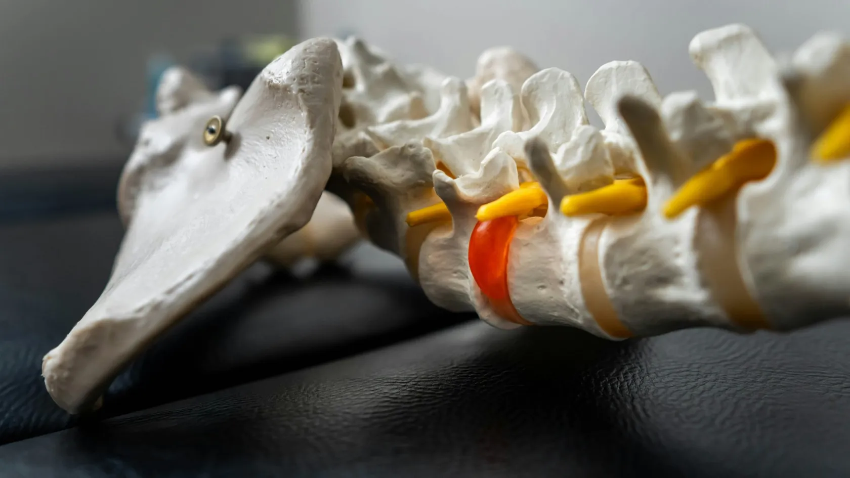
Endoscopic Lumbar Discectomy - Disc Surgery
Twenty-one cases to clear most of the learning curve, around sixty to reach proficiency, and a one in four complication risk on a surgeon's very first. The technique is excellent. The vetting matters more.
About This Department
Twenty-one cases to get through most of the learning curve, around sixty to reach proficiency, and a one in four risk of a complication on a surgeon's very first attempt. Those numbers come from a 2024 meta-analysis of 864 patients and they explain why this page spends as much time on how to vet a surgeon as on how the operation works. Endoscopic lumbar discectomy is an excellent technique in trained hands. What follows covers the two routes in, the awake option, what the BMJ trial actually found, and how to tell whether the person offering it has done enough of them.
Free consultation
Send the MRI and find out whether your herniation is reachable through an endoscope
The review costs nothing and commits you to nothing. Send the lumbar MRI images with the radiologist's report, the side and distribution of the leg pain, how long it has lasted, and any weakness or numbness. A spine surgeon reads the films and answers three things. Whether the fragment sits somewhere an endoscope can reach at all, which of the two corridors would be used, and whether an open microdiscectomy would serve this particular herniation better than either endoscopic route.
Two ways in, and why the level decides
Full-endoscopic surgery reaches the disc through a single working channel of around eight millimetres carrying a camera, a light source, continuous saline irrigation and the instruments, all in one tube. Two corridors lead to it. Neither is new.
Transforaminal, from the side
Entry sits eight to twelve centimetres out from the midline, angled forwards through the muscle towards the foramen, which is the bony window where the nerve root leaves the spine. Nothing in the back muscles gets cut. No bone comes off the lamina in the standard version. The tube passes through a natural corridor between the exiting and the traversing nerve roots, a triangular space surgeons call Kambin's triangle, and arrives at the side of the disc. Getting there needs X-ray guidance and a needle first, then a guidewire, then a series of dilators over that wire, each one a millimetre or two wider than the last, until the working sheath sits where the surgeon wants it. That sequence is why the transforaminal operation looks so undramatic from outside the sterile drapes and why it demands so much of the person doing it, since every one of those steps is judged on a two-dimensional X-ray image of a three-dimensional problem.
L5 to S1 is where this route runs into trouble. The pelvis gets in the way, and in patients whose iliac crest sits high the approach angle simply does not exist, which is an anatomical fact visible on the scan and not a matter of surgical preference. A far lateral herniation, sitting outside the canal beside the exiting root, is the reverse case, because the transforaminal corridor lands almost on top of it while an open midline operation has to travel a long way round.
Interlaminar, from behind
Entry sits close to the midline, and the endoscope passes through the same gap between the laminae that an open microdiscectomy uses, which makes this corridor the natural answer at L5 to S1 where that gap is widest and the pelvis is irrelevant. A little bone comes off the laminar edge in many cases. The nerve root gets retracted directly, the way it would be in open surgery, so the surgeon works with the same anatomical picture through a smaller window. Familiarity is the practical advantage of this corridor, and it explains why units adopting endoscopic surgery frequently start here before attempting anything through the foramen. A surgeon who has spent fifteen years looking at that anatomy down a microscope sees the same view down an endoscope, whereas the transforaminal corridor arrives from an angle nobody trained in open surgery has ever worked from, which is a large part of why the two techniques carry different learning curves.
Neither corridor wins outright.
Level and herniation position choose between them, and a surgeon who performs only one of the two is limited to the herniations that route can reach, and that limitation belongs in your first message to the coordinator.
Awake on the table
Transforaminal endoscopic surgery can be done with local anaesthetic and light sedation, with the patient awake enough to answer questions and to say when something near the nerve hurts. Conscious sedation appears in the great majority of published transforaminal series.
That changes the risk profile for a specific group of patients. Somebody with significant heart or lung disease, somebody who has had a bad reaction to a general anaesthetic before, somebody elderly and frail enough that the anaesthetic is the largest single risk in the whole plan, all gain something real here that no comparison of scar sizes captures. Feedback from an awake patient adds a safety layer too, since a sudden shooting pain as an instrument passes the root is information arriving in real time, hours before a deficit would otherwise be discovered in recovery. There is a practical benefit as well that patients notice more than surgeons do. Nobody spends two hours in a recovery bay waiting for a general anaesthetic to clear, nobody feels sick from it, and a patient who walked into the theatre suite frequently walks out of the ward the same evening, which for somebody who has flown in for a single procedure changes the shape of the whole trip.
It is not for everybody. Lying face down and still for an hour while somebody works in your back is tolerable for many people and intolerable for some, and anxiety is a perfectly good reason to choose a general anaesthetic instead of talking yourself into being brave. Interlaminar surgery is more often done asleep in any case, because retracting the nerve root directly is uncomfortable in a way the transforaminal corridor is not.
What the BMJ trial found
One trial dominates this subject and it deserves reading properly. Published in the BMJ in 2022, it randomised 613 patients across four Dutch hospitals to percutaneous transforaminal endoscopic discectomy or to conventional open microdiscectomy, and it was designed as a non-inferiority study, meaning the question asked was whether the endoscopic route was at least as good rather than whether it was better. The design carried an unusual and creditable feature. The investigators deliberately included 125 patients operated on by surgeons performing their first endoscopic cases, and reported them separately, so that the learning curve appeared in the data instead of being hidden by recruiting only expert centres.
Endoscopic patients did better, and by less than the marketing implies.
Twelve month leg pain came in at a median of 7 on a hundred-point scale for the endoscopic group against 16 for open microdiscectomy, a between-group difference of 7.1 with a confidence interval running from 2.8 to 11.3. Blood loss was lower, hospital admission shorter, and patients got moving sooner. Disability scores, back pain, quality of life and self-perceived recovery all leaned the same way. Repeat surgery within the year happened in 5 percent of the endoscopic group and 6 percent of the open group. The authors said plainly that these differences, while statistically significant, were small and might not reach clinical relevance, and including the learning-curve patients in a sensitivity analysis did not change the conclusion.
A companion economic analysis from the same trial went further. Surgery itself cost more in the endoscopic arm, and every other category of cost came out lower, including the costs falling outside the hospital on patients and employers, so that total societal cost favoured the endoscopic route with better outcomes attached. The probability that it was simultaneously cheaper and more effective came out above 99 percent on both leg pain and quality-adjusted life years.
So the position this page takes is straightforward. Where a surgeon is properly trained and the herniation is reachable, full-endoscopic discectomy is the better operation, on evidence rather than on enthusiasm, and the margin is real if modest.
One portal or two
Uniportal endoscopy uses one tube. Camera and instruments go down it together. Biportal endoscopy, known as UBE, makes two separate small incisions a few centimetres apart, one for the camera and one for the working instruments, so the surgeon moves each independently instead of steering both down one channel. The second arrangement handles more like arthroscopy of a knee and it is markedly easier to learn for surgeons who already work that way.
Nine studies covering 1,001 patients were pooled in the European Spine Journal in 2024 to compare biportal endoscopic discectomy against open microdiscectomy. Scores for leg pain came out the same at every time point measured. Back pain ran lower in the biportal group for three days. After that, level. Disability scores favoured biportal surgery at final follow-up. Operating time was shorter, hospital stay significantly shorter, blood loss lower, and complication rates showed no difference between the two.
This table scrolls sideways on a narrow screen. Swipe or drag to see every column.
| Variant | Access | Where it fits |
|---|---|---|
| Uniportal transforaminal | One 8 mm channel entering from the side through the foramen, frequently under local anaesthetic and sedation | Far lateral and foraminal herniations, patients unsuited to general anaesthesia. Blocked by a high pelvis at L5 to S1 |
| Uniportal interlaminar | One 8 mm channel entering near the midline through the interlaminar gap, usually under general anaesthesia | L5 to S1 and other levels with a wide interlaminar window, axillary herniations sitting against the dural sac |
| Biportal, UBE | Two small incisions, camera and instruments moved independently, saline irrigation throughout | Migrated fragments and cases needing more room to work. Shorter stay and less blood loss than open surgery in pooled data |
Choosing between these three matters far less to a patient than choosing the surgeon who will perform whichever one they do best.
The learning curve, in numbers
Most articles about endoscopic spine surgery mention a learning curve and move on, which leaves a patient with a warning and no way to act on it. A 2024 systematic review in the Global Spine Journal quantified the thing instead, pooling individual participant and aggregated data from thirteen studies covering 864 patients operated on by fifteen different surgeons, and applying mixed-effects regression to work out how performance changed with case number. The resulting figures are the most useful thing a patient can carry into a consultation, because they convert a vague anxiety about experience into a specific question with a specific number attached, and because a surgeon who knows their own numbers answers that question in about four seconds.
Here is what they found. For transforaminal endoscopic discectomy, the estimated operating time on a surgeon's first case was 95 minutes against a plateau of 66 minutes, with 21 cases needed to close 80 percent of that gap and 59 cases before performance approached the plateau. The complication risk on the very first case was estimated at 25 percent, with a confidence interval running from 11 to 46 percent, and an 80 percent reduction in that relative risk took around 41 cases. Leg pain outcomes took longest of all to mature, with an estimated 96 cases needed for an 80 percent improvement on the first-case result. Similar figures applied to the interlaminar and biportal techniques. The reviewers concluded that around 60 cases are needed for proficiency, while the bulk of the curve is behind a surgeon by case 20. Twenty cases is a few months of regular practice. Sixty is a couple of years.
Read those numbers again from the patient's side of the table.
Somebody on case number three is a different proposition from somebody on case number three hundred, and no amount of equipment closes that gap. Every argument about whether the endoscope beats the microscope matters less than the question of who is holding either one. All of which is knowable before you book. Surgeons keep logbooks, units keep numbers, and a hospital that treats international patients answers volume questions in writing all day long.
Which herniations suit it
Contained herniations and moderately extruded ones at a single level are the bread and butter of endoscopic surgery. They do very well. Recurrent herniations at a previously operated level are a genuine strength too, because the endoscope enters through virgin tissue at the side and avoids the scar sitting in the old midline corridor, an advantage of pure geometry. Anybody who has watched a surgeon spend forty minutes separating a nerve root from scar during a revision operation understands immediately why coming in from somewhere else appeals. Foraminal and far lateral herniations belong in the same category of endoscopic strength, since the transforaminal corridor arrives at them directly while an open operation has to remove part of the facet joint to see round the corner, and removing facet joint is how a stable segment becomes an unstable one. That single anatomical fact is the clearest case anywhere in spinal surgery for choosing an endoscope over a microscope, and it has nothing to do with scar size.
Four situations push a surgeon towards an open operation instead. A fragment that has migrated far up or down behind the vertebral body sits outside the reach of a single fixed channel. Severe central canal stenosis needs more decompression than an eight millimetre tube provides. Segmental instability, where the level has started to slip, needs stabilising rather than clearing. And a calcified herniation, hardened over years into something closer to bone than to gel, resists endoscopic instruments in a way that wastes time and risks the root.
This table scrolls sideways on a narrow screen. Swipe or drag to see every column.
| Finding on the scan | Why it matters | Usual answer |
|---|---|---|
| Far migrated fragment | Sits beyond the arc a fixed 8 mm channel can sweep | Open microdiscectomy, or a biportal approach with more working room |
| Severe central stenosis | Needs decompression across a wider area than the herniation itself | Open or tubular decompression |
| Segmental instability | The level has started to slip, so clearing the fragment leaves the problem | Decompression with fusion |
| Calcified herniation | Hardened towards bone and resistant to endoscopic instruments | Open microdiscectomy |
| High iliac crest at L5 to S1 | The pelvis blocks the transforaminal approach angle entirely | Interlaminar endoscopic corridor instead |
Being told that your particular herniation would be better served by an open microdiscectomy tells you something valuable about the surgeon's judgement.
Complications the endoscope brings with it
Pooled randomised data put overall complications lower for full-endoscopic surgery than for open discectomy, and dural tears in particular occur less frequently, which is the strongest single safety argument for the technique and the one clinic pages quote most. Three problems belong specifically to endoscopic surgery, and those rarely appear anywhere. None of them is dangerous in the way a nerve injury is dangerous, all three are manageable, and every one of them is far less frightening to a patient who was told about it in clinic than to a patient meeting it for the first time at home in week two with a phone in one hand and a search engine in the other.
Dysaesthesia from the exiting root
Transforaminal corridors pass close to the dorsal root ganglion, a swelling on the nerve packed with sensory cell bodies, and brushing past it can leave a burning, hypersensitive patch in the leg that appears within days of an otherwise perfect operation. Sufferers describe it as different in character from the sciatica that brought them in, more electrical, more surface, and provoked by clothing or bedsheets in a way the old pain never was. It settles across weeks to months in the great majority, helped by the nerve pain medicines used for any neuropathic problem, and it leaves nothing permanent behind in all but a very small number of people. Knowing about it beforehand converts a terrifying development into an expected one, and a patient who has been warned rings the coordinator to report it calmly instead of concluding at two in the morning that the operation has failed and that nobody abroad will answer the phone.
Fragments left behind
A narrow fixed channel shows the surgeon a small circle of anatomy at a time, so a second fragment sitting outside that circle is easier to miss than it would be under a microscope with a wider field. Cohort data show residual fragments several times more common after endoscopic surgery, and that figure tracks surgeon experience closely. The consequence is leg pain that never went away, and a second operation.
Pressure from the irrigation
Saline runs continuously through the working channel to keep the view clear, and without it there would be no endoscopic surgery at all, since blood clouds an eight millimetre field within seconds. Run at too high a pressure for too long, that same fluid raises pressure inside the spinal canal and transmits it upwards, and the classic sign is neck pain or a headache developing during the operation or in the hours after it. Careful pressure control and a sensible operating time prevent it, both of which come with experience, so this complication is the learning curve appearing in yet another guise. It resolves once the pressure comes off, and it is the reason an awake patient complaining of neck stiffness partway through gets taken seriously rather than reassured.
None of these three ends a recovery.
Infection stays rare. Nerve root injury stays rare. Both match open surgery. Recurrence at the operated level runs at broadly similar rates to open discectomy, since the hole in the disc wall behaves the same way whatever route the surgeon took to reach it.
The day of surgery and the fortnight after
Mobilisation came earlier in the endoscopic arm of the BMJ trial, hospital admission was shorter, and blood loss was lower, which is a randomised trial confirming what anybody standing in the theatre already suspected. Patients sit up within two hours. Many walk that afternoon, and those who had the procedure under local anaesthetic with sedation skip the grogginess, the nausea and the two hours in recovery that a general anaesthetic adds to everybody else's day. Appetite comes back the same evening. The single most common thing patients say in the first twelve hours concerns the absence of the leg pain they arrived with, and the second most common concerns how strange it feels to have had spinal surgery and be eating dinner sitting up.
Wounds close with a single stitch, or two for a biportal procedure.
Some units use a plaster and nothing else.
What does not shrink along with the incision is the six week restriction on bending, lifting and twisting, and this is the single most misunderstood thing about endoscopic disc surgery. The hole in the outer wall of the disc, through which the fragment escaped, is the same hole regardless of how the surgeon reached it, and it seals with scar on its own timetable rather than on the timetable of the skin. Patients who feel excellent at ten days and lift a suitcase at three weeks recur at the same rate as anybody else. A smaller scar buys a more comfortable first fortnight and buys nothing at all in the disc.
Desk work restarts inside one to three weeks for many people, sooner than after open surgery. Physiotherapy starts at week four. Manual work and sport follow the same schedule as any discectomy, which means eight to twelve weeks depending on how heavy the work is and a conversation with the surgeon about anything involving impact.
Arranging it from another country
Six to nine days in the country covers a single-level endoscopic discectomy. Assessment takes a day. The procedure and the ward stay take one night in published Turkish programmes, and the balance is recovery near the hospital until a surgeon has seen the wound and cleared you to fly. Booking the trip a few days longer than the minimum costs a hotel room and buys the ability to absorb a delay, and that matters more than it sounds when the alternative is rebooking an inflexible ticket from a foreign city with a sore back. Choose accommodation within walking distance of the hospital, and leave the scenic districts for another trip, since the two journeys that matter are the one to the wound check and the one to the airport.
The flight
Take the aisle seat. Stand every half hour. An aircraft chair holds the lumbar spine in exactly the flexed position that the first six weeks exist to avoid, and the small incision changes nothing about that. Compression stockings and any prescribed clot prevention apply as after any operation. Somebody else lifts the bags.
Documents and follow-up
Three documents matter, all in English. The operation note naming the level, the side, the corridor used and whether the procedure was uniportal or biportal. The discharge summary. The written restrictions. A doctor at home reading L4 to L5 right transforaminal uniportal endoscopic discectomy knows exactly what happened. Once you are back home the follow-up runs on a photograph of the wound at two weeks, a note on walking distance and leg pain, and a physiotherapy report at six weeks. See a local doctor the same day for new weakness, severe returning leg pain, a discharging wound, fever, or any bladder or bowel change.
What moves the quotation
Endoscopic sets carry single-use components. The disposables cost per case therefore sits above a microscope-based operation before anybody has discussed the surgeon, the level or the length of the stay. Uniportal and biportal kit price differently again. After that come the number of levels, whether the plan is a discectomy alone or a discectomy with fusion, the number of ward nights, whether the case is a first operation or a revision, and the patient's own medical picture, since weight, diabetes and previous spinal surgery all change what the operation involves.
Turkish packages published for disc surgery cover the procedure, the ward nights, pre-operative tests, medications, transfers, an interpreter and the appointments before departure, while flights, insurance, extra nights and the treatment of any complication sit outside. Two questions settle most of it. Which technique does the quotation assume, and what happens to it if the surgeon converts to open surgery during the case.
One number means something, and it arrives after a surgeon has read your MRI.
Frequently asked questions
Is endoscopic discectomy better than open microdiscectomy?
How many operations should my surgeon have done?
Can endoscopic disc surgery be done awake?
Which herniations cannot be treated endoscopically?
Do the six week restrictions still apply after endoscopic surgery?
References
- Gadjradj PS, Rubinstein SM, Peul WC, Depauw PR, Vleggeert-Lankamp CL, Seiger A, van Susante JL, de Boer MR, van Tulder MW, Harhangi BS. Full endoscopic versus open discectomy for sciatica. Randomised controlled non-inferiority trial. BMJ. 2022;376:e065846. doi:10.1136/bmj-2021-065846
- Gadjradj PS, Broulikova HM, van Dongen JM, Rubinstein SM, Depauw PR, Vleggeert C, Seiger A, Peul WC, van Susante JL, van Tulder MW, Harhangi BS. Cost-effectiveness of full endoscopic versus open discectomy for sciatica. British Journal of Sports Medicine. 2022;56(18):1018-1025. doi:10.1136/bjsports-2021-104808
- Koh CH, Booker J, Choi D, Khan DZ, Layard Horsfall H, Sayal P, Marcus HJ, Prezerakos G. Learning curve of endoscopic lumbar discectomy. A systematic review and meta-analysis of individual participant and aggregated data. Global Spine Journal. 2025;15(2):1435-1444. doi:10.1177/21925682241289901
- Feng Z, Zhao Z, Cui W, Meng X, Hai Y. Unilateral biportal endoscopic discectomy versus microdiscectomy for lumbar disc herniation. A systematic review and meta-analysis. European Spine Journal. 2024;33(6):2139-2153. doi:10.1007/s00586-023-08116-2
Editor's note
Written by the Biruni Hospital medical editorial team. Reviewed by Assistant Professor Şafak SAYAR, Orthopedics and Traumatology.
Medically reviewed by
Assistant Professor Şafak SAYAR
Orthopedics and Traumatology
Related Treatments
View All-
Achilles Tendon Repair
Achilles tendon repair stitches a torn tendon back together and cuts the risk of a second rupture, while a boot with early movement gives the same function at one year in trials. This guide covers the signs of a tear, surgery or a boot, open and minimally invasive repair, rehabilitation week by week and the stay.
-
Bone Tumor Resection
Most people arrive expecting to lose the limb. For the large majority of bone tumours today the limb is saved, the bone is rebuilt with an implant or a graft, and survival is no worse for it. What decides the answer is the margin, and the margin is planned from an MRI of the whole bone before anything is cut.
-
Chondrosarcoma Surgery and Bone Tumor Resection
Chemotherapy and radiation barely touch this tumor, which sounds like the worst news on the page and is the reason a single operation cures it outright. For a grade 1 chondrosarcoma in an arm or a leg the bone is scraped clean from the inside and the natural joint stays where it is.
-
Complex Knee Revision Surgery
Infection now causes more failed knee replacements than any other single reason, 21.6 percent across registries in seven countries, and it can turn a revision into two operations with weeks of antibiotics between them. This page explains how the cause is found before surgery, when cones or a hinge are needed, and how a two-stage revision fits around travel.
-
Complex Trauma Reconstruction
A fracture that has not healed after a year, healed crooked or became infected can still be repaired, and the treatment often runs for months in an external frame. This page explains nonunion, malunion and bone loss repair, the choice between bone transport and the Masquelet technique, and how a long treatment fits around travel to Istanbul.