Most people arrive expecting to lose the limb. For the large majority of bone tumours today the limb is saved, the bone is rebuilt with an implant or a graft, and survival is no worse for it. What decides the answer is the margin, and the margin is planned from an MRI of the whole bone before anything is cut.
About This Department
ORTHOPEDIC ONCOLOGY
Everything about a bone tumor operation reduces to one measurement. How far from the tumor the surgeon actually cut.
Thirteen studies covering 1559 patients with osteosarcoma agree on one thing. Where the surgeon cuts, measured afterward under a microscope, predicts whether the tumor comes back in the same place.
5 mm
A planned bone margin reported as safe in the published planning literature
90 percent
The tumor necrosis threshold that counts as a good response to chemotherapy
Under 1 mm
The error achieved in half of resections guided by 3D printed cutting guides
Free
Written opinion on your imaging and biopsy report before you travel
A bone tumor resection removes a segment of bone containing a tumor, together with whatever soft tissue around it the surgeon judges to be at risk. The word resection carries a specific meaning here. It describes taking the tumor out in one piece with uncut tissue surrounding it, which surgeons call an en bloc resection, and it stands apart from curettage, where a lesion gets scraped out of the inside of the bone and the cavity is then filled.
That distinction is oncological, and it has nothing to do with how the limb looks afterward. Scraping a malignant tumor opens it and spills cells into the wound. Removing it inside an intact envelope of healthy tissue does not.
Curettage remains entirely correct for many benign lesions, where opening the tumor causes no harm and preserving bone matters more. For a sarcoma it is the wrong operation, and the difference between the two decisions rests on the tissue diagnosis, so the pathology has to be settled before a date is set. A tumor scraped out by curettage that then turns out on pathology to have been malignant almost always needs a wider second operation.
Margins decide whether it worked
Margin describes the layer of normal tissue between the tumor and the surface of the specimen the surgeon hands to the pathologist. Surgeons grade it by how much healthy tissue separates the two. Those grades carry real consequences.
Narrow screens scroll this table sideways. Swipe or drag to reach every column.
How surgeons classify what was removed
Margin
What it means
Where it belongs
Intralesional
The surgeon entered the tumor itself and left tumor behind
Acceptable for many benign lesions, inadequate for a sarcoma
Marginal
The cut passed through the reactive tissue immediately around the tumor
Carries a measurably higher local recurrence rate in malignant disease
Wide
A continuous cuff of normal tissue surrounds the tumor on every surface
The standard aim for a high-grade bone sarcoma
Radical
The entire compartment containing the tumor comes out
Rarely needed now, since wide margins achieve the same control with far less loss
According to PubMed, a systematic review and meta-analysis of thirteen studies covering 1559 patients with osteosarcoma of the limbs and pelvis found that tumors removed with inadequate margins recurred locally significantly more often than those removed with adequate margins, and that marginal excisions fared significantly worse than wide ones (He et al, 2016). Of the whole group, 175 patients developed a local recurrence.
Millimeters matter here in a way patients rarely expect.
Why the pelvis behaves differently from the thigh
Pelvic osteosarcomas removed with exactly the same margin as limb tumors still recurred locally at a significantly higher rate in that same meta-analysis. Same margin. Different outcome. Bone in the pelvis sits against the bladder, the bowel, the major vessels and the nerve roots, so the cuff of healthy tissue a surgeon would like on every surface simply does not exist in some directions, and the anatomy imposes compromises that a femur in the middle of a thigh never forces. A patient told their tumor sits in the pelvis or the sacrum should expect a longer operation, a longer stay and a more guarded conversation about local control than the figures quoted for limb tumors suggest.
Planning the cut before theater day
Accuracy is mostly decided before the patient reaches the operating table.
Four things happen in order, and the order is not negotiable.
An MRI covering the whole femur, tibia or humerus, not simply the visible lump, since the tumor reaches further inside the marrow cavity than the plain x-ray suggests and the same scan picks up skip lesions, meaning separate deposits in the same bone at a distance from the main one. Missing a skip lesion turns a well-executed operation into an inadequate one.
Staging scans of the chest and, in many cases, a bone scan or PET scan, which settle whether the disease has already traveled elsewhere and therefore whether this operation is the right one at all.
A decision, taken off that imaging, on how many centimeters of bone to take beyond the visible edge of the tumor.
Reproduction of that decision with a saw, on a wet operative field, through an approach that hides most of the anatomy. This is the step where plans go wrong.
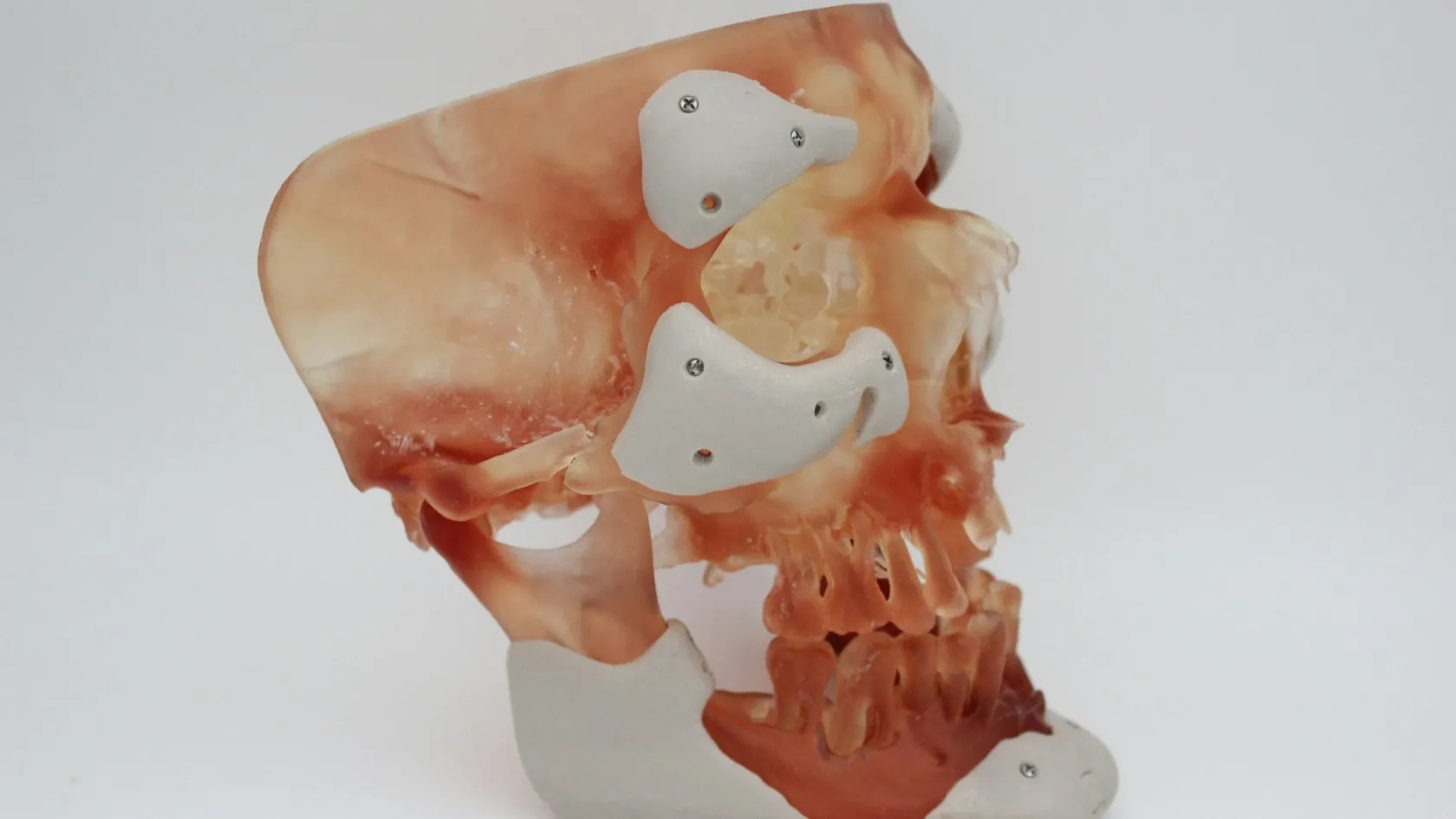
Two technologies now help with the reproduction. Intraoperative navigation tracks the instruments against the preoperative scan in real time, in much the same way a satellite navigation system tracks a car against a map. Patient-specific instrumentation takes the opposite approach, using the scan to design and 3D print a guide that only sits on the patient's bone in one position and carries slots that direct the saw exactly where the plan intended. One systematic review on PubMed compared the two across 81 studies and reported positive margin rates between zero and 19 percent and bone cut accuracy within 0.3 to 4 mm of the plan for both methods, with both outperforming freehand cutting, and it found a planned bone margin of at least 5 mm to be safe (Bruschi et al, 2023). The two differ mainly in logistics. A printed guide takes two to four weeks to plan and manufacture but adds only one to five minutes in theater, while navigation can be set up at short notice with no lead time yet adds fifteen to sixty five minutes to the operation.
Neither one removes the need for judgment. A separate series of 29 resections using 3D printed guides, also reported on PubMed, found the largest error in the amount of bone removed to be 2 mm, with just over half of cases accurate to within 1 mm, and the largest guide-fitting error 3 mm, while noting that the achieved margin lengths still differed from the planned ones in some cases and that careful review of the imaging remains necessary even when a guide is used (Lee et al, 2023). Complex resections produced larger errors than straightforward ones.
Inside the operating room
The operation runs in a fixed sequence, and every stage exists to protect the margin established in the planning.
1
The approach and the old biopsy track. Following the line of the previous biopsy, the incision takes the contaminated track out attached to the specimen. Skin, track and tumor leave the body as one piece.
2
Freeing the nerves and vessels. Working under direct vision, the surgeon traces the main artery, vein and nerve of the limb and separates each one from the tumor. This stage takes the longest, and it determines whether the limb stays.
3
The osteotomy. Guided freehand, by navigation or through a printed cutting guide, a saw divides the bone at the planned level. A sliver of marrow from the cut end goes to the laboratory immediately, so the surgeon knows before closing whether the cut was clear.
4
Delivery of the specimen, then reconstruction. Out comes the specimen, intact and oriented for the pathologist. Instruments that touched the tumor bed are exchanged, the field is redraped, and the reconstruction begins with clean equipment.
That last detail surprises people. Changing gloves, gowns and instruments between the resection and the reconstruction is standard practice, because a cell carried on a blade from the tumor bed into the space where an implant is about to sit would defeat the entire operation.
What the pathologist finds in the specimen
Removed bone goes to the laboratory whole, and what comes back over the following days matters more to your future than anything recorded on the operation note.
Two findings dominate the report. Margin status on every surface comes first, answering whether the resection achieved what it set out to achieve. The second finding applies to anyone who had chemotherapy before surgery, and it gives the proportion of the tumor found dead when the specimen is sectioned.
The 90 percent line
More than 90 percent tumor necrosis counts as a good histological response to chemotherapy and predicts a better outcome. Below that line, patients are classed as poor responders, and the finding changes what gets prescribed after surgery. In one reported series good response was achieved in 71 percent of patients on a four-drug regimen against 42 percent on a three-drug one (Kang et al, 2019), according to PubMed.
Waiting for that report tests people. It takes a week or two, because decalcifying bone before it can be sectioned is slow chemistry that cannot be hurried, and the specimen has to be sampled in many places before anyone can speak about the whole of it. If you traveled for surgery, plan for the final histology arriving after you have flown home, and make sure it reaches your own oncologist directly.
Saving the joint next to the tumor
Many bone tumors sit near the end of a long bone, close to the knee, the shoulder or the ankle, and the classical operation takes the neighboring joint out along with the tumor. Given enough clearance on the imaging, that joint can sometimes stay. What comes out instead is a segment from the middle of the shaft, leaving a gap to be bridged and the joint surfaces at both ends untouched, which surgeons call an intercalary resection.
Keeping a patient's own joint avoids everything that comes with replacing it, which for a young patient means avoiding a lifetime of implant revisions.
The evidence supports doing this where the anatomy genuinely allows it. According to PubMed, a multicentre series of 73 patients with tumors of the distal tibia compared intercalary resection preserving the epiphysis against joint replacement and against fusing the ankle, and reported that local recurrence occurred in eight patients without any correlation to the reconstruction chosen, while functional scores were highest in the joint-preserving group (Sambri et al, 2020). Preserving the joint end proved safe and effective, and the authors were explicit that it depends on rigorous preoperative planning built on accurate analysis of the imaging.
The question to put to your surgeon
Preserving the joint end proved safe and effective, and it depends on rigorous preoperative planning built on accurate analysis of the imaging.
The conclusion of the distal tibia series, and the reason the scan matters more than the surgeon’s confidence.
Put the question to your surgeon directly. Can the joint be preserved in my case, and if it cannot, what on the scan makes that impossible?
What can go wrong
Resections run long, leave a large exposed surface, cost significant blood, and frequently happen to patients whose marrow recent chemotherapy has suppressed. Wound healing problems and deep infection follow from exactly those conditions and sit at the top of the list. Damage to the nerve or artery being dissected off the tumor is uncommon but real, and it is the risk that grows as the tumor sits closer to those structures. Blood clots in the leg deserve attention after any long limb operation in a patient with cancer, since the disease raises clotting risk on its own.
Two problems belong specifically to the resection itself.
A positive margin reported afterward means tumor reached the cut surface, and it usually forces a decision about going back for more tissue, adding radiotherapy, or both. Local recurrence is the longer-term version of the same problem, appearing months or years later at the operated site, and it is the outcome the whole margin discussion exists to prevent. Bone bridged with a donor graft brings a mechanical problem on top of the oncological ones, since that graft can fail to unite with the patient's own bone at either end.
Ask any unit for its own figures. Published averages describe a whole field of surgery, and the person operating on you is one surgeon within it.
The weeks after the resection
What the first weeks look like depends almost entirely on how the gap in the bone was filled, and patients get handed a single generic timeline when two very different ones apply.
Columns run past the edge on a small screen. Slide the table across to read them.
Two recoveries that get handed the same leaflet
Gap bridged with metal
Gap bridged with bone
Weight through the limb
Protected weight-bearing with crutches or a frame inside the first days
Nothing through the limb until the graft unites at both ends
What sets the timetable
The implant carries load from the moment it is fitted, so the limit is the wound and the muscle
Biological union, which runs on its own schedule of months and cannot be hurried
Where the waiting sits
At the far end, in the revisions a young patient will need across a lifetime
At the beginning, in a protected limb through most of the first year
What is watched
Wound healing, infection, and the mechanics of the implant itself
Union at each end of the graft on serial imaging, and any sign the graft is not taking
A metal implant carries load from the day it is fitted, so protected weight-bearing with crutches or a frame starts within days of the operation. Bone bridged with a graft has to heal to the host bone at both ends before it takes any weight, and that biological union runs on its own schedule of months, during which the limb stays protected. Neither route is a shortcut. They simply distribute the waiting differently, one at the beginning and one across the following year. Drains come out in the first days, stitches or clips at around two weeks, and the wound gets watched closely throughout, particularly where chemotherapy is due to restart, since resuming it over an unhealed wound invites trouble. Physiotherapy begins in hospital and continues for months. If the resection took muscle as well as bone, some strength never fully returns, and rehabilitation aims at a limb that works well for the life you want to lead.
Length of stay and getting home
Inpatient stay after a resection with reconstruction commonly runs one to two weeks, and a curettage for a benign lesion is far shorter. Patients traveling from abroad need to add time at both ends of that. Arrival brings blood tests, imaging review and the pre-operative consultation. Departure waits on the wound, on safe mobility with whatever walking aid the reconstruction requires, and on a fitness-to-fly assessment.
Patients underestimate the flight home.
Long flights combine immobility with a raised clotting risk that surgery and cancer have each already increased, so clearance depends on how the wound looks, how you are moving and whether you are on anticoagulation, and it gets decided at a review before departure. Nobody can honestly promise you a number of days when the trip is booked. Request a fitness-to-fly letter before discharge, since airlines sometimes ask for one after major surgery, and tell the airline in advance if you will be traveling with a frame or crutches, because assistance and seating have to be arranged ahead of the flight.
What changes the price of a resection
Two people can both be told they need a bone tumor resection and receive quotes that look nothing like each other, because the phrase covers operations of wildly different size.
How much bone comes out is the first driver, since resection length dictates what has to bridge the gap and a longer custom implant costs more than a short one. Preserving the neighboring joint changes the reconstruction entirely. Site matters, because a pelvic or sacral resection takes far longer, uses more blood products and usually needs a second surgical team, while a mid-shaft femoral resection does not. Planning technology carries its own cost, since a custom 3D printed cutting guide involves design work and manufacturing that a freehand resection avoids. Soft tissue coming out alongside the bone adds another layer, because closing that defect may need a plastic surgical flap and the theater time that goes with it. And a resection through previously operated tissue takes longer than a first operation through clean planes. Patient factors move the figure as well, among them age, nutritional state, whether recent chemotherapy has left blood counts too low to operate on schedule, and any heart, lung or kidney condition that changes what anesthesia and postoperative care involve.
What a published package usually covers
Published packages in this market for musculoskeletal tumor surgery cover the surgeon and anesthesiologist fees, theater time, the hospital stay, pre-operative imaging and blood work, the histopathology on the specimen and airport transfers with interpreting. Flights, accommodation past the stated nights, treatment of a complication, a return to theater and any systemic treatment given outside the package period sit outside them by default. Reading two quotes against those lists is the only way to know whether they describe the same operation. Before accepting any figure, establish how much bone the plan assumes will be removed and what will bridge the gap, whether the implant or graft is named and included, whether navigation or a printed guide is part of the quote, whether the pathology on the specimen sits inside it, and what happens financially if the surgeon has to take more than planned once the tumor is exposed. That last question separates a real quote from a hopeful one, and only a surgeon who has read your own imaging can answer it. That review costs nothing.
This review costs nothing and does not commit you to anything. Send the MRI of the whole affected bone, any staging scans, and the biopsy report if tissue has been taken. A surgeon will tell you how much bone a safe resection would need to remove in your case, whether the neighboring joint could stay, and what would have to be settled before a date could be discussed.
Going home, and what follows the resection
Resections generate documents that determine what happens next, and flying home without them creates problems that are difficult to fix at a distance.
Four things belong in the folder, and each of them answers a question somebody will ask you later.
The operation note, describing exactly what was removed and how the gap was bridged.
The full histopathology report, giving margin status on each surface and, where chemotherapy came first, the necrosis percentage that the next phase of treatment depends on.
The postoperative imaging, so a future scan has something to be compared against.
The make, model and reference numbers of any implant, because a surgeon revising it in fifteen years cannot easily find that information afterward.
Ask for all of it in a language your own doctors read.
Once you are home, follow-up splits between the two places now caring for you.
What belongs near where you live
Wound checks and stitch removal fall at around two weeks, so book that appointment near home before you fly. Your own oncologist takes over the systemic treatment as soon as the histology lands, so the report has to go to them directly. Imaging of the operated site and the chest continues on a schedule that starts frequent and stretches out over years, and it belongs near where you live, since nobody flies back every three months for a scan.
What only the operating team can answer
Keep the operating surgeon reachable through all of it, because remote follow-up earns its keep at exactly the awkward moments. When a wound looks wrong in the third week, or a scan taken back home shows something ambiguous at the resection site, the person who cut that margin can say whether it matches what they deliberately left behind, and no written report conveys that judgment on its own. Agree before discharge who you write to. Here the coordinator who has handled your case since your first message stays on the same WhatsApp number after you fly home, and imaging or reports your own doctors produce are reviewed without charge, which is what makes a third-week question answerable at all.
Frequently asked questions
How long do I need to stay in the country after a bone tumor resection?
Plan on one to two weeks as an inpatient after a resection with reconstruction, plus additional days at each end for the pre-operative assessment on arrival and for wound review, early mobilization and a fitness-to-fly check before departure. A curettage for a benign lesion needs a fraction of that. The final histology report frequently arrives after patients have flown home, so arrange for it to be sent directly to your own oncologist.
What happens if the pathology report says the margin was positive?
A positive margin means tumor reached the cut surface of the specimen, and it usually leads to a discussion about returning to theater to remove more tissue, adding radiotherapy, or both, depending on the tumor type and where the involved surface sits. It does not automatically mean the disease will return, but it does raise the local recurrence risk, which is why the decision is taken by the whole treating team rather than by the surgeon alone.
Does a 3D printed cutting guide make the operation safer?
Printed guides and intraoperative navigation both improve the accuracy of the bone cut compared with cutting freehand, with published bone cut accuracy for both techniques falling within 0.3 to 4 mm of the plan. Neither replaces surgical judgment, and studies report that achieved margins can still differ from planned ones, particularly in complex resections. A guide also needs two to four weeks of design and manufacturing time, so it suits a case with a date some weeks away.
Can the joint next to my tumor be saved?
Sometimes, and it depends on how much normal bone separates the tumor from the joint surface on the MRI. Where enough clearance exists, an intercalary resection removes a segment from the shaft and leaves the joint intact, which published series report as safe and as giving better function than replacing the joint. Where the tumor reaches the joint end, preserving it would mean accepting an inadequate margin, and that trade is not worth making.
References
He F, Zhang W, Shen Y, et al. Effects of resection margins on local recurrence of osteosarcoma in extremity and pelvis. Systematic review and meta-analysis. Int J Surg. 2016;36(Pt A):283-292.
Bruschi A, Donati DM, Di Bella C. What to choose in bone tumor resections? Patient specific instrumentation versus surgical navigation. A systematic review. J Bone Oncol. 2023;42:100503.
Lee SH, Kim W, Lee JS. What are the resection accuracy and guide-fitting errors associated with 3D-printed, patient-specific resection guides for bone tumor resections? Bone Joint J. 2023;105-B(2):190-197.
Sambri A, Dalla Rosa M, Scorianz M, et al. Different reconstructive techniques for tumors of the distal tibia. Bone Joint J. 2020;102-B(11):1567-1573.
Kang JM, Ju HY, Joo J, et al. Histologic Response and Toxicity following Interval-Compressed Four-Drug Therapy Given Preoperatively in Children and Young Adults with Osteosarcoma. A Retrospective Study. Oncology. 2019;98(2):81-90.
Editor's note
Written by the Biruni Hospital medical editorial team. Reviewed by Assoc. Prof. Dr. Suat BATAR, Orthopedics and Traumatology.