
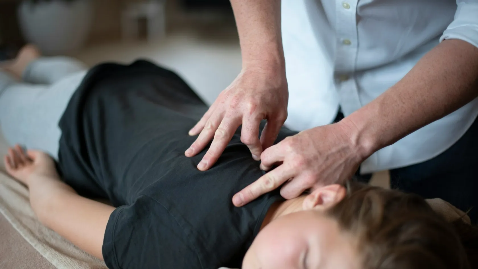
Scoliosis Surgery - Spinal Deformity Correction
Under 25 degrees a curve gets watched. Past 45 or 50 in a spine that is still growing it gets an operation. Everything families agonise over sits in the gap between those two sentences.
About This Department
A radiologist has written a number on your report, and everything now turns on it. Curves under 25 degrees get watched, curves past 45 or 50 in a spine that still has growing to do get an operation, and the space between those two sentences is where families spend an anxious year. This article covers how surgeons read that number, what correction surgery does to a spine, what the trials say when fusion is set against the fusionless alternative, and how the whole thing is arranged from another country.
Free consultation
Send the standing spine X-rays and find out whether this curve has reached surgical range
The review costs nothing and commits you to nothing. Send standing full-spine X-rays from the front and the side, any spine MRI you hold, the date and measurement of an earlier film so the two can be compared, and for a teenager the age, the height and whether growth has finished. A spinal deformity surgeon reads them and answers three things. Whether the curve is in surgical range. Roughly how much of the spine an operation would involve. Whether waiting another six months is safe.
Reading the number on the report
Radiologists measure a curve by drawing a line along the top edge of the most tilted vertebra at one end of it, a second line along the bottom edge of the most tilted vertebra at the other end, and then measuring where those two lines cross. That angle carries the surgeon's name, Cobb, and it governs the entire decision. Under 10 degrees the spine is called normal, because everybody has some asymmetry and nobody treats it. From 10 to 25 degrees the plan is a repeat film in six or twelve months and nothing else. Between 25 and 40 degrees in a child with growth left, a brace goes on. Once the angle passes 45 or 50, surgery comes onto the table, and that threshold is mechanical rather than cosmetic. A curve that large keeps creeping by a degree or so a year for the rest of the patient's life, because the spine is now leaning far enough off centre that gravity works on it, and a 50 degree curve at seventeen becomes a 70 degree curve at fifty.
Growth changes the arithmetic completely.
Take two patients with 42 degree curves. One is twelve and has four years of spinal growth ahead. The other is nineteen and finished growing two years ago. The twelve year old faces a curve that will very likely cross the surgical threshold before she stops growing, so bracing starts now and the family plans for the possibility of an operation. The nineteen year old has a curve that will creep, slowly, over decades. Same number on the report, entirely different conversations. Surgeons judge remaining growth from the Risser sign, which grades how far the growth plate along the rim of the pelvis has fused, and from menarche in girls.
Bracing deserves more credit than families give it. The evidence is not soft. In 2013 the New England Journal of Medicine published BrAIST, a multicentre study of 242 adolescents with curves in the high-risk band, and the finding was strong enough that the trial was stopped early for efficacy. Seventy-two percent of braced patients avoided progressing to 50 degrees, against 48 percent of those simply observed. In the randomised arm alone the gap widened, 75 percent against 42. Wear time drove the result, and the more hours the brace was on, the better the outcome, which is a difficult thing to sell to a fourteen year old and worth every argument at the kitchen table. A brace never straightens a spine. It holds the line during the years when a curve would otherwise run away, and for three teenagers in four it takes surgery off the table permanently.
One caution. The angle says nothing at all about how much the curve bothers the person carrying it, and two patients at 55 degrees can arrive with completely different complaints, from a rib hump that ruins swimming lessons to nothing at all beyond an odd hemline.
Four kinds of scoliosis, four operations
Scoliosis names a shape, and four very different diseases produce it. Getting the label right matters to you as a reader because the published outcome figures belong to one of the four, and reading the wrong set will either frighten you or reassure you for no reason.
Adolescent idiopathic scoliosis covers the great majority of cases. It appears between ten and eighteen, runs in families, affects girls far more than boys once curves get large, and has no identified cause after a century of looking. Nearly all the research quoted anywhere on this page describes this group.
Congenital scoliosis begins before birth, when vertebrae fail to form or fail to separate from each other, and it turns up in toddlers rather than teenagers. Operations here are early, short and focused on the malformed segment.
Neuromuscular scoliosis is a different order of problem. Children with cerebral palsy, Duchenne muscular dystrophy, spina bifida and spinal muscular atrophy lose the muscular control that holds a trunk upright, and the spine collapses into a long C-shaped curve that drags the pelvis with it. These curves keep progressing after growth stops, which idiopathic curves largely do not. The operations run longer, involve more levels, extend down to the pelvis in many cases, and carry complication rates several times those quoted for adolescent idiopathic surgery. Families comparing a neuromuscular case against the reassuring numbers in this article are reading figures that do not apply, and any surgeon offering the same risk profile for both is being careless with you.
Adult scoliosis splits again. Some adults carry an adolescent curve that nobody treated, now stiff and painful in the fifth or sixth decade. Others develop a new curve from asymmetric wear in the discs and facet joints, which surgeons call degenerative or de novo scoliosis, and those patients rarely mention appearance at all. They come in with leg pain, with a back that gives out after twenty minutes of standing, with a trunk that has started to tip forwards. Restoring that forward-and-back balance, the sagittal profile, drives satisfaction in adult surgery far more than the sideways correction visible on the front view.
Kyphosis sits in the same clinic. Scheuermann kyphosis in adolescents and osteoporotic vertebral collapse in older adults both get corrected with the same screws and rods, which is why departments file all of it under spinal deformity.
Watching, bracing, tethering, fusing
Four things can be done with a curve. Curve size and remaining growth decide between them, and the table below sets out how each option is matched.
This table scrolls sideways on a narrow screen. Swipe or drag to see every column.
| Option | Who it suits | What it delivers |
|---|---|---|
| Observation | Under 25 degrees, or any curve in a skeleton that has finished growing and causes no symptoms | A repeat film every 6 to 12 months. Nothing is corrected, and the great majority of these curves never need anything further |
| Bracing | 25 to 40 degrees with growth remaining | Cuts the chance of reaching the surgical threshold from roughly one in two to roughly one in four, with wear hours driving the result. Useless once growth has stopped |
| Vertebral body tethering | Younger patients with substantial growth left and flexible curves in a moderate range | Keeps the spine unfused and bending, and steers growth to reduce the curve. Correction is smaller and less certain, and unplanned second operations run one in four beyond three years |
| Spinal fusion | Past 45 to 50 degrees, stiff curves, and adult deformity with pain or imbalance | Corrects the curve and stops it progressing for good, with a 2 percent pooled complication rate in adolescent series. The instrumented levels lose their movement permanently |
Schroth physiotherapy and the other scoliosis-specific exercise programmes have a real place beside all four. They build trunk strength, they help with pain, they teach a patient to feel where their spine sits in space. No exercise regime has ever been shown to reverse a structural curve, and anybody promising to straighten a 50 degree spine without surgery is selling something.
Inside a correction
Posterior spinal fusion has been the workhorse operation for four decades and it earns that position. The patient lies face down under general anaesthesia. One incision runs down the midline of the back over the length of the curve. Screws go into the pedicles, the two short bony bridges that connect the back of each vertebra to its body, and into those screws the surgeon locks two rods that have been bent to the shape the spine should take. Rotating and pulling on those rods brings the vertebrae round, and the correction happens over a few minutes of careful, incremental force. Bone graft then goes along the stripped joint surfaces so that the instrumented segment knits into one continuous block of bone during the following year.
Titanium holds nothing in the long run. It holds everything still while the bone fuses, and the bone is what lasts.
Choosing the top and bottom levels
Every level inside the fusion stops moving, so where the construct ends shapes the rest of the patient's life. Surgeons classify the curve pattern, for adolescent idiopathic scoliosis by the Lenke system, and then work to leave as many lumbar segments mobile as the correction allows. Ten to twelve levels covers a large thoracic curve. Five or six covers a short lumbar one. That difference decides whether a twenty-two year old can still bend to pick something off the floor without stopping to plan the movement. A surgeon who spends twenty minutes explaining the level choice has understood what he is deciding on that patient's behalf, and that conversation tells you more about a unit than any brochure.
Anterior surgery, approaching the spine from the side of the chest or abdomen, still holds ground for single lumbar curves. A 2021 meta-analysis in Spine Deformity, covering nine comparative studies and 583 patients, found the anterior route achieved equal correction using significantly fewer instrumented levels, which is exactly the trade a young patient with a short lumbar curve wants. The posterior route gave a shorter hospital stay and better restored the sagittal profile.
Neuromonitoring, and the blood
Electrodes track signals running down the spinal cord to the legs and back up to the brain, continuously, from before the first cut until the wound is closed. When a correction pulls the cord harder than it will tolerate, those signals drop within seconds, the surgeon releases the force, and the trace recovers. This single technology is what turned large corrections from a gamble into routine work, and no unit should be operating on a deformity without it.
Long fusions bleed. Three measures handle it in every competent theatre, namely tranexamic acid to slow the breakdown of clot, a cell saver that washes the patient's own lost blood and returns it, and a transfusion plan agreed before the case starts. Ask about all three.
Fusion against tethering
Vertebral body tethering is the question families raise most, and it deserves a straight answer. Screws enter the sides of the vertebrae through the chest, a flexible cord runs between them under tension, and the tethered side of the spine grows more slowly than the free side, so the curve unwinds across the remaining growing years. Nothing fuses. The spine keeps bending.
Two meta-analyses put numbers on what that flexibility costs, and the numbers are not close. Pooling eleven comparative studies and 1,112 patients, a 2025 analysis in Spine Deformity found fusion delivered significantly greater curve correction both immediately and at two years, with significantly lower odds of unplanned revision surgery, and it recorded tether breakage in 13 to 23 percent of tethering cases. An earlier analysis in JB and JS Open Access set 211 tethering patients against 1,069 fusion patients and reported pooled complication rates of 26 percent against 2 percent, with reoperation at 14.1 percent against 0.6 percent. Restrict that comparison to studies following patients three years or longer and the reoperation figures move to 24.7 percent against 1.8. One tethering patient in four back in theatre, against one fusion patient in fifty. Quality of life scores came out similar in both groups, which tells you the extra operations bought no measurable advantage in how these young people felt.
This table scrolls sideways on a narrow screen. Swipe or drag to see every column.
| Measure | Spinal fusion | Vertebral body tethering |
|---|---|---|
| Curve correction at 2 years | Significantly greater | Smaller and less predictable |
| Pooled complication rate | 2 percent | 26 percent |
| Reoperation beyond 3 years | 1.8 percent | 24.7 percent |
| Hardware failure | Rod or screw breakage is uncommon once fusion is solid | Tether breakage reported in 13 to 23 percent |
| Spinal movement kept | Instrumented levels stop moving permanently | Lumbar flexibility largely preserved |
| Quality of life at mid-term | Similar in both groups | Similar in both groups |
Tethering suits a narrow group. Young, growing, flexible curve, moderate size, and a family that has understood and accepted a real chance of a second operation. Outside that group fusion is the better operation, and for a stiff 60 degree curve in someone who has stopped growing it is the only sensible one. A clinic offering a tether for that spine is worth leaving.
What can go wrong
Every rate quoted below comes from adolescent idiopathic series, the group with the best data and the lowest risk. Neuromuscular scoliosis and adult deformity run higher across every category.
Nerve injury
Permanent spinal cord injury is the fear that keeps parents awake, and modern series with monitoring in place put it well under one percent in adolescent idiopathic surgery. Transient changes in the monitoring trace during correction happen far more frequently than actual injury, and catching them is precisely the point of the technology. Leg weakness or numbness on waking gets assessed within minutes.
Infection around the implants
Deep infection reaches one to two percent in idiopathic cases and considerably more in neuromuscular scoliosis, and it declares itself in the first weeks through a wound that keeps draining, spreading redness, or fever. Treatment means a return to theatre for a washout plus a course of antibiotics chosen once the organism is known, and occasionally removal of the metalwork after the fusion has become solid enough to hold the correction without it. What makes this complication different from the others on the list, and what makes it the single strongest argument for staying in the country until the wound has properly closed, is that it declares itself in exactly the window when a family flying home on day ten would be somewhere over Europe with a child who has started running a temperature and a discharge letter in a language the nearest emergency department cannot read. Prevention is unglamorous and effective. Antibiotics on induction, meticulous closure, and a dressing left alone rather than peeled back by everyone who walks past the bed.
Failed fusion and broken hardware
Pseudarthrosis means the bone never knitted. The rods then carry a load they were designed to carry for a year and not for a decade, and eventually something breaks. It shows up as new pain months or years after the patient had stopped thinking of themselves as a surgical patient, and it needs revision. Screws can also sit a millimetre or two outside a pedicle and cause nothing at all. Keep the post-operative scan in your own file so the next surgeon sees that from the start.
Trouble at the ends of the construct
The vertebra immediately above a long fusion carries load it never carried before and can tip forwards over years, which surgeons call proximal junctional kyphosis. A 2024 review in the European Journal of Medical Research, pooling 1,115 patients followed for a mean of 22.6 years, found degeneration of the segments next to the fusion to be the commonest mechanical finding at long-term follow-up. Its effect on how those patients actually felt stayed unclear in the same data, and that gap between what a film shows and what a person feels explains why a radiology report at year fifteen can read alarmingly while the patient walks in with no complaint at all. Level selection is the main lever surgeons have against this, and it is the reason two surgeons will argue for an hour about whether to stop at L3 or L4.
Blood clots, chest infection, an ileus where the bowel sulks for a couple of days, and the ordinary hazards of a long general anaesthetic all belong on the list too. Nearly all of them get sorted on the ward before anybody goes home.
The first year afterwards
Comfort comes back in weeks. The fusion takes a year. Holding both facts at once is what stops a patient panicking in month four when the back still aches after a long day.
On the ward
Standing and walking a few steps happens on the first day after surgery, which startles families who had pictured a fortnight flat on a bed. Pain relief runs as a combination of drugs rather than one heavy opiate. Drains come out, the catheter comes out, the bowel wakes up, and discharge follows once walking, stairs and getting out of bed can all be done safely. Published series put that at three to seven days.
Weeks one to six
Three movements are out for six weeks. Bending, lifting, twisting. Nothing heavier than two kilos gets picked up in the first fortnight, walking is the whole exercise programme, and the distance grows day by day from a corridor to a street to a park. Households adapt around those three prohibitions faster than they expect, by raising the height of whatever the patient sits on, putting the things they reach for daily between waist and shoulder level, and accepting for a month and a half that somebody else picks up what falls on the floor. School restarts part time somewhere between week three and week six, on a normal chair, with somebody else carrying the bag. Sleep is the hard part of this stretch. Rolling over needs a technique, the back aches at four in the morning, and a fortnight of broken nights wears down the parent sleeping in the same room quite as much as the patient.
Three to six months
Restrictions come off in stages from three months, guided by films showing the graft maturing. Swimming and cycling return first. Running and gym work follow near the six month mark for the majority of surgeons. The scar is still firm and pink through all of this and keeps softening for another year or two.
Six months to a year
Full clearance, contact sport included where the surgeon permits it, lands between six and twelve months once the fusion reads as solid. Height goes up by two to four centimetres, because a straightened spine is a longer one, and patients notice their clothes hanging differently long before they notice anything in their back. Stamina is the slowest thing to return, and a teenager who was fit before surgery is generally back to that level inside a year.
Nights on the ward, days in the country
Published Turkish hospital programmes for scoliosis correction plan a minimum of 12 to 15 days in the country. One or two days go on pre-operative assessment, four to seven nights on the ward, and the rest in a hotel nearby until the wound check that units want on day 10 to 12.
Allow two more days than the minimum. Stitches come out around day twelve to fourteen, and having the operating team remove them beats handing that job to a doctor at home who has never seen the wound.
Flying is the part patients underestimate. A long fusion means sitting still in a fixed seat with limited mobility and a fresh wound, at the exact point in recovery when clot risk peaks, and the wound check therefore has to happen before departure. Surgeons want the incision dry and the patient walking comfortably before clearing even a short hop, and they ask for longer before a long haul flight. An aisle seat, standing every hour, compression stockings and whatever clot prevention has been prescribed cover the rest.
Book a changeable return ticket.
Why no two quotes match
Implants drive the cost of deformity surgery, and implant cost scales with levels. A six level construct carries roughly half the screw and rod bill of a twelve level one, so the same diagnosis produces very different figures depending on what the films show. Theatre time follows the same curve. Then come the questions of whether the case needs an anterior stage as well as a posterior one, how many nights on the ward, whether any of those nights are in intensive care, how much inpatient physiotherapy is included, and whether this is a first operation or a revision on a spine somebody has already instrumented.
Patient factors move the same number. Age, weight, bone quality, diabetes, heart and lung disease, blood thinning medication, and the amount of rehabilitation the case will need all change the plan and therefore the bill.
None of that is visible from a keyword search.
A package quote from a Turkish hospital normally covers the pre-operative imaging, which for this operation means standing films, MRI and a planning CT in many cases, along with the surgeon and anaesthesia fees, the implants, the nights on the ward, in-hospital physiotherapy, an interpreter, airport transfers and a set number of hotel nights. Outside the quote sit the flights, travel insurance, extra hospital nights, unplanned intensive care and the treatment of any complication.
Five questions separate a real quote from a headline one. How many levels does this figure assume. What happens financially if the surgeon opens and finds twelve where the plan said eight. Are the implants named. How many physiotherapy sessions are counted. Are the pre-operative scans inside the figure or billed separately.
Any figure that means anything comes from a spinal deformity surgeon reading these particular films and this particular history. That review costs nothing.
Arranging it from abroad
Hospitals in Turkey that handle international patients run the opening stage the same way. A coordinator takes in the films and reports, routes them to the spine unit, and a surgeon reads them. Back should come a written opinion, a plan naming the likely top and bottom levels of the fusion, and a costed estimate broken into lines, in a language you read, before a single booking is made. Most Turkish hospitals charge nothing for that review, and a quote arriving without a written clinical opinion behind it deserves a question.
Most of these patients are teenagers, and that changes every practical arrangement around the operation. A parent stays on the ward through the whole admission, which means one adult is committed for two to three weeks rather than the two to three days a knee or a hernia would ask for, and the second parent at home carries the other children through that fortnight alone. School needs a plan for the term that gets interrupted, and the school needs it in writing from the surgeon, because a fourteen year old returning part time on a normal chair with somebody else carrying the bag is a different arrangement from a fourteen year old on sick leave. The hotel needs a lift and a step-free bathroom, since nobody in the first fortnight after a fusion manages a flight of stairs while carrying anything at all. Book the room within walking distance of the hospital rather than the pretty part of the city.
That last one sounds trivial until day nine.
Follow-up once you are back home
Four documents matter, and you should leave with all of them as files you can keep, in English. The discharge summary. The operation note naming every level instrumented. The implant identification labels. The post-operative films. A surgeon at home assessing this patient in five years starts from those four, and a family that never asked for them is in a poor position. Get the out-of-hours contact route in writing as well.
Remote follow-up after a fusion runs on photographs and questions. A picture of the wound at two weeks and six weeks, a note on walking distance and what the patient manages at school, a report from any local physiotherapist, and the surgical team can see whether recovery is tracking. Standing films at six months and a year are the standard checks after that, taken locally and sent on.
Read the travel insurance wording before booking flights, since ordinary policies exclude anything arising from planned treatment abroad, and if complications sit outside the cover, ask what an extended stay would cost.
Twenty years with a fused spine
Long-term data on scoliosis fusion is thinner than the volume of these operations suggests, and what exists reads well. That 2024 review of 1,115 patients, followed for an average of 22.6 years, found satisfactory scores on the standard questionnaires two decades out, with some measurable gap against people who never had scoliosis at all.
Movement after a thoracic fusion restricts daily life far less than the X-ray suggests, because the thoracic spine barely bends to begin with. Fusions running well down into the lumbar spine are a different matter, and touching the floor with straight legs may never come back. Patients adapt by bending at the hips and knees, and they describe the limit as something they meet while gardening or tying a shoelace, and something that leaves the rest of the day untouched.
Pregnancy after fusion is safe. The same review noted a modestly higher caesarean rate, which the authors attributed to changes in the shape of the spine and pelvis. Epidural anaesthesia is harder to place through fused segments. Give the anaesthetist the operation note well before the due date.
The metalwork stays in for life unless it causes trouble, and for the great majority of patients it never does. It sets off some airport scanners and not others, so a letter from the surgeon saves an argument at security. It does not block an MRI of the rest of the body, though the images taken immediately around the implants degrade badly enough that a radiologist looking for something subtle at those levels will say so in the report. Keep the operation note, the implant details and the post-operative films somewhere you can lay hands on them in twenty years, in a form that survives three changes of phone and two house moves, because the doctor who eventually needs to read them will not be the surgeon who put them in and will have no way of getting them from anyone else.
Print a copy. Give it to the patient when they turn eighteen.
Frequently asked questions
At what curve size does scoliosis need surgery?
How long is the stay in hospital and in the country?
When can you fly home after spinal fusion?
Is vertebral body tethering better than fusion?
Can a teenager go back to sport after scoliosis surgery?
References
- Weinstein SL, Dolan LA, Wright JG, Dobbs MB. Effects of bracing in adolescents with idiopathic scoliosis. The New England Journal of Medicine. 2013;369(16):1512-1521. doi:10.1056/NEJMoa1307337
- Al-Naseem AO, Al-Naseem A, Al Balushi B, Marwan Y, Leong J, Shafafy R. Posterior spinal fusion versus vertebral body tethering for paediatric scoliosis. A meta-analysis of comparative studies. Spine Deformity. 2025;13(3):681-694. doi:10.1007/s43390-025-01050-x
- Shin M, Arguelles GR, Cahill PJ, Flynn JM, Baldwin KD, Anari JB. Complications, reoperations, and mid-term outcomes following anterior vertebral body tethering versus posterior spinal fusion. A meta-analysis. JB and JS Open Access. 2021;6(2):e21.00002. doi:10.2106/JBJS.OA.21.00002
- Pishnamaz M, Migliorini F, Blume C, Kobbe P, Trobisch P, Delbrueck H, Hildebrand F, Herren C. Long-term outcomes of spinal fusion in adolescent idiopathic scoliosis. A literature review. European Journal of Medical Research. 2024;29(1):534. doi:10.1186/s40001-024-02052-7
- Hirase T, Ling JF, Haghshenas V, Thirumavalavan J, Dong D, Hanson DS, Marco RAW. Anterior versus posterior spinal fusion for Lenke type 5 adolescent idiopathic scoliosis. A systematic review and meta-analysis of comparative studies. Spine Deformity. 2021;10(2):267-281. doi:10.1007/s43390-021-00436-x
Editor's note
Written by the Biruni Hospital medical editorial team. Reviewed by Assistant Professor Şafak SAYAR, Orthopedics and Traumatology.
Medically reviewed by
Assistant Professor Şafak SAYAR
Orthopedics and Traumatology
Related Treatments
View All-
Achilles Tendon Repair
Achilles tendon repair stitches a torn tendon back together and cuts the risk of a second rupture, while a boot with early movement gives the same function at one year in trials. This guide covers the signs of a tear, surgery or a boot, open and minimally invasive repair, rehabilitation week by week and the stay.
-
Bone Tumor Resection
Most people arrive expecting to lose the limb. For the large majority of bone tumours today the limb is saved, the bone is rebuilt with an implant or a graft, and survival is no worse for it. What decides the answer is the margin, and the margin is planned from an MRI of the whole bone before anything is cut.
-
Chondrosarcoma Surgery and Bone Tumor Resection
Chemotherapy and radiation barely touch this tumor, which sounds like the worst news on the page and is the reason a single operation cures it outright. For a grade 1 chondrosarcoma in an arm or a leg the bone is scraped clean from the inside and the natural joint stays where it is.
-
Complex Knee Revision Surgery
Infection now causes more failed knee replacements than any other single reason, 21.6 percent across registries in seven countries, and it can turn a revision into two operations with weeks of antibiotics between them. This page explains how the cause is found before surgery, when cones or a hinge are needed, and how a two-stage revision fits around travel.
-
Complex Trauma Reconstruction
A fracture that has not healed after a year, healed crooked or became infected can still be repaired, and the treatment often runs for months in an external frame. This page explains nonunion, malunion and bone loss repair, the choice between bone transport and the Masquelet technique, and how a long treatment fits around travel to Istanbul.