
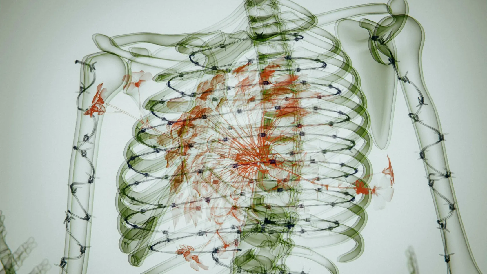
Sarcoma and Bone Cancer Surgery
Almost everyone sent to a sarcoma unit arrives braced for the word amputation. The decision actually being made is a narrower one, about where the tumour ends and how much healthy tissue has to leave with it.
About This Department
A biopsy report has come back with the word sarcoma on it, and the next conversation is about whether the arm or the leg stays. Bone sarcomas are rare, they behave nothing like the common cancers, and the operation that treats them is judged on one measurement above all others, which is how much normal tissue came out around the tumour. This page covers how that decision gets made, what the surgery removes and rebuilds, and what having it done abroad actually involves.
Free consultation
Send the pathology and the scans before anyone operates
An orthopaedic oncology team will read the biopsy report, the MRI of the whole affected bone and the staging scans, then tell you whether limb salvage is realistic in your case, how much bone the resection would take, and what reconstruction would follow. The review costs nothing and carries no obligation.
Can the limb be saved?
Limb salvage surgery removes the tumour together with a continuous cuff of healthy tissue around it and then rebuilds the gap, so the arm or the leg stays attached and does work. It has displaced amputation as the default operation for bone sarcoma in the extremities, and two developments did that. Chemotherapy given before the operation shrinks the tumour and attacks the microscopic disease that used to seed the lungs no matter how much limb had been removed, which is the reason amputation alone never cured osteosarcoma in the era before drugs. Magnetic resonance imaging then made the tumour's true edge visible, mapping how far the disease has travelled inside the marrow, whether it has crossed the growth plate, and exactly how it sits against the artery, the vein and the main nerve. A surgeon planning a margin needs all four of those answers before choosing where to cut, and none of them can be obtained from a plain X ray or from feeling the lump.
Outcome data support saving the limb wherever the anatomy allows it. A 2023 meta-analysis in Oncology Letters pooling studies of adolescent osteosarcoma found overall survival significantly longer after limb salvage than after amputation, with a hazard ratio of 0.71 and a confidence interval of 0.63 to 0.80, and progression free survival longer as well at a hazard ratio of 0.60. Distant metastasis rates came out no different between the two operations. The lungs do not care which operation was done.
Local recurrence is the trade.
In that same analysis the tumour came back at the original site more than twice as often after limb salvage, at an odds ratio of 2.68. Read those survival numbers with one caveat attached, because the comparison is not a randomised one and never will be, and surgeons offer limb salvage to the patients whose tumours are smaller, better placed and further from the neurovascular bundle, so some part of the survival gap belongs to the tumours themselves and not to the operations performed on them. What the data do establish is that saving a limb in a properly selected patient costs nothing in survival, and that is the question a patient is asking.
A broken bone at the tumour site changes the arithmetic less than most people expect. A systematic review in The Bone and Joint Journal covering eight studies and 1,713 patients with extremity osteosarcoma found a pathological fracture in 303 of them, and five year event free survival of 49.3 percent in that group against 66.8 percent without a fracture. Local recurrence rose from 11.4 to 14.4 percent, a difference the analysis could not separate from chance. The authors then compared amputation against limb salvage inside the fracture group and found no significant difference in local recurrence between them, concluding that a fracture through a bone sarcoma is not by itself a reason to amputate. Many units still treat it as one.
Why the first biopsy decides so much
Biopsy of a bone sarcoma is a surgical act with consequences, and the team that will perform the resection should plan it. The needle or the incision contaminates every tissue plane it passes through with tumour cells. Whatever track it takes has to be excised in one piece with the tumour later, which means a biopsy placed across a compartment, through a muscle belly that was going to be spared, or anywhere near the femoral vessels can turn an operation that would have saved the leg into one that cannot.
The single most consequential finding in this field
Twenty five surgeons across 21 institutions reviewed 597 patients biopsied for a suspected bone tumour. The diagnosis was wrong in 17.8 percent. A problem created by the biopsy itself forced a different and more complex operation, or added radiotherapy or chemotherapy that would not otherwise have been needed, in 19.3 percent, and altered the outcome in 10.1 percent. Eighteen patients in that series had an amputation they did not need because of how the biopsy was done. Errors and complications ran two to twelve times higher when the biopsy was carried out at the referring hospital rather than at the treating centre.
That paper was published in 1996 and the pattern it described has not gone away.
Soft tissue sarcomas suffer the same problem in a different form, because they present as a painless lump that gets shelled out at a general hospital on the assumption it is a lipoma. A Mayo Clinic review of 246 patients with synovial sarcoma found that 87 of them, more than a third, reached the sarcoma service only after such an unplanned excision. Forty six still had residual tumour in the specimen taken at the second, proper operation. Those patients did substantially worse, with a hazard ratio of 8.17 for metastasis free survival and 7.66 for disease specific survival against the ones whose re-excision came back clear.
So the practical instruction for anyone reading this before treatment has started is short. Send the imaging to a sarcoma unit and let that unit decide who takes the biopsy, where the track goes and what is done with the specimen. If a lump deeper than the fascia is larger than five centimetres, or is growing, or hurts at night, it belongs in that pathway until proven otherwise. Nothing later in this article recovers what a badly placed biopsy costs.
If someone has already operated, the situation is retrievable and needs assessing properly. Re-excision of the tumour bed, with radiotherapy where the histology and the margins call for it, remains the standard response, and roughly half of these patients turn out to have no viable tumour left in the re-excised specimen.
What the operation removes
Wide resection is the operation, and the word wide is doing the work. The surgeon takes the tumour inside an unbroken envelope of normal tissue, never cutting into the mass itself, never seeing its surface during the dissection, and removing the biopsy track and any previously operated scar in the same specimen. Fascia, periosteum and muscle become the barriers that define the envelope, because sarcoma spreads along the path of least resistance. Healthy fascia resists. Bone margins get planned off the MRI, with a measured length of normal marrow left beyond the furthest signal change, and the pathologist checks the cut end during the operation itself, so that when the check comes back involved the surgeon takes more bone there and then, instead of learning about it three weeks later.
Everything else in the operation serves that envelope.
What the surgeon is protecting while doing it. Three structures decide whether a limb can be saved once the tumour leaves. The major artery and vein have to be preserved or reconstructed, because a limb with no blood supply is not a limb. The main motor nerve determines whether the reconstructed leg will lift the foot or the reconstructed arm will bend the elbow, and a sciatic nerve encased by tumour is one of the few genuine reasons to amputate a leg that could otherwise be rebuilt. Enough muscle has to survive to move the joint and to cover the implant with living tissue, so plastic surgeons join these operations for flap coverage far more often than patients expect. Pelvic and spinal primaries sit at a different scale of undertaking from a knee or a shoulder, taking longer, bleeding more and carrying a higher complication rate at every stage, and the pelvis in particular has no compartment boundaries worth the name, so the margin has to be built out of whatever anatomy is available.
Operating time for a knee or shoulder resection with an implant reconstruction runs to several hours. Blood loss is significant enough that cross-matched units are prepared as routine. The specimen goes to pathology whole, and the report that comes back two to three weeks later carries two numbers that shape everything afterwards, being the margin status and the percentage of tumour cells killed by the pre-operative chemotherapy.
Rebuilding the gap
Removing fifteen centimetres of femur leaves a problem that has to be solved in the same anaesthetic. Bone does not grow back. Five reconstructions cover almost all of this work, and the choice turns on which bone went, how old the patient is, how much muscle survived and whether chemotherapy is still to come.
This table scrolls sideways on a narrow screen. Swipe or drag to see every column.
| Reconstruction | What it is | The trade |
|---|---|---|
| Endoprosthesis | A modular metal implant replacing the removed bone and the joint at its end, cemented or press fitted into what remains. | Walking starts within days. The implant is mechanical, so it wears, loosens and occasionally becomes infected, and a young patient should expect revision surgery in their lifetime. |
| Bone allograft | A matched segment of donor bone from a tissue bank, fixed with plates and screws to bridge the defect. | Biological where the implant is mechanical, and it lasts once it unites. Union takes a year or more, and fracture or non-union of the graft is the common failure. |
| Vascularised fibula | The patient's own fibula moved with its artery and vein and plumbed into the recipient site by a microsurgeon. | Living bone that heals and thickens over years, and the best option in a child. It needs a microsurgical team and a longer operation. |
| Extendable implant | A growing prosthesis for a child whose growth plate was resected, lengthened by an external magnetic field or a small procedure. | Keeps the legs level as the child grows. Lengthenings continue for years and each one is a hospital visit. |
| Rotationplasty | The knee region is removed, the lower leg is rotated 180 degrees and reattached, and the ankle becomes a working knee inside a prosthesis. | Outstanding function and durability in an active child, with no implant to fail. The appearance is confronting and families need time and counselling to decide. |
Endoprosthetic replacement dominates adult practice because it restores a working limb immediately and does not depend on biology that chemotherapy is about to suppress. That last point matters more than it sounds. Cytotoxic drugs resumed three weeks after surgery interfere with bone union and with wound healing, so a reconstruction that needs a graft to knit is being asked to do it under the worst possible conditions, whereas a metal implant is load bearing on the day it goes in.
Children are the exception and their reconstructions are chosen differently, because an eight year old with a fixed length implant in the femur finishes growing with one leg substantially shorter than the other.
When amputation is the better operation
Surgeons who never recommend amputation are not being kind. Limb salvage that leaves tumour behind, or that produces a painful useless limb after four operations and a year of complications, is worse than a well made amputation with a well fitted prosthesis, and the reasons to choose it are specific.
Five findings make it the right call.
- Tumour encasing the main nerve, with no plane between them.
- Tumour wrapped around the artery and the vein in a way that no bypass graft can rescue, which happens in the pelvis and the popliteal fossa more than anywhere else.
- Too little muscle left after a wide margin to cover an implant or move the joint.
- Infection already established in the tumour bed.
- A fungating tumour that has broken through the skin, contaminating the field beyond what any plane of dissection can contain.
Recurrence at the operated site makes a sixth, and it produces the hardest conversation of all, because by then the patient has spent a year keeping the limb. Salvaging a salvage is possible in selected cases. It is also the setting in which insisting on the limb starts to cost survival rather than function.
Patients who reach that decision properly informed do well with it. Prosthetic limbs have improved enormously. Modern below knee and above knee designs restore walking, work and sport, the operation itself is shorter and safer than a complex reconstruction that may need revising three times, and no implant sits waiting to loosen in fifteen years. The choice has to come from a team that does both operations and holds no stake in either.
The chemotherapy either side of surgery
High grade osteosarcoma and Ewing sarcoma are treated as systemic diseases that happen to present in a bone. Chemotherapy comes first, for roughly ten weeks, then surgery, then more chemotherapy once the wound has healed. Methotrexate, doxorubicin and cisplatin carry the standard osteosarcoma regimen and are known together as MAP, while Ewing sarcoma takes a different and more intensive combination of drugs spread across the better part of a year. Chondrosarcoma is the outlier. It answers poorly to drugs and to radiotherapy, so the surgery has to do all of the work at the first attempt, which puts more weight on the margin in that histology than in any other. The pre-operative course does two jobs at once. It attacks micrometastases too small to appear on any scan, and it hands the team a measurement no imaging can provide, because the pathologist counts what proportion of the tumour cells in the resected specimen have been killed. Under ten percent viable tumour counts as a good response and it remains the strongest prognostic marker in the disease, which is also why nobody can give you a prognosis on the day of the operation.
EURAMOS-1, the largest randomised trial ever run in osteosarcoma, registered 2,260 patients and found a good histological response in 1,041 of them. Among the 716 good responders who agreed to randomisation, three year event free survival was 76 percent. The trial tested whether adding maintenance interferon alfa-2b to MAP improved on that and it did not, returning a hazard ratio of 0.83 with a confidence interval crossing one, while 133 of the 268 patients who started interferon and reported toxicity had grade three or worse side effects. That result left MAP as the backbone, and nothing has been added to it since.
Nothing has beaten those three drugs since.
Radiotherapy plays little part in bone sarcoma surgery of the limbs. Ewing sarcoma is the exception, where radiation treats an unresectable primary or supplements a close margin, and soft tissue sarcomas around the bone frequently receive it before or after the resection.
Practically, this means the surgery is one appointment inside a treatment that runs the better part of a year. Any plan that offers you an operation without saying who is giving the chemotherapy, where, and on what schedule, is not a plan.
Risks and how often they happen
Complication rates after tumour resection and reconstruction sit well above those of ordinary orthopaedic surgery, and any page implying otherwise is not describing this operation, because the tissue envelope has been stripped, the implant is large, the patient has been immunosuppressed by chemotherapy, and the whole thing takes hours. Every item below gets managed. Knowing the early signs is what gets it managed in time.
Volume affects all of this. Bone sarcomas are rare enough that a general orthopaedic surgeon may see two in a career, and the biopsy series quoted earlier measured exactly what happens when the rare thing is handled where it is rare.
Recovery, week by week
The first fortnight. Physiotherapy starts the day after surgery. With a cemented endoprosthesis in the femur or the tibia, weight bearing through the operated leg begins within the first few days, because the construct is stable from the moment it is implanted. An uncemented stem or a graft reconstruction is protected for six to twelve weeks instead, and the surgeon sets that instruction from what was actually done in theatre, so a general protocol does not override it. Drains come out as they stop draining, the ward stay runs five to ten nights for a limb resection and considerably longer after a pelvic one, and by the time you leave the ward you should be walking a corridor with a frame or crutches under supervision.
Weeks two to six. Wound healing takes priority in this window, because chemotherapy cannot restart until it is closed and every week of delay costs something. Sutures or clips come out at two to three weeks. Range of movement work continues daily, quadriceps strength after a distal femoral replacement rebuilds slowly enough to be genuinely frustrating, and the pathology report lands during this period and gets discussed with the oncologist.
Swelling outlasts pain by months.
Three months to a year. Most patients walk without aids by three months after a lower limb reconstruction, and an upper limb reconstruction reaches useful function around the same point, though overhead reach after a proximal humeral replacement stays limited permanently. The rest of the year belongs to chemotherapy as much as to the limb, and strength returns unevenly around it. Full confidence in the reconstruction takes six to twelve months.
Running, jumping and contact sport stay off the list for good with a joint replacing endoprosthesis. Swimming, cycling, walking and gym work on a machine are all encouraged, and a rotationplasty or a fibular reconstruction in a young patient allows considerably more than an implant does.
How long you stay and when you fly
Sarcoma treatment does not fit the short surgical trip most medical travel is built around, and any arrangement pretending otherwise will fail somewhere in the middle of it, usually at the point where chemotherapy has to start and nobody has agreed who gives it. Plan around three phases.
Nobody does this in ten days.
Assessment and biopsy take a week to ten days on the ground, covering the MRI of the whole bone, a chest CT and a bone scan or PET, the biopsy itself, and the multidisciplinary meeting where the plan is set. Pre-operative chemotherapy then runs about ten weeks, and it can be given at home under a shared protocol if your local oncology service will accept it, which is the arrangement that makes the whole pathway affordable in time as well as money.
Surgery and the recovery around it need four to six weeks in the country. That covers the pre-operative workup, the operation, five to ten nights on the ward, the wound check at two to three weeks, the start of physiotherapy and the pathology discussion.
Flying depends on the wound and on the clotting risk more than on the calendar. Three to four weeks after a major limb reconstruction is the usual clearance, given a healed wound, no fever and an agreed thromboprophylaxis plan for the flight. Aisle seat, hourly walking, compression stocking, and a supply of the injections you have been prescribed.
Bring somebody with you. A patient on crutches after a femoral resection cannot manage luggage, an airport transfer and a hotel room alone, and the invitation letter for the visa can name a companion if you ask for it when the appointment is booked.
What drives the cost
No figure on any website applies to a bone sarcoma, because the treatment is not one thing. It is a diagnostic workup, a course of chemotherapy of uncertain length, an operation whose extent is not fixed until the imaging is read, an implant chosen off a shelf of very different implants, and a rehabilitation programme sized to the damage. Six variables move the total, and they move it a long way.
Which bone and how much of it. A distal femoral replacement, a total femoral replacement and a hemipelvectomy are three different operations at three scales of cost. The reconstruction chosen. A modular endoprosthesis, a custom implant made to your own CT, an allograft from a tissue bank and a free vascularised fibula each carry different device and theatre costs, and the custom implant adds manufacturing lead time as well. The histology. Chondrosarcoma is treated with surgery alone, while osteosarcoma and Ewing sarcoma add months of inpatient chemotherapy cycles. The number of cycles, which the response decides and nobody fixes in advance. Whether radiotherapy joins the plan, which it does in Ewing sarcoma and in many soft tissue cases and does not in most bone ones. And whether a plastic surgical flap is needed for cover, which adds a second surgical team and a longer operation. Those six do not move the total by ten percent each. Between a straightforward distal femoral replacement in a chondrosarcoma and a custom hemipelvic reconstruction in a Ewing sarcoma needing eighteen cycles and a free flap, the two treatments have almost nothing in common except the word surgery.
Your own medical history moves it too. Age, diabetes, heart and lung disease, previous surgery at the same site, blood thinning medication and the amount of inpatient rehabilitation your case will need all change the plan and therefore the number.
Packages published in this market normally cover the transfers, the pre-operative tests, the surgeon and anaesthesia fees, the implant, the ward nights allowed for, inpatient physiotherapy, an interpreter, a set number of hotel nights and the follow up appointments before departure. They normally exclude flights, travel insurance, extra hospital nights, intensive care beyond what was planned, and the treatment of a complication. Chemotherapy cycles are frequently quoted separately from the surgical package, and that is the single most common reason two quotations for the same disease look nothing alike. Five questions turn a headline into something you can compare. What extent of resection does this figure assume, and what happens to it if the MRI shows more disease. Is the implant named, and is it modular or custom made to your own CT scan. Are the chemotherapy cycles inside the figure or outside it. How many ward nights does the figure allow before extra nights start being charged separately. And who pays when a complication needs a second operation.
Any usable figure comes from a surgeon and an oncologist who have read your own reports and imaging. That review costs nothing, and the contact details at the top of this page are where it starts.
After you fly home
Sarcoma follow up runs intensively and for years, because recurrence and lung metastasis are both treatable when caught small, and the schedule below, which follows the pattern used across sarcoma centres, needs two things from you once you are home, being a local oncologist or orthopaedic surgeon willing to order the scans and a working route back to the operating team who will read them.
Follow up here runs for a decade.
This table scrolls sideways on a narrow screen. Swipe or drag to see every column.
| Period | How often | What is done |
|---|---|---|
| Years 1 and 2 | Every 3 months | Examination of the operated limb, chest imaging for lung metastases, and X rays of the reconstruction. |
| Years 3 to 5 | Every 4 to 6 months | The same set, with MRI of the site added whenever a symptom or an X ray raises a question. |
| Years 6 to 10 | Yearly | Chest imaging, implant X rays and a functional review. Late recurrence and late implant loosening are both found here. |
| Beyond 10 years | Yearly or as agreed | Implant surveillance mainly, plus screening for the late effects of anthracycline and platinum chemotherapy. |
Ask before you leave for the operative note, the pathology report, the implant details with catalogue and serial numbers, the chemotherapy record and the discharge imaging, in a language your own doctors read. An implant card matters more than patients realise, because a surgeon at home cannot revise a device they cannot identify.
Four things need a doctor the same day, and a message will not do. Fever with a hot, swollen or discharging wound. A new lump or new pain at the operated site. Sudden inability to bear weight on a leg that had been fine the day before. Breathlessness or chest pain.
Remote check ins between scans run by message and video call, with imaging sent as files. That is how the operating team stays involved once you are home.
Frequently asked questions about sarcoma surgery
Will I lose my leg?
A local hospital has already biopsied or removed the lump. Is it too late?
How long do I need to stay in Turkey?
When can I fly home after limb salvage surgery?
What happens if the implant fails years later, when I am at home?
Can someone travel with me?
References
- Mankin HJ, Mankin CJ, Simon MA. The hazards of the biopsy, revisited. Members of the Musculoskeletal Tumor Society. The Journal of Bone and Joint Surgery. American Volume. 1996;78(5):656-663.
- Wang L, Fan Y, Zhou Y, Zhong G. Prognosis of limb-salvage treatment of osteosarcoma in adolescent patients, a meta-analysis. Oncology Letters. 2023;26(5):466.
- Salunke AA, Chen Y, Tan JH, Chen X, Khin LW, Puhaindran ME. Does a pathological fracture affect the prognosis in patients with osteosarcoma of the extremities? A systematic review and meta-analysis. The Bone and Joint Journal. 2014;96-B(10):1396-1403.
- Bielack SS, Smeland S, Whelan JS, et al. Methotrexate, doxorubicin, and cisplatin plus maintenance pegylated interferon alfa-2b versus MAP alone in patients with resectable high-grade osteosarcoma and good histologic response to preoperative MAP. Journal of Clinical Oncology. 2015;33(20):2279-2287.
- Broida SE, Arguello AM, Sullivan MH, et al. Unplanned excision of synovial sarcoma, factors associated with recurrence and survival. Cancers. 2024;16(18):3157.
Editor's note
Written by the Biruni Hospital medical editorial team. Reviewed by Assistant Professor Şafak SAYAR, Orthopedics and Traumatology.
Medically reviewed by
Assistant Professor Şafak SAYAR
Orthopedics and Traumatology
Related Treatments
View All-
Achilles Tendon Repair
Achilles tendon repair stitches a torn tendon back together and cuts the risk of a second rupture, while a boot with early movement gives the same function at one year in trials. This guide covers the signs of a tear, surgery or a boot, open and minimally invasive repair, rehabilitation week by week and the stay.
-
Bone Tumor Resection
Most people arrive expecting to lose the limb. For the large majority of bone tumours today the limb is saved, the bone is rebuilt with an implant or a graft, and survival is no worse for it. What decides the answer is the margin, and the margin is planned from an MRI of the whole bone before anything is cut.
-
Chondrosarcoma Surgery and Bone Tumor Resection
Chemotherapy and radiation barely touch this tumor, which sounds like the worst news on the page and is the reason a single operation cures it outright. For a grade 1 chondrosarcoma in an arm or a leg the bone is scraped clean from the inside and the natural joint stays where it is.
-
Complex Knee Revision Surgery
Infection now causes more failed knee replacements than any other single reason, 21.6 percent across registries in seven countries, and it can turn a revision into two operations with weeks of antibiotics between them. This page explains how the cause is found before surgery, when cones or a hinge are needed, and how a two-stage revision fits around travel.
-
Complex Trauma Reconstruction
A fracture that has not healed after a year, healed crooked or became infected can still be repaired, and the treatment often runs for months in an external frame. This page explains nonunion, malunion and bone loss repair, the choice between bone transport and the Masquelet technique, and how a long treatment fits around travel to Istanbul.