
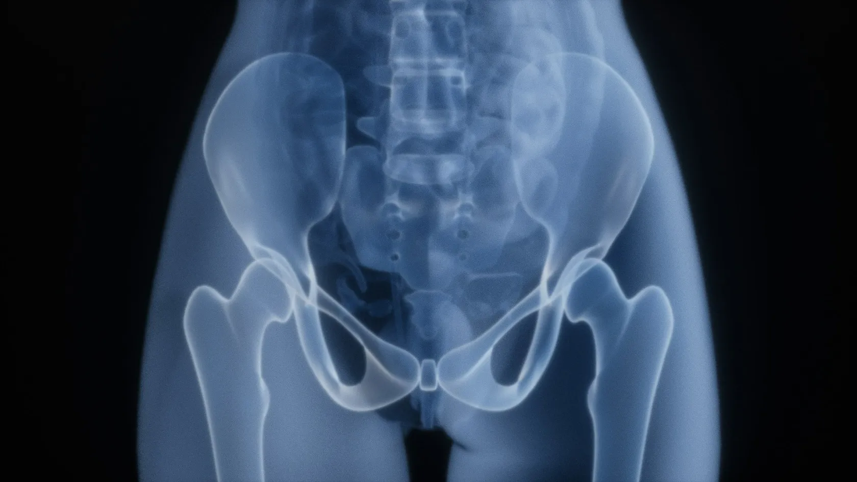
Sacral Tumor Surgery
Sacrectomy takes the pain away and it does not give the nerves back. In a cohort followed for six years, pain fell significantly after surgery while weakness, numbness and bladder problems stayed exactly where they were. Knowing which of those you are trading is the point of this page.
About This Department
Chordoma grows from a piece of tissue that should have vanished before you were born. That rod of cells, the notochord, lays down the spinal column in an embryo and then disappears, except that in a small number of people a few cells survive inside the sacrum and, decades later, start dividing again. That origin explains almost everything about the disease, including why it grows so slowly, why chemotherapy has nothing to offer it, and why the operation has to take it out in one unopened piece. This page covers what sacral tumor surgery involves, how high the resection goes and what each level costs, and how treatment like this is arranged from another country.
Free consultation
Send the sacral MRI before anyone puts a needle in
Specialists in spine and orthopaedic oncology read the MRI of the whole sacrum with the pelvis, work out the highest nerve root the tumour has reached, and tell you which level of sacrectomy that implies, whether radiotherapy should come before the operation, and what you would be trading. Image files, please, and not a report. The review costs nothing and carries no obligation.
What grows in the sacrum
Chordoma accounts for more than half of all primary tumours of the sacrum. This page is mostly about it. Chordoma behaves malignantly and slowly at the same time, spreading by pushing outward and not by travelling in the bloodstream, so patients frequently stay well for years while the mass quietly enlarges behind them. Chemotherapy does nothing. Ordinary radiotherapy doses do very little either, and those two facts together put the entire burden of cure onto a single operation, which is a position no other bone tumour puts a surgeon in quite so completely.
Giant cell tumour comes second and behaves quite differently, being locally aggressive without being malignant in the usual sense, and it now has a drug that works.
The rest of the list matters because it changes the plan completely. Metastatic deposits from breast, lung, kidney, thyroid and prostate cancer reach the sacrum often, and those are treated to control pain and preserve function rather than to cure. Ewing sarcoma and osteosarcoma occur here and bring chemotherapy with them. Chondrosarcoma behaves like chordoma in that surgery has to do the work alone. Benign lesions turn up too, including aneurysmal bone cysts and the small osteoid osteoma that causes pain out of all proportion to its size, and both are managed far more simply than anything above. The biopsy that separates these is planned by the surgeon who would perform the resection, and it goes through a track that the resection can later remove in one piece. Get it wrong and options disappear. A needle passed through the wrong plane, or through the rectum, seeds tumour cells the whole length of its path, and every one of those cells then has to be removed with the specimen or left behind to grow, which is how a curable tumour quietly becomes an incurable one before treatment has even started.
Why it takes years to find
Almost every symptom a sacral tumour produces belongs to something commoner. Buttock ache becomes sciatica. Difficulty emptying the bowel becomes constipation. Bleeding at the back passage becomes haemorrhoids, and numbness across the saddle area gets put down to a trapped nerve, and each of those explanations is reasonable on the day it is offered. Meanwhile the tumour grows forward into the pelvis, where there is nothing to stop it and nothing to feel. So these tumours are large by the time somebody scans them, and size is not a cosmetic detail. In a Johns Hopkins series of 101 patients followed over twenty years, a chordoma of 100 cubic centimetres or more carried nearly six times the risk of local recurrence compared with a smaller one.
Four symptoms deserve an MRI instead of another course of treatment. Buttock or low back pain that wakes you at night and does not vary with position. A change in bowel habit that no laxative shifts. Numbness across the perineum, the buttocks or the backs of the thighs, in the pattern a saddle would cover, which is the finding that should stop a clinician mid-consultation and send the patient for a scan the same week. And any mass a doctor can feel on rectal examination, which is how a great many sacral tumours are eventually detected.
Plain X rays miss most of them. Bowel gas hides it. Bowel gas obscures the sacrum on a standard film, so the test that answers the question is an MRI of the whole sacrum and pelvis, and it costs very little.
En bloc, and why the word matters
Two operations exist for a sacral chordoma and only one of them treats it. En bloc resection removes the tumour inside an unbroken envelope of normal tissue. The surgeon never enters the mass, never sees its surface, and takes the biopsy track and any previous scar out in the same specimen. Intralesional surgery goes into the tumour and scoops it out, which relieves pain quickly and leaves cells scattered through the wound.
That difference shows up in the numbers, and never in how the patient feels during the first month.
The finding this whole page rests on
Researchers at Massachusetts General followed 126 patients with spinal and sacral chordoma treated with high dose proton based radiotherapy and reported five year local control of 72 percent after en bloc resection against 55 percent after intralesional surgery. Among the 28 patients who had a primary chordoma, received radiotherapy before the operation, and then underwent en bloc resection, zero local recurrences were seen. The authors put it plainly, which is that intralesional and incomplete resections carry higher local failure and are to be avoided.
Recurrence here comes back local and late, so a return of the tumour at four years counts as ordinary. Each recurrence proves harder than the last. Tissue planes have gone, the anatomy has been rearranged, and the margin has to be built out of scar tissue that gives the surgeon none of the landmarks the first operation had, in a patient who has already spent a year recovering from that first operation and has less reserve for a second. Surgeons therefore accept a larger operation at the first attempt in exchange for a smaller chance of needing a second.
Ask whether the plan is en bloc. Fair question. One word answer.
How high the resection goes
Surgeons describe a sacrectomy by the highest level at which the bone is divided, and that single number carries most of what a patient wants to know. Surgeons divide the bone half a vertebra to a whole vertebra above the top of the tumour, so the disease sets the level and nobody chooses it.
This table scrolls sideways on a narrow screen. Swipe or drag to see every column.
| Level | What is removed | What it usually means afterwards |
|---|---|---|
| Low, below S3 | The coccyx and the bottom of the sacrum, through a posterior approach only. | Bladder, bowel and sexual function are generally preserved. Numbness around the scar and discomfort sitting are the usual complaints. |
| Mid, through S2 or S3 | The lower two thirds of the sacrum, sacrificing the roots below the cut on both sides. | Continence becomes unreliable and is managed with a bladder and bowel programme. Walking is unaffected and the pelvic ring stays stable. |
| High, through S1 | Almost the whole sacrum, usually needing a combined front and back approach. | Bladder and bowel control is lost or heavily impaired, and the link between spine and pelvis needs metal fixation. |
| Total sacrectomy | The entire sacrum with both sacroiliac joints, in two stages or one long combined operation. | Spinopelvic reconstruction is mandatory, walking takes months to rebuild, and continence is not retained. |
Nothing on that table is guesswork on the day. That level comes off the MRI weeks in advance, which means the conversation about what you are trading can be had properly, with time, and with somebody from the continence team in the room rather than after the event.
Sacrifice one side and keep the other, where the tumour allows it, and a great deal is preserved. Nerve roots work in pairs, and unilateral loss is tolerated far better than the same loss on both sides, so a tumour sitting off the midline sometimes permits an operation that a central one does not.
Radiotherapy, before and after
Chordoma resists conventional radiotherapy, and for years that was taken to mean radiotherapy did not work. What it actually meant was that the dose needed to kill it exceeds what ordinary X ray beams can deliver to a sacrum without wrecking the bowel, the bladder and the nerve roots sitting in the same field. Particle beams changed that arithmetic, because a proton or a carbon ion stops at a depth the physicist chooses instead of carrying on through the patient, so the dose can be pushed far higher with the exit dose largely removed. A meta-analysis of eleven carbon ion studies, in which 552 of the treated lesions sat in the sacrum, reported local control of 96 percent at one year, 83 percent at three, 76 percent at five and 54 percent at ten, with overall survival of 85 percent at five years and 69 percent at ten. Acute toxicity ran mostly at grade one and two, and late toxicity mostly at grade one to three.
Sequencing brings the surgical and the radiation plan together, and it goes wrong more often than anything else here.
Radiotherapy given before the operation treats tissue that still has its normal blood supply, sterilises the surface of the tumour so that any spillage during surgery matters far less, and goes into a field that has not yet been scarred by a scalpel, which together explain why the same dose does more work before an operation than it can ever do afterwards. The Massachusetts General series found local control of 85 percent among primary chordomas that had preoperative radiotherapy against 56 percent among those that did not. The Johns Hopkins cohort found the same direction of effect, with neoadjuvant radiotherapy associated with roughly an eleven fold reduction in the risk of local recurrence. Booking the operation first and thinking about radiotherapy afterwards forfeits that advantage, and it cannot be recovered later. Proton and carbon ion facilities are scarce and are not available in every country, which is a practical planning problem rather than a medical one. Ask early where the radiotherapy would be delivered, in what order, and who is talking to whom about it.
Giant cell tumour and denosumab
Sacral giant cell tumour used to be a miserable diagnosis. Giant cell tumour rarely turns malignant, yet it destroys bone locally, sits in exactly the place where a wide resection costs the most function, and comes back after curettage often enough to be a real problem. Then a drug arrived that blocks the signalling pathway the tumour depends on. Denosumab goes in as a monthly injection under the skin. In the spinal and sacral subgroup of an international phase two study, 132 patients were assessed, most of them with disease that could not be removed surgically at all. The estimated probability of progression or recurrence in that unsalvageable group was 3 percent at one year and 7.4 percent at three and five years. Among the patients whose planned surgery was expected to cause severe harm, fewer than half ended up having an operation at all. Investigators recorded clinical benefit in 83 percent across the whole study.
So the modern sequence for a sacral giant cell tumour frequently starts with the drug and not the knife, shrinking and calcifying the lesion until either a smaller operation becomes possible or none is needed at all. Neither denosumab nor embolisation replaces the biopsy, since everything downstream depends on knowing what the tumour actually is.
This table scrolls sideways on a narrow screen. Swipe or drag to see every column.
| Approach | What it does | Where it fits |
|---|---|---|
| Denosumab | Monthly injection blocking the signalling pathway the tumour depends on, shrinking and calcifying it. | First, in most sacral cases. It makes a smaller operation possible, or removes the need for one. |
| Arterial embolisation | A radiologist blocks the feeding vessels through a catheter, over several sessions. | An alternative where the drug is unsuitable, and a preparation before surgery where bleeding is the worry. |
| Curettage | The lesion is scraped out from inside and the cavity treated, keeping the sacrum and the nerve roots. | Where function must be preserved and some recurrence risk is acceptable in exchange. |
| En bloc resection | The same wide removal used for chordoma, taking the involved sacrum out in one piece with a cuff of normal bone around it. | Reserved for aggressive or recurrent disease, because the functional price at this site is high. |
Closing the wound
Patients never think about the wound and surgeons think about little else. A sacrectomy leaves a large cavity where bone used to be, covered by skin that has to survive over a dead space, in a region that sits against the bed, close to the perineum, and frequently in tissue that has already been irradiated. Wound problems after this operation are common in every published series that reports them honestly.
The plastic surgical team joins the operation from the start and scrubs for the closure, instead of being telephoned a fortnight later when the wound opens.
Muscle does the filling. A vertical rectus abdominis flap comes down from the abdomen on its own blood supply, or gluteal tissue rotates in from the side, and either way the dead space ends up filled with tissue that has a circulation of its own.
Drains, a pressure relieving mattress and a period spent off your back. None of that is optional, and the nursing plan for the first fortnight is written around the flap.
A prospective series of 53 sacropelvic resections with immediate flap reconstruction recorded exudative wounds in 50 percent and 31.5 percent of its two groups, dehiscence in 41.1 percent and 31.5 percent, and seroma in around 29 percent and 26 percent. Those figures run high and they also happen to be the truth about this operation. The same study answered a useful question along the way, namely whether giving radiotherapy in theatre harms flap survival. It did not.
What does not come back
Nerve function does not recover after a sacrectomy, and a page written to reassure would stop telling the truth at exactly this point. The Johns Hopkins cohort of 101 patients tracked function over an average of six years and found that pain fell significantly after surgery while sensory loss, weakness and bladder and bowel dysfunction stayed exactly where they were. Cut nerve roots never regrow. The deficit a patient wakes up with on the first morning is the deficit they will be describing to a doctor twenty years later, and the conversation before the operation therefore carries far more weight here than any conversation after it. Whatever function gets traded on the day of the operation is traded permanently, and no amount of physiotherapy alters that.
Stating it plainly does not amount to discouragement. Knowing the trade beforehand is what makes the decision a real one, because a patient who understands it copes far better than one who spends the first year waiting for a recovery that was never coming.
What can be built around the deficit is considerable. Intermittent self catheterisation, taught properly, keeps the kidneys safe and lets people work and travel. A structured bowel programme built from diet, timing, suppositories or irrigation turns unpredictable days into predictable ones, which is the difference between planning a journey and not leaving the house. Good units discuss sexual function openly, and medical and mechanical options exist for both men and women. Foot drop from an L5 or S1 injury is managed with an ankle brace, and walking survives even high resections.
Meet a continence specialist before the operation, never after it.
Holding the spine to the pelvis
Your entire upper body transmits its weight through the sacrum into the two hip bones, so removing enough of it breaks that chain. Below S1 the ring holds and no metalwork is needed. Take the resection through or above S1 and the sacroiliac joints go with it, at which point the spine is sitting on nothing and has to be bolted to the pelvis. Spinopelvic reconstruction does that with rods running from the lumbar spine down into screws placed in the iliac bones, sometimes with additional rods or a graft bridging the gap where the sacrum used to be. Rods and screws on that scale go into a patient who has just had a long operation and who will be irradiated, so hardware problems come with the territory here. Revision to adjust or replace part of the metalwork happens.
Sitting up starts within days. Slowly. Standing and walking come back over weeks to months, depending on how much of the load the fixation is carrying, and the physiotherapy plan gets written around that construct and around the bone it is anchored in rather than around any standard timetable printed on a ward wall.
Risks worth knowing about
Sacrectomy takes hours in a difficult place, and the risk list runs correspondingly long. Every item here is anticipated and planned for rather than merely accepted, and knowing the early signs is what gets each of them treated in time.
Volume matters here. Sacral chordoma occurs rarely enough that most spinal surgeons will finish a career without operating on one.
Recovery and daily life
Ward stays after a sacrectomy run longer than after most cancer operations, commonly two to four weeks and longer after a total resection or a combined approach, and the length is driven by the wound and by bladder and bowel retraining rather than by pain. Physiotherapy starts within days, sitting is restricted or cushioned to protect the flap, and a urinary catheter stays in until the bladder programme is established.
Then comes the part nobody schedules. Learning the new routine. Catheterisation, bowel timing and skin care over numb areas all have to become automatic, and they take weeks of practice with a specialist nurse before they stop dominating the day. Patients who leave hospital with that training in place do far better than those sent home with a leaflet. Pain improves markedly. That is the reliable win. Most people walk independently within weeks after a low or mid resection, and within a few months after a high or total one, and sitting tolerance builds slowly with a pressure relieving cushion. Numbness across the buttocks and the back of the thighs is permanent and needs a skin check every day, because an area that cannot feel pressure will develop a sore without warning and the first sign a patient notices is frequently a stain on the clothing rather than any sensation at all. Driving comes back once you can sit comfortably and react normally, typically somewhere between six and twelve weeks.
Swimming, cycling on an upright bike and gym work on machines are all encouraged once the wound is sound. Heavy lifting and impact sport are restricted permanently after spinopelvic fixation, because the construct is carrying a load the sacrum used to carry and it was never designed to do that under repeated shock.
Staying and flying home
Sacral tumour treatment does not fit a two week surgical trip, and any plan built as though it does will come apart in the middle.
Assessment and biopsy take about a week on the ground, covering the MRI of the sacrum and pelvis, a staging CT, the planned biopsy and the multidisciplinary meeting where the level and the sequence are decided. Where radiotherapy comes first, that course and the recovery from it occupy the following weeks and can sometimes be delivered nearer home if a suitable particle centre exists there. The surgical block itself needs eight to ten weeks in the country, covering the operation, two to four weeks on the ward, the transfer to rehabilitation, the wound reviews and the continence training that has to be finished before anybody flies anywhere. Flying gets cleared at around six to eight weeks after the resection, later than for limb surgery, and the wound and the clot risk are why. Clearance needs a healed and stable flap, no fever, an agreed plan for clot prevention, and a bladder routine you can manage inside an aircraft toilet, which is a genuine planning problem rather than a detail and one that the continence nurse should walk through with you before you book the seat. Book an aisle seat near the toilet, request airport assistance, carry catheters and dressings in hand luggage with a doctor's letter, and take a pressure relieving cushion for the flight.
Travel with somebody, for the whole stay. Have that person named on the invitation letter when the appointment is booked.
What drives the cost
Any published figure for sacral tumour surgery describes somebody else's operation. The level of the resection alone separates a short posterior procedure from a two stage total sacrectomy with spinopelvic reconstruction, and those are not variations on one treatment.
Seven things move the total. Start with the level of the sacrectomy, which outweighs everything else. Whether the approach is posterior only or combined front and back, which changes theatre time, teams and critical care. Whether spinopelvic instrumentation is required, which brings an implant cost and a longer operation. Whether radiotherapy joins the plan and in what form, since proton and carbon ion treatment carries quite different pricing from conventional photon therapy and may be delivered at a separate centre. Whether a flap reconstruction is needed, which adds a plastic surgical team. How many surgical specialties are scrubbed, counting colorectal, urology and vascular colleagues where the tumour has reached the rectum or the iliac vessels. And the length of inpatient rehabilitation and continence training, which for this operation is measured in weeks.
Your own history moves it further, through age, kidney and heart function, previous surgery or radiotherapy in the same field, blood thinning medication and anything that raises the transfusion requirement.
Packages published in this market ordinarily include the transfers, the tests before admission, the surgical and anaesthetic fees, implants, a stated number of ward nights, inpatient physiotherapy, interpreting, a block of hotel nights and the appointments before departure. Left outside, ordinarily, are the flights, the insurance, nights beyond the stated allowance, unplanned critical care and the treatment of any complication. For sacral work the lines that move most are the critical care allowance, the rehabilitation weeks and whether radiotherapy sits inside or outside the quotation, so read those three before anything else. Six questions make a quotation comparable. Where does this figure assume the sacrum is divided. Is the approach posterior only or combined. Is spinopelvic instrumentation included. Is radiotherapy inside the figure, and if so which modality, and delivered where. How many weeks of rehabilitation and continence training does it cover. And who pays for a return to theatre months later when a wound breaks down.
No number means anything until a surgeon and a radiation oncologist have read the actual scans side by side and agreed on the level and the sequence between them. That reading costs nothing. The contact details at the top of this page are where it starts.
Surveillance once you are home
Late recurrence is the rule with chordoma, so the follow up schedule runs longer than patients expect and ten years is the working horizon. MRI of the sacrum and pelvis is the test that matters, since a plain film shows the metalwork and tells you nothing whatever about the soft tissue in front of it where a recurrence would actually sit, and chest imaging runs alongside because chordoma does eventually metastasise to the lungs in a minority of patients. Examination and imaging every four to six months for the first two years, then every six to twelve months, is a reasonable pattern for a local team to follow.
This needs a local doctor willing to order the scans and a working route back to the operating team who will read them.
Take everything home with you, in a language your own doctors read.
- The operative note, stating how high the bone was divided and exactly which nerve roots were cut.
- The pathology report with the margin status.
- The implant card with catalogue and serial numbers, where metalwork was used.
- The radiotherapy record with the doses and the fields treated.
- The discharge imaging and the written continence plan.
Those first two items are what every future clinician will ask about, and neither can be worked out from a scar.
Four things need a doctor the same day. Fever with a wound that is hot, swollen or discharging. Clear fluid leaking from the wound. New weakness, new numbness or a sudden change in bladder or bowel control. Breathlessness or chest pain.
Between scans the team stays reachable by message and video call. Send the image files themselves.
Frequently asked questions about sacral tumour surgery
Will I lose control of my bladder and bowel?
What does en bloc resection mean and why does everyone insist on it?
Should radiotherapy come before or after the operation?
Can a sacral giant cell tumour be treated without major surgery?
How long do I need to be in Turkey, and when can I fly home?
Will the numbness and weakness improve with physiotherapy?
References
- Rotondo RL, Folkert W, Liebsch NJ, et al. High-dose proton-based radiation therapy in the management of spine chordomas, outcomes and clinicopathological prognostic factors. Journal of Neurosurgery. Spine. 2015;23(6):788-797.
- Xia Y, Papali P, Al-Mistarehi AH, et al. Outcomes after definitive surgery for spinal and sacral chordoma in 101 patients over 20 years. Neurosurgery. 2024;96(3):494-504.
- Dong M, Liu R, Zhang Q, et al. Efficacy and safety of carbon ion radiotherapy for chordomas, a systematic review and meta-analysis. Radiation Oncology. 2023;18(1):152.
- Bukata SV, Blay JY, Rutkowski P, et al. Denosumab treatment for giant cell tumor of the spine including the sacrum. Spine. 2021;46(5):277-284.
- Lasso JM, Pinilla C, Vasquez W, Asencio JM. The effect of intraoperative radiotherapy on healing and complications after sacrectomy and immediate reconstruction. Annals of Plastic Surgery. 2021;86(6):688-694.
Editor's note
Written by the Biruni Hospital medical editorial team. Reviewed by Assoc. Prof. Dr. Suat BATAR, Orthopedics and Traumatology.
Medically reviewed by
Assoc. Prof. Dr. Suat BATAR
Orthopedics and Traumatology
Related Treatments
View All-
Achilles Tendon Repair
Achilles tendon repair stitches a torn tendon back together and cuts the risk of a second rupture, while a boot with early movement gives the same function at one year in trials. This guide covers the signs of a tear, surgery or a boot, open and minimally invasive repair, rehabilitation week by week and the stay.
-
Bone Tumor Resection
Most people arrive expecting to lose the limb. For the large majority of bone tumours today the limb is saved, the bone is rebuilt with an implant or a graft, and survival is no worse for it. What decides the answer is the margin, and the margin is planned from an MRI of the whole bone before anything is cut.
-
Chondrosarcoma Surgery and Bone Tumor Resection
Chemotherapy and radiation barely touch this tumor, which sounds like the worst news on the page and is the reason a single operation cures it outright. For a grade 1 chondrosarcoma in an arm or a leg the bone is scraped clean from the inside and the natural joint stays where it is.
-
Complex Knee Revision Surgery
Infection now causes more failed knee replacements than any other single reason, 21.6 percent across registries in seven countries, and it can turn a revision into two operations with weeks of antibiotics between them. This page explains how the cause is found before surgery, when cones or a hinge are needed, and how a two-stage revision fits around travel.
-
Complex Trauma Reconstruction
A fracture that has not healed after a year, healed crooked or became infected can still be repaired, and the treatment often runs for months in an external frame. This page explains nonunion, malunion and bone loss repair, the choice between bone transport and the Masquelet technique, and how a long treatment fits around travel to Istanbul.