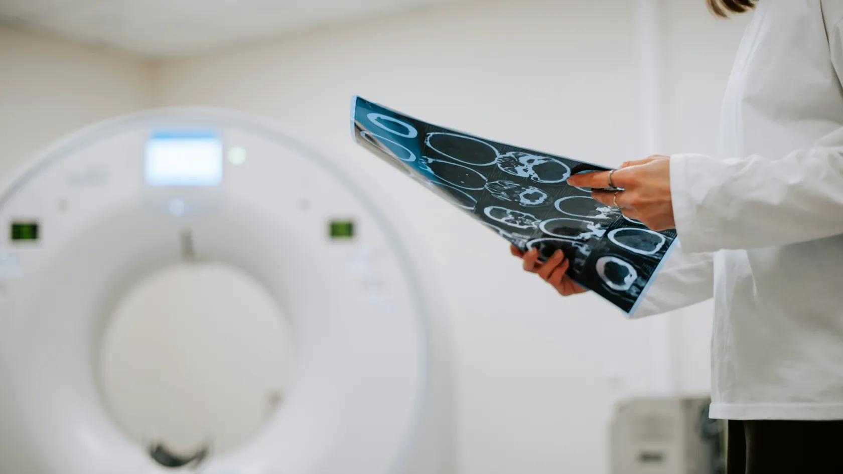
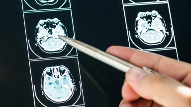
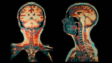
A PSMA PET/CT scan uses a tracer that binds to prostate cancer cells to show spread anywhere in the body, with far greater accuracy than CT and bone scan. It stages high-risk prostate cancer, locates recurrence when PSA rises after surgery or radiotherapy, and selects patients for PSMA radioligand therapy. The scan takes about two hours, the report follows within two working days, and you can fly home the same day.
About This Department
Nuclear Medicine
A PSMA PET/CT scan finds prostate cancer cells that CT and bone scans miss. What it shows can change the treatment plan.
A PSMA PET/CT scan is a nuclear medicine test for prostate cancer. A small amount of a radioactive tracer that binds to PSMA, prostate-specific membrane antigen, a protein found in large amounts on prostate cancer cells, is injected into a vein, and a PET/CT scanner maps where it collects throughout the body. It is used to stage high-risk prostate cancer before surgery or radiotherapy, to find the source of a rising PSA after treatment, and to select patients for PSMA radioligand therapy. In a randomized trial of 302 men with high-risk prostate cancer, PSMA PET/CT was 92 percent accurate in detecting spread to lymph nodes or distant sites, against 65 percent for CT and bone scan combined, with less than half the radiation dose (Hofman and colleagues, Lancet, 2020). This page explains how the scan works, who needs one, results in staging and in recurrence, its role before radioligand therapy, preparation and the scan day, reading the report, safety, and how patients from abroad arrange a scan at Biruni Hospital in Istanbul.
92
Percent accuracy of PSMA PET/CT for spread in high-risk prostate cancer, against 65 percent for CT and bone scan
75
Percent of men with rising PSA after treatment in whom the scan located the recurrent cancer
8.4
Millisieverts of radiation for PSMA PET/CT, against 19.2 for CT and bone scan together
Free
Review of your PSA history and earlier imaging by our nuclear medicine team
Cancer cells anywhere in the body, down to a few millimeters
Some prostate cancers make little PSMA
PSMA sits on the surface of most prostate cancer cells at levels hundreds of times higher than in normal tissue. The tracer seeks it out wherever the cancer has gone.
PSMA tracers
Tracer
Label
Note
Ga-68 PSMA-11
Gallium-68
The most studied, used in the main trials
F-18 PSMA-1007, piflufolastat and others
Fluorine-18
Longer half-life, sharp images, less bladder activity with some agents
The PET part detects the tracer and the CT part provides the anatomy, so the report can say exactly which lymph node or which bone holds the cancer. Some centers combine PSMA PET with MRI instead of CT.
Who needs one
PSMA PET/CT is part of European and American prostate cancer guidelines for specific situations. It is most useful when finding or excluding spread changes the treatment.
Main reasons for a PSMA PET/CT scan
Situation
Question answered
Effect on treatment
High-risk or unfavorable intermediate-risk cancer at diagnosis
Has it spread beyond the prostate
Surgery or radiotherapy planned correctly, or systemic treatment
Rising PSA after prostatectomy or radiotherapy
Where is the cancer coming back
Targeted salvage radiotherapy or surgery
Advanced cancer before radioligand therapy
Do the tumors carry enough PSMA
Eligibility for lutetium-177 PSMA
Suspicious findings on other scans
Is this prostate cancer
Avoids wrong treatment
1
Low-risk cancer
Men with low-risk or favorable intermediate-risk prostate cancer have a very small chance of spread and do not need a PSMA scan.
Not a screening test
PSMA PET/CT is not used to look for prostate cancer in men without a diagnosis. A PSA blood test, MRI and biopsy remain the route to diagnosis.
Staging high-risk cancer
The proPSMA trial
PSMA PET-CT had a 27 percent greater accuracy than that of conventional imaging, 92 percent against 65 percent, and conventional imaging had more equivocal findings, 23 percent against 7 percent (Hofman and colleagues, Lancet, 2020).
proPSMA results, 302 men with high-risk prostate cancer
Measure
CT and bone scan
PSMA PET/CT
Accuracy
65 percent
92 percent
Sensitivity
38 percent
85 percent
Management changed by first scan
15 percent
28 percent
Radiation dose
19.2 millisieverts
8.4 millisieverts
Finding spread before treatment spares some men an operation that could not cure them, and it lets radiotherapy include lymph nodes that would otherwise be missed. A PSMA scan can also show disease that a CT and bone scan would have shown only years later, which is why treatment decisions based on PSMA findings are discussed at the uro-oncology tumor board.
Rising PSA after treatment
After prostatectomy, PSA should fall to an undetectable level. A rise above 0.2 nanograms per milliliter signals recurrence. After radiotherapy, a rise of 2 above the lowest point does the same. Finding where the cancer has returned decides whether salvage treatment can still cure it.
1
When to scan
Guidelines recommend a PSMA scan once PSA rises after surgery, ideally while it is still below 1, because salvage radiotherapy works best at low PSA levels. Waiting for a higher PSA makes the scan more likely to be positive but the cancer harder to cure.
Detection rate by PSA level, 635 men with recurrence
PSA before scan, ng/mL
Scans showing cancer
Note
Below 0.5
38 percent
Still worth scanning when salvage treatment is planned
0.5 to under 1.0
57 percent
Good balance of detection and curability
1.0 to under 2.0
84 percent
High detection
5.0 or above
97 percent
Nearly always positive
2
Targeted treatment
A PSMA scan that shows cancer only in the prostate bed or a few lymph nodes allows radiotherapy or surgery aimed at those sites. In the same trial, PET-directed focal therapy alone lowered PSA by half or more in 80 percent of treated men.
For PSA categories 0 to 0.19, 0.2 to 0.49, 0.5 to 0.99, 1 to 1.99 and 2 or more ng/mL, the percentages of positive scans were 33, 45, 59, 75 and 95 percent (Perera and colleagues, European Urology, 2020).
Before PSMA radioligand therapy
Lutetium-177-PSMA-617 plus standard care prolonged overall survival to a median of 15.3 months against 11.3 months with standard care alone in PSMA-positive metastatic castration-resistant prostate cancer (Sartor and colleagues, New England Journal of Medicine, 2021).
Selecting patients
Radioligand therapy uses the same PSMA target to deliver radiation directly to cancer cells. It works only when the tumors carry enough PSMA, and a PSMA PET/CT scan confirms this before treatment. In the VISION trial, patients qualified only when their tumors took up more tracer than the liver and no significant deposit lacked PSMA.
Monitoring
Repeat scans during and after radioligand therapy show how the disease responds.
1
Our approach
The nuclear medicine physician reviews the PSMA scan with the oncologist and decides together with the patient whether radioligand therapy is suitable.
Preparing for the scan
1
The day before
No special diet is needed in most cases. Drink plenty of water the day before and on the day of the scan. Bring earlier scans, pathology reports and PSA results.
Hormone therapy changes how much PSMA the cancer shows, sometimes markedly within weeks of starting, so tell the team if you have started or stopped hormone treatment. The scan is sometimes timed before it begins.
A recent PSA result, within the last four weeks, helps interpret the scan.
Patients who find it hard to lie still for 20 to 30 minutes, because of pain or anxiety, can take painkillers or a mild sedative beforehand. Claustrophobia is rarely a problem, since the PET/CT ring is short and open at both ends.
What to bring and do
Item
Why
Note
PSA history with dates
Interpreting the result
Shows the trend
Earlier scans on disc
Comparison
MRI, CT, bone scan, earlier PET
List of medicines
Hormone therapy affects uptake
Include start dates
Comfortable clothes without metal
Avoids artifacts
A gown can be provided
Scan day
1
Step by step
A small cannula is placed in an arm vein and the tracer injected. You rest quietly for about 60 minutes while it spreads through the body. You empty your bladder just before the scan, then lie on your back on the scanner bed for 20 to 30 minutes.
Timeline
A typical scan day
Step
Time
Note
Arrival and check-in
15 minutes
Weight, PSA and history reviewed
Injection and rest
About 60 minutes
Quiet room, drink water
Scan
20 to 30 minutes
Lie still, breathe normally
Leave
Right after
Normal activity
After the scan
Drink water and empty your bladder every hour or two for the rest of the day to clear the tracer. Eat, drive and work as usual.
Reading the report
The nuclear medicine physician reads the images and writes a report within one to two working days. The report lists each site of tracer uptake, its size, how intense it is, and how likely it is to be prostate cancer.
Terms in a PSMA report
Term
Meaning
Why it matters
SUVmax
Intensity of tracer uptake
Higher values point to more PSMA-rich cancer
PSMA-RADS or PROMISE score
Standard scale of how likely a finding is cancer
Helps decide whether to act on it
miTNM
Staging based on molecular imaging
Local, nodal and distant disease
Can the scan show something that is not cancer?
Yes. Normal structures such as salivary glands, kidneys and nerve ganglia take up the tracer, as do some healed fractures and benign bone lesions. Experienced readers recognize these patterns, and when a single uncertain spot would change treatment, a biopsy or another scan may be advised.
What a negative scan means
A clear scan is good news, with a caveat.
The scanner cannot see deposits smaller than a few millimeters, and up to one in ten prostate cancers make little PSMA. After prostatectomy, a negative scan with a low but rising PSA still supports early salvage radiotherapy to the prostate bed, since microscopic cancer there is the likeliest source. The PSA trend over time remains the guide.
Repeat the scan later if PSA keeps rising. Detection climbs with every doubling of PSA, and a scan that was negative at 0.3 may show a single treatable lymph node at 0.8, still early enough for targeted radiotherapy with a real chance of long-term control.
Timing matters more than anything.
Safety
The radiation dose from PSMA PET/CT, about 8 millisieverts, is less than half that of a CT and bone scan combined and similar to a few years of natural background radiation.
Allergic reactions to the tracer are extremely rare.
The tracer does not affect kidney function or blood sugar.
The CT part is done without contrast in most cases, or with contrast if requested for diagnosis.
The small amount of radioactivity decays within hours. Most of the tracer leaves the body in the urine on the same day.
No special precautions are needed after you leave the department, apart from drinking water and emptying the bladder often.
Contact with young children and pregnant women
Avoid holding babies or sitting close to pregnant women for the rest of the scan day. No other steps apply.
For patients from abroad
Length of stay
One coordinator from the international patients office handles arrangements from the first message through discharge and answers the same WhatsApp number once you are back home. The office works in English, Arabic, French, Russian, Serbian, Romanian and Spanish and books interpreters for other languages, and it arranges the visa invitation letter, airport transfers, daily transport and accommodation near the hospital. Halal, vegetarian and diabetic meals, a prayer room on site, and a female physician on request wherever the rota allows.
1
Length of stay and fitness to fly
Two to three days are enough for the scan, the report and a consultation with the urologist or oncologist. Patients are fit to fly the same day as the scan.
Send your PSA history, pathology report and earlier scans in advance so the team can confirm the scan is appropriate and book it. The report and images are given to you on disc and electronically for your doctors at home. Many patients combine the scan with a consultation on treatment options, such as salvage radiotherapy on our Elekta Versa HD linear accelerator.
Cost
What a quote for PSMA PET/CT covers
Item
Included
Priced separately
The scan
Tracer, PET/CT imaging, report
Additional MRI or biopsy
Consultation
Nuclear medicine review of the result
Urology or oncology treatment consultation
Copies
Images on disc and electronic report
Translation of the report into another language
The price depends on the tracer used and whether diagnostic contrast CT is added. It is quoted as a single fee.
Comparing quotes
Ask which tracer is used, whether the report follows a standard scoring system, and how quickly it is ready.
Insurance
Many international insurers cover PSMA PET/CT for guideline indications. The coordinator can provide a quote and a medical justification letter in advance.
References
Hofman MS, Lawrentschuk N, Francis RJ, et al. Prostate-specific membrane antigen PET-CT in patients with high-risk prostate cancer before curative-intent surgery or radiotherapy (proPSMA). A prospective, randomised, multicentre study. Lancet. 2020;395(10231):1208-1216.
Fendler WP, Calais J, Eiber M, et al. Assessment of 68Ga-PSMA-11 PET accuracy in localizing recurrent prostate cancer. A prospective single-arm clinical trial. JAMA Oncol. 2019;5(6):856-863.
Perera M, Papa N, Roberts M, et al. Gallium-68 prostate-specific membrane antigen positron emission tomography in advanced prostate cancer. Updated diagnostic utility, sensitivity, specificity, and distribution of prostate-specific membrane antigen-avid lesions. A systematic review and meta-analysis. Eur Urol. 2020;77(4):403-417.
Sartor O, de Bono J, Chi KN, et al. Lutetium-177-PSMA-617 for metastatic castration-resistant prostate cancer. N Engl J Med. 2021;385(12):1091-1103.
Editor's note
Written by the Biruni Hospital medical editorial team. Reviewed by Prof. Dr. Burçak YILMAZ, Nuclear Medicine.