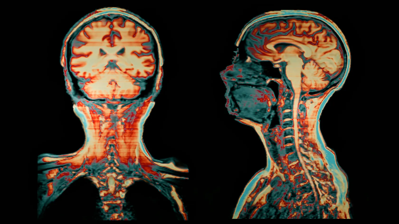
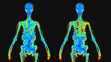
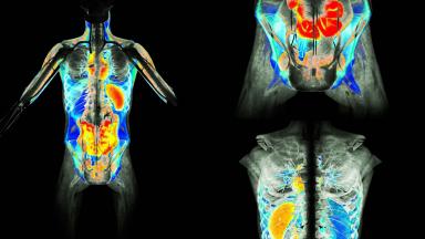
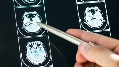
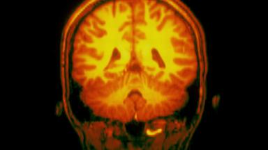
Ga-68 FAPI PET/CT images the cancer-associated fibroblasts that surround most carcinomas, showing tumors against a quiet background with no fasting or blood sugar control. It helps where FDG PET struggles, in signet ring stomach cancer, peritoneal spread, pancreatic and bile duct cancer, liver metastases and brain tumors. At Biruni Hospital in Istanbul, our nuclear medicine team reviews your case first, and the scan and report take one to two days.
About This Department
Nuclear Medicine
Many cancers build a scaffold of fibroblasts around themselves. Ga-68 FAPI PET makes that scaffold visible.
Ga-68 FAPI PET/CT uses a small molecule, a fibroblast activation protein inhibitor, labeled with gallium-68, to find the cancer-associated fibroblasts that surround and support a tumor. Those fibroblasts appear in the great majority of carcinomas, while healthy organs carry almost none, so tumors stand out against a quiet background. The scan needs no fasting and no blood sugar control. In stomach, duodenal and bowel cancers it detected every primary tumor, against 53 percent for FDG PET, and it found more lymph node, bone and organ spread (Pang and colleagues, Radiology, 2021). It helps most where FDG struggles, in signet ring stomach cancer, peritoneal spread, bile duct and pancreatic cancer, liver metastases and brain tumors. This page explains how the tracer works, how it compares with FDG, the cancers where it changes decisions, false positives, regulatory status, the scan day, radiation dose, FAP-targeted therapy and how patients from abroad arrange a FAPI scan at Biruni Hospital in Istanbul.
100
Percent of primary stomach, duodenal and bowel tumors detected by FAPI PET, against 53 percent with FDG
86.4
Percent of lymph node metastases detected by FAPI PET across cancers, against 45.5 percent with FDG
28
Cancer types showing FAPI uptake in the first clinical series of 80 patients
Free
Review of your scans and pathology by our nuclear medicine team
A tumor consists of more than cancer cells. Around them lies a supporting tissue, the stroma, full of fibroblasts that the cancer has reprogrammed to help it grow, invade and resist treatment. These cancer-associated fibroblasts produce large amounts of fibroblast activation protein, FAP, on their surface. In some cancers, such as pancreatic, breast and colorectal cancer, stroma makes up most of the tumor mass.
Normal adult organs express very little FAP, so a tracer that binds it shows tumors with high contrast and little background.
1
The tracer
FAPI tracers are small molecules that block FAP. Linked to a chelator holding gallium-68, a positron emitter with a 68-minute half-life, they show where FAP sits on a PET/CT scan. The versions in clinical use include FAPI-04 and FAPI-46, and fluorine-18 versions such as FAPI-74 are in development.
Why fibroblasts matter
The idea turns cancer imaging around.
For two decades PET scanning of cancer has relied almost entirely on FDG, a radioactive sugar taken up by cells that burn glucose fast, and that works well for lymphoma, lung cancer and many others. Yet several cancers burn little glucose, and some organs, the brain and to a lesser degree the liver and bowel, take up so much sugar themselves that tumors disappear against them. FAPI does not look at the cancer cell at all. It looks at the fibroblasts the cancer recruits, which behave in much the same way across tumor types, and so a single tracer shows cancers of the stomach, pancreas, bile ducts, breast, head and neck, and connective tissue with a clarity FDG rarely reaches in those sites.
That breadth explains the attention. Some researchers have called FAPI a candidate for the next pan-cancer tracer.
It will not replace FDG. It fills gaps.
FAPI compared with FDG
Ga-68 FAPI PET and F-18 FDG PET
Feature
Ga-68 FAPI
F-18 FDG
Fasting before the scan
Not needed
Four to six hours
Blood sugar control
Not needed
Needed, difficult in diabetes
Brain background
Very low
Very high
Liver and bowel background
Low
Moderate
Signet ring and mucinous tumors
Clear uptake in many
Weak uptake
Inflammation and healing tissue
Uptake in fibrosis and scars
Uptake in active inflammation
In the first dosimetry study, brain background on FAPI measured 0.32 against 11.01 on FDG, expressed as maximum standardized uptake. Tumors near the brain, the liver and the mouth therefore stand out more clearly (Giesel and colleagues, Journal of Nuclear Medicine, 2019).
FDG measures how hungry the cancer cells are for sugar. FAPI measures the tissue the cancer has built around itself. The two scans answer different questions.
1
When both are done
In some patients we perform both scans within a few days. Aggressive deposits bright on FDG and fibrotic deposits bright on FAPI together give the fullest picture before a major operation or radiotherapy plan.
Cancers where it helps
Where FAPI PET adds most
Cancer
Why FAPI helps
Typical question
Stomach cancer, including signet ring type
Low FDG uptake in diffuse types
Extent of spread, peritoneum
Pancreatic cancer
Dense stroma, high FAP
Staging before surgery
Bile duct cancer, cholangiocarcinoma
High uptake, low liver background
Local extent, nodes
Colorectal cancer and liver metastases
Low liver background
Number and site of deposits
Head and neck cancer
Low uptake in normal mouth and throat
Tumor edge for radiotherapy
Sarcoma
Among the highest uptake
Extent, recurrence
Breast and lung cancer
High uptake
Selected staging questions
The first clinical series, 80 patients with 28 kinds of cancer, found the highest uptake, a maximum standardized uptake value above 12, in sarcoma, esophageal, breast, bile duct and lung cancer. Uptake stayed lowest, below 6, in pheochromocytoma, kidney, differentiated thyroid, adenoid cystic and some stomach cancers (Kratochwil and colleagues, Journal of Nuclear Medicine, 2019).
Where it adds little
Prostate cancer has a better tracer in PSMA, neuroendocrine tumors in DOTATATE, and differentiated thyroid cancer in iodine. Our PSMA PET/CT and PRRT pages describe those scans.
A FAPI scan earns its place when FDG leaves a real question unanswered.
Stomach and bowel cancers
FAPI PET detected more primary tumors, lymph node metastases and bone and organ metastases than FDG PET in 35 patients with stomach, duodenal and colorectal cancer, and changed the stage in a fifth of those not yet treated (Pang and colleagues, 2021).
Detection in stomach, duodenal and bowel cancers
Finding
Ga-68 FAPI
F-18 FDG
Primary tumors detected
100 percent
53 percent
Lymph node metastases detected
79 percent
54 percent
Bone and organ metastases detected
89 percent
57 percent
Uptake in peritoneal deposits, average SUVmax
8.4
3.6
1
Upstaging
In 21 percent of the untreated patients, FAPI found disease that raised the stage and would change treatment, most commonly peritoneal or distant spread missed by FDG.
Results across cancer types
Head-to-head comparison in 75 patients with 12 cancer types
Finding
Ga-68 FAPI
F-18 FDG
Primary tumors detected
98.2 percent
82.1 percent
Lymph node metastases detected
86.4 percent
45.5 percent
Bone and organ metastases detected
83.8 percent
59.5 percent
1
Where the gap was largest
Chen and colleagues found the clearest advantage in liver metastases, peritoneal carcinomatosis and brain tumors, the three sites where FDG background is high or tumor sugar uptake low (European Journal of Nuclear Medicine and Molecular Imaging, 2020).
These are single-center studies of a few dozen patients each, mostly from China and Germany. Larger prospective trials are under way, and the figures may settle lower once more varied patients are scanned.
Peritoneal spread
1
Why it matters
Cancer seeding the lining of the abdomen, peritoneal carcinomatosis, decides whether surgery with curative intent is possible, and it hides from CT and FDG PET in its early, thin form.
Peritoneal deposits carry a dense fibrotic stroma. On FAPI PET they show as sharp spots and plaques, and in the studies above the tracer found peritoneal spread that FDG missed in stomach and colorectal cancer.
Peritoneal assessment
Method
Strength
Weakness
CT
Available everywhere
Misses small deposits
FDG PET/CT
Whole-body picture
Weak in mucinous and signet ring tumors
FAPI PET/CT
High contrast in fibrotic deposits
Uptake also in scars from earlier surgery
Diagnostic laparoscopy
Direct view and biopsy
Invasive
FAPI does not replace laparoscopy before major surgery. It guides where to look and helps avoid an operation that cannot help.
HIPEC candidates
For patients considered for cytoreductive surgery with heated chemotherapy, a FAPI scan helps estimate the peritoneal cancer index before the operation.
Choosing between the scans
We start with the question.
A scan is useful when its answer changes what happens next. In newly diagnosed lung cancer, lymphoma or melanoma, FDG PET/CT remains the standard, and FAPI adds nothing worth the extra cost and travel. For a patient with signet ring stomach cancer whose FDG scan looks clean, for pancreatic cancer before a Whipple operation, for a bile duct cancer whose extent decides between resection and transplant assessment, or for a rising tumor marker after bowel surgery with a negative CT, FAPI may show what the other tests do not. Our nuclear medicine physicians review the pathology and previous imaging first and advise on which tracer fits.
Sometimes neither scan earns its cost.
False positives and limits
Benign causes of FAPI uptake
Cause
Where
How we tell it apart
Scars and healing wounds
Surgical sites, biopsy tracts
Timing, CT appearance
Fibrosis and cirrhosis
Lungs, liver, pancreas
Pattern, earlier scans
Arthritis and degenerative joints
Spine, shoulders, knees
Location on CT
IgG4-related disease and pancreatitis
Pancreas, bile ducts
Blood tests, biopsy
Atherosclerotic plaque
Arteries
Linear shape along vessels
Normal uterus
Endometrium in women before menopause
Physiological pattern
FAP appears wherever tissue repairs itself. Any recent surgery, radiotherapy or inflammation can light up and mimic cancer.
Scars stay FAPI-avid for months, so we record the date of every operation and course of radiotherapy before reading the scan.
A bright spot needs CT, MRI or biopsy correlation.
Uptake varies between cancers and between patients, and a negative FAPI scan does not exclude cancer.
Interpretation standards
What reporting takes into account
Factor
Why
Source
Treatment history
Scars and radiation fibrosis
Your records
CT findings
Shape and density
Same scan
Earlier FDG or MRI
Comparison
Imaging you send
Regulatory status
Is FAPI PET an approved scan?
Not yet in most countries. At the time of writing, no FAPI tracer holds marketing approval from the United States Food and Drug Administration or the European Medicines Agency, and phase 2 and 3 trials of FAPI-46 and FAPI-74 are ongoing. Hospitals prepare the tracer in-house under national rules for radiopharmaceuticals and use it on the judgment of the nuclear medicine physician with written patient consent. Insurers rarely pay for it, and guidelines do not yet list it as standard.
Results therefore guide decisions alongside standard tests and never on their own, and before you book we tell you in writing what the scan can and cannot answer in your case.
The scan day
No fasting, no diet and no special preparation. Take your usual medicines and drink water freely.
1
Injection and wait
A nurse places a small cannula and injects the tracer. Images can be taken ten minutes later, and we scan at one hour, the standard time in most published studies.
The scan itself takes 20 to 30 minutes lying on the PET/CT table. A contrast CT may be added for detail. Diabetic patients eat and take insulin as usual, since blood sugar does not affect the tracer.
Timeline of a FAPI scan
Step
Duration
Note
Registration and cannula
15 minutes
Bring earlier scans on a disc
Tracer injection
A few seconds
Painless
Waiting period
60 minutes
Rest, drink water
PET/CT imaging
20 to 30 minutes
Lie still, breathe normally
Report
Next working day
Reviewed with your oncologist
Radiation and safety
A standard injection of about 200 megabecquerels gives an effective dose of 3 to 4 millisieverts from the tracer, similar to or below the tracer dose of an FDG scan. The CT part adds a further dose that depends on the protocol (Giesel and colleagues, 2019).
No side effects from the tracer have been reported in the published studies. The amount of molecule injected is tiny.
Radiation in context
Source
Effective dose
Note
FAPI tracer, 200 MBq
3 to 4 mSv
Giesel 2019 estimate
Low-dose CT for PET
1 to 4 mSv
Varies with protocol
Natural background, one year
2 to 3 mSv
Varies with place
1
Pregnancy and breastfeeding
Tell us if you might be pregnant. Breastfeeding mothers pause feeding for several hours and express and discard milk in that window, since gallium-68 decays fast.
After the scan
Situation
Advice
Duration
Driving and work
No restriction
None
Close contact with babies
Keep short
Rest of the day
Flights
Allowed
Same day
What the result changes
A scan matters only through the decision it informs.
When FAPI shows the cancer confined to one area, surgery or radiotherapy with curative intent stays on the table, and the radiation oncologist can use the images to draw the target on our Elekta Versa HD linear accelerator more precisely, especially in head and neck and pancreatic cancer. When it shows spread the other tests missed, the plan shifts toward systemic treatment and spares the patient an operation that could not have cured. Both outcomes help.
The report goes to the tumor board. The board decides.
FAP-targeted therapy
1
The theranostic idea
The same FAP target can carry a therapeutic isotope such as lutetium-177 or yttrium-90, as PSMA does in prostate cancer with Lu-177 PSMA therapy.
2
Where research stands
Small series and early trials, including LuMIERE with Lu-177 FAP-2286, are testing FAP-targeted radioligands in sarcoma, pancreatic and other cancers. The main challenge so far has been that FAPI tracers leave tumors quickly, which limits the radiation dose delivered.
FAP-targeted therapy remains experimental and is available only within clinical trials. We do not offer it outside a trial, and a FAPI scan today serves diagnosis.
3
Clinical trials
Our cancer clinical trials office can check whether a trial open to international patients fits your case.
For patients from abroad
The length of stay for a FAPI scan is one to two days in Istanbul, the scan on the first and a review of the report with our nuclear medicine physician on the next. You can fly the same day.
1
What to send
Your pathology report, recent CT, MRI and FDG PET images on a disc or by secure link, a summary of surgery and radiotherapy with dates, and the question your oncologist wants answered.
We reply with an assessment of whether FAPI suits your case, or whether FDG, PSMA or DOTATATE PET would serve better, before you book travel. After you return home, your oncologist receives the report and images. Our physician answers follow-up questions by video call.
One coordinator from the international patients office handles arrangements from the first message through discharge and answers the same WhatsApp number after you return. The office works in English, Arabic, French, Russian, Serbian, Romanian and Spanish and books interpreters for other languages, and it arranges the visa invitation letter, airport transfers, daily transport and accommodation near the hospital. Halal, vegetarian and diabetic meals, a prayer room on site, and a female physician on request wherever the rota allows.
Cost
What a quote for FAPI PET/CT covers
Item
Included
Priced separately
Scan
Ga-68 FAPI tracer, PET/CT imaging
Contrast CT if added
Reporting
Written report, physician review
Second tracer scan such as FDG
Consultation
Review of prior imaging
Biopsy if needed
The tracer is produced for each patient, so scans are booked on set days and priced as a single package.
Compare quotes for the same tracer and protocol, and ask whether the report includes review by a physician who reads FAPI scans regularly.
References
Kratochwil C, Flechsig P, Lindner T, et al. 68Ga-FAPI PET/CT. Tracer uptake in 28 different kinds of cancer. J Nucl Med. 2019;60(6):801-805.
Giesel FL, Kratochwil C, Lindner T, et al. 68Ga-FAPI PET/CT. Biodistribution and preliminary dosimetry estimate of 2 DOTA-containing FAP-targeting agents in patients with various cancers. J Nucl Med. 2019;60(3):386-392.
Chen H, Pang Y, Wu J, et al. Comparison of [68Ga]Ga-DOTA-FAPI-04 and [18F]FDG PET/CT for the diagnosis of primary and metastatic lesions in patients with various types of cancer. Eur J Nucl Med Mol Imaging. 2020;47(8):1820-1832.
Pang Y, Zhao L, Luo Z, et al. Comparison of 68Ga-FAPI and 18F-FDG uptake in gastric, duodenal, and colorectal cancers. Radiology. 2021;298(2):393-402.
Editor's note
Written by the Biruni Hospital medical editorial team. Reviewed by Specialist Dr. Sedat OFLUOĞLU, Nuclear Medicine.