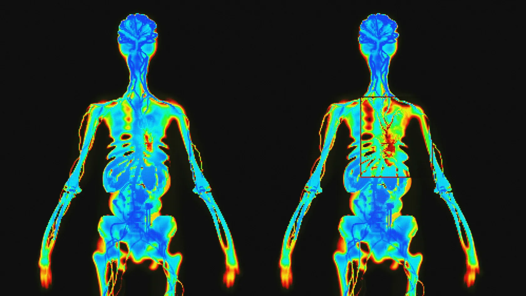
A study of 44 men with high-risk prostate cancer compared four ways of imaging the skeleton on the same day. Planar bone scan images alone found 70 percent of the men with bone metastases, adding SPECT raised that to 92 percent, and fluoride PET/CT found all of them with no false positives. Half of the individual lesions seen on fluoride PET/CT had looked normal on the planar bone scan. The lesson is to add SPECT/CT to the bone scan when the question matters.
How does it compare with PET, MRI and CT?
Three studies measured the bone scan against its rivals, in general and in prostate cancer.
This table scrolls sideways on a narrow screen. Swipe or drag to see every column.
Detecting bone metastases, per patient, pooled across 67 studies
| Test |
Sensitivity |
Specificity |
| Bone scintigraphy |
86.0 percent |
81.4 percent |
| FDG PET |
89.7 percent |
96.8 percent |
| MRI |
90.6 percent |
95.4 percent |
| CT |
72.9 percent |
94.8 percent |
PET and MRI were found to be comparable and both significantly more accurate than CT and bone scintigraphy for the diagnosis of bone metastases.
Meta-analysis of 67 articles, European Radiology, 2011
This table scrolls sideways on a narrow screen. Swipe or drag to see every column.
High-risk prostate cancer, four skeletal tests in the same 44 men
| Test |
Sensitivity |
Specificity |
| Planar bone scan |
70 percent |
57 percent |
| Bone scan with multi-field SPECT |
92 percent |
82 percent |
| Fluoride PET |
100 percent |
62 percent |
| Fluoride PET/CT |
100 percent |
100 percent |
This table scrolls sideways on a narrow screen. Swipe or drag to see every column.
proPSMA, 302 men with high-risk prostate cancer
| Measure |
CT and bone scan |
PSMA PET/CT |
| Overall accuracy |
65 percent |
92 percent |
| Sensitivity |
38 percent |
85 percent |
| Equivocal findings |
23 percent |
7 percent |
| Radiation dose |
19.2 mSv |
8.4 mSv |
When PSMA PET replaces the bone scan
For men with newly diagnosed high-risk prostate cancer, and for rising PSA after treatment, PSMA PET/CT finds spread to lymph nodes and bone that CT and a bone scan miss, at a lower radiation dose. Where PSMA PET is available and covered, it has replaced the combination for these questions. The bone scan remains useful for follow-up on certain drugs, where trials have used it to measure response, and where PSMA PET is not available.
PSMA PET-CT is a suitable replacement for conventional imaging, providing superior accuracy, to the combined findings of CT and bone scanning.
proPSMA trial, The Lancet, 2020
An unexpected use, the heart
The same tracer settles in heart muscle infiltrated by transthyretin amyloid, a protein that stiffens the heart and causes heart failure, mostly in older men. In a multicenter study of 1,217 patients referred with suspected cardiac amyloidosis, heart uptake on a bone scan was more than 99 percent sensitive for this form. Combined with blood and urine tests that ruled out a related blood disorder, it was 100 percent specific, which allows the diagnosis without a heart biopsy. New drugs slow the disease, so an early diagnosis now matters.
Other benign uses
A three-phase bone scan can find a stress fracture days after pain begins, when the X-ray is still normal, and it helps separate a loose joint replacement from an infected one, in many cases together with a white cell scan. In children with limping or unexplained bone pain, it images the whole skeleton in a single session.
Reading a bone scan takes experience.
Arthritis, old fractures and dental problems all light up, and a single spot in a patient with cancer turns out benign in the majority of cases. SPECT/CT, the CT image and earlier scans settle the question. Two patterns deserve mention. A superscan, in which the whole skeleton is so evenly bright that the kidneys hardly show, can mean widespread metastases and is easy to miss because nothing looks focal. A flare, in which spots grow brighter in the first months of successful hormone or chemotherapy as the bone heals, may look like progression, so a scan within three months of starting treatment is read with caution and compared with the next one.
Pregnancy, breastfeeding and children
Pregnancy postpones the scan unless it is essential. Breastfeeding is paused for a few hours after the injection, and close contact with babies and pregnant women is limited for the rest of the day. Children receive a dose adjusted to their weight.
Bone scintigraphy enables the diagnosis of cardiac transthyretin amyloidosis to be made reliably without the need for histology in patients who do not have a monoclonal gammopathy.
Multicenter study of 1,217 patients, Circulation, 2016
Days in Istanbul, cost and travel
The length of stay is short. Half a day covers the scan. The written report follows in one to two working days, so a two-day stay allows a consultation with the result. The tracer leaves the body within a day. Fly the same evening if you wish, and carry the letter we give you, since airport radiation detectors can pick up the small remaining activity for a day or two after the injection.
Cost covers the tracer, the whole-body images and the specialist report. SPECT/CT of one or more regions, a three-phase study and a consultation add to it. Your own figure follows the free review of your file.
One coordinator handles your visit. The team works in seven languages, with interpreters for the rest. Ten days before travel, an invitation letter naming the hospital and the physician goes out, and the international patients office books the transfer, a hotel for you and a companion, and the appointment times that fit your flights. Halal, vegetarian and diabetic meals are available, there is a prayer room, and a request for a female doctor is passed to the department and met wherever the rota allows. Back home, the coordinator stays reachable on the same WhatsApp number, and the images go to your own doctors in a standard format.
Bone scan FAQ
What does a bone scan show?
Areas where bone is being repaired or rebuilt, from cancer spread, fractures, infection, arthritis or metabolic bone disease. It also shows amyloid deposits in the heart.
How long does a bone scan take?
Two to four hours between injection and imaging, and 30 to 45 minutes on the camera, plus 15 to 30 minutes if SPECT/CT is added. Plan half a day.
Is a bone scan safe?
Yes. The radiation dose is similar to a year of natural background radiation, allergic reactions are extremely rare, and pregnancy is the main reason to postpone the test.
Is a bone scan or PET scan better for cancer?
PET and MRI detect bone metastases more accurately in pooled studies. For high-risk prostate cancer, PSMA PET/CT reached 92 percent accuracy against 65 percent for CT and bone scan. The bone scan remains useful, inexpensive and widely available.
Do I need to fast before a bone scan?
No. Eat normally, take your usual medicines and drink plenty of water between the injection and the images.
Can my family come with me?
Yes. The international patients office arranges a hotel for you and a companion.
References
- Van den Wyngaert T, Strobel K, Kampen WU, et al. The EANM practice guidelines for bone scintigraphy. European Journal of Nuclear Medicine and Molecular Imaging. 2016;43(9):1723-1738.
- Yang HL, Liu T, Wang XM, Xu Y, Deng SM. Diagnosis of bone metastases: a meta-analysis comparing 18FDG PET, CT, MRI and bone scintigraphy. European Radiology. 2011;21(12):2604-2617.
- Even-Sapir E, Metser U, Mishani E, et al. The detection of bone metastases in patients with high-risk prostate cancer: 99mTc-MDP planar bone scintigraphy, single- and multi-field-of-view SPECT, 18F-fluoride PET, and 18F-fluoride PET/CT. Journal of Nuclear Medicine. 2006;47(2):287-297.
- Hofman MS, Lawrentschuk N, Francis RJ, et al. Prostate-specific membrane antigen PET-CT in patients with high-risk prostate cancer before curative-intent surgery or radiotherapy (proPSMA): a prospective, randomised, multicentre study. Lancet. 2020;395(10231):1208-1216.
- Gillmore JD, Maurer MS, Falk RH, et al. Nonbiopsy Diagnosis of Cardiac Transthyretin Amyloidosis. Circulation. 2016;133(24):2404-2412.