
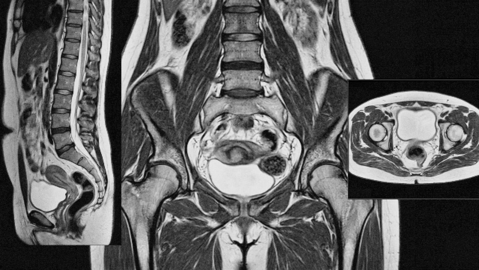
PLIF - Posterior Lumbar Interbody Fusion
Across 990 patients, complications ran at 17.0 percent with PLIF and 8.7 percent with TLIF. What the bilateral route buys for that gap is the whole question.
About This Department
Pool nine studies covering 990 people operated on for a slipped vertebra and the complication rate lands at 8.7 percent for one posterior fusion technique and 17.0 percent for the other. The one at 17.0 percent is PLIF. Disability scores between the two differed by three and a half points on a hundred point scale, which no patient would feel, and pain scores did not differ at all. That is an uncomfortable opening for a page about posterior lumbar interbody fusion, and it is also the honest place to start, because the operation still has situations that belong to it and no other route reaches as directly. Knowing which situations those are is the entire decision.
Free consultation
Ask whether your level needs this route or a simpler one
No charge, no obligation. Send the MRI images as files, and add standing x-rays taken bending forward and backward if anyone has ordered them, since movement between two films is what separates a slipped vertebra that needs holding from one that does not. Mention your age, whether you smoke, whether you have diabetes and whether this level has been operated on before. Those four answers change the plan more than anything else you can tell us.
What this page does not re-argue
Whether your spine should be fused at all is an earlier question
Everything below assumes that question has already been asked properly and answered yes. Our spinal fusion page takes it on directly, including the indications that rest on thin evidence. Settle it there first, because if the answer turns out to be no, none of the acronyms on this page matter at all.
What the four letters describe
PLIF is an abbreviation that unpacks into a full description of the operation, which is unusual and useful. Each letter answers a question a patient would otherwise have to ask.
| Letter | What it settles |
|---|---|
| P for posterior | The surgeon works from behind, through an incision down the midline of the back. Nothing is opened in the abdomen or the flank. |
| L for lumbar | The lower back, and in practice most often one of the bottom two levels, where load and degeneration both concentrate. |
| I for interbody | The graft goes into the emptied disc space between the two vertebral bodies, which is where roughly eighty percent of the load through a spinal segment travels. |
| F for fusion | The result being aimed at is living bone bridging that gap. Screws and rods hold the position while it happens, and the biology takes six to twelve months. |
The distinctive part is the third letter combined with the first. Getting a cage into the disc space from directly behind means passing between and around the nerve roots that leave the canal at that level, on both sides, because a PLIF cage is usually placed in pairs. Everything this operation does well, and everything it does badly, follows from that one sentence.
Where PLIF sits among the routes
Surgeons can reach a lumbar disc space from four directions, and each direction has an abbreviation attached to it. Reading them as a family makes the choice legible.
From the front, through the abdomen, comes ALIF. The disc is fully exposed, the biggest cage fits, and the correction of a flattened lower back is the best any route achieves. The price is that a vascular or general surgeon usually opens the approach, the great vessels sit directly over the target, and men are counseled about a small risk to ejaculatory function. From the side, through the flank, come LLIF and OLIF. These slip past the muscle that runs down the front of the spine and place a wide cage without ever entering the spinal canal, at the cost of numbness or weakness in the thigh in a proportion of patients. Then there is plain posterolateral fusion, which uses no cage at all and lays graft along the back of the spine instead.
The two that share one incision
PLIF and TLIF both go in from the back through the same skin incision, and this is where nearly every real-world choice is made. PLIF removes bone on both sides, retracts the nerve root and the covering of the nerve sac inward on each side in turn, and slides a cage in on each side. TLIF removes the small joint on one side entirely, comes in through the wider corridor that opens up, sweeps around the front of the nerve sac, and places one larger cage angled across the space. One side is opened instead of two. The nerve sac is pulled on less.
So the difference between them is not really the cage. It is how many times the neural structures are handled, and it is the reason the two techniques produce the numbers further down this page.
Who the operation is for
The commonest reason a surgeon proposes PLIF is a vertebra that has slipped forward on the one below and moves further when the spine bends, causing both back pain and leg symptoms. Restoring height in the disc space opens the tunnels the nerve roots travel through, and holding the level still stops the slip progressing. A second common reason is a level that has already been operated on, where the disc has collapsed and the nerve is being squeezed by the loss of height alone.
Where the posterior route earns its extra reach
The bilateral exposure that costs PLIF its complication margin also buys something specific. Both sides of the canal come into view at once and both nerve roots can be freed under direct vision, which matters when compression is genuinely on both sides rather than one. There are levels and body shapes where a surgeon can honestly say the transforaminal corridor would not have given a safe angle. Those are the cases where the technique justifies itself.
- Compression on both sides needing direct decompression. Central and bilateral recess narrowing where each root has to be seen and freed individually under direct vision.
- A previously operated level with heavy scarring. Working from both sides gives more room to separate scar from the nerve sac without dragging on it.
- A collapsed disc space that has to be reopened. Two cages placed in parallel share the load evenly across the endplate, which is an argument in bone that is not strong.
- Anatomy that closes the transforaminal corridor. A high iliac crest, an unusually oriented facet joint or a conjoined nerve root can all make the one-sided route the harder of the two.
Outside those, the burden of proof shifts. A surgeon proposing PLIF for a straightforward one-sided problem should be able to say why in a sentence you can follow.
PLIF measured against TLIF
The two posterior techniques have been pooled against each other in patients with lumbar spondylolisthesis. Nine studies qualified, one of them randomized and eight case series, covering 990 patients split 450 to the transforaminal route and 540 to PLIF. The comparison is worth reading line by line, because the pattern is not a simple win.
| Measure | TLIF | PLIF |
|---|---|---|
| Overall complication rate | 8.7 percent, range 0 to 25 | 17.0 percent, range 4.7 to 28.8 |
| Disability score after surgery | Better by 3.46 points | Reference |
| Pain score after surgery | No measurable difference | No measurable difference |
| Average duration of surgery | 169 minutes | 190 minutes |
| Estimated blood loss | 350 milliliters | 418 milliliters |
Three and a half points of disability on a hundred point scale is below what most people notice in daily life, and the pain scores were flat. Twenty minutes and seventy milliliters are real but small. The complication rate is the line that carries weight, at roughly twice the frequency, with a pooled odds ratio of 0.47 favoring the transforaminal route.
Two cautions belong with those figures. Eight of the nine studies were case series, which is a design that cannot rule out surgeons choosing the harder technique for the harder spines. And the ranges are wide, from zero to twenty five percent in one group and 4.7 to 28.8 in the other, which says as much about differences between surgical teams as about differences between techniques. What the pooling supports is that the bilateral route carries a higher price when nothing about the case demands it. What it does not support is abandoning the technique.
Does the cage earn its place
Step back one level of abstraction and a harder question appears. Interbody fusion is more work than laying bone graft along the back of the spine and leaving the disc space alone, so it should deliver something in return. Six studies covering 721 patients with degenerative spondylolisthesis were pooled to test exactly that, 458 of them having posterolateral fusion alone and 263 having posterior interbody fusion.
Disability scores did not separate. Neither did pain scores, blood loss, reoperation rate, hospital stay, complication rate or the proportion of spines that actually fused. On every outcome the authors examined, adding the cage failed to show a measurable advantage over graft laid along the back of the spine.
That result should be held carefully rather than waved about. The pooled studies were few, the follow-up periods were short by the standards of a fusion, and degenerative spondylolisthesis with a modest slip is only one of the reasons anyone reaches for an interbody cage. Restoring collapsed height to open a nerve tunnel, and correcting a lower back that has lost its curve, are mechanical goals that a posterolateral graft cannot address at all, and neither was the outcome being measured.
Still, the finding is a legitimate question to put to a surgeon who has proposed a cage. Ask what the cage is expected to accomplish in your spine specifically. Restoring height, correcting alignment and creating a fusion surface that sits under compression are all defensible answers, and a surgeon who has thought about your films will give one of them without hesitating. The absence of an answer tells you something too.
Inside the operating room
A single-level PLIF runs around three hours in the pooled figures, and two levels or a heavily scarred revision extend that substantially. You lie face down on a frame that lets the abdomen hang free, which lowers pressure in the veins around the spine and reduces bleeding. Nerve monitoring runs throughout in most modern practice.
The five things that happen, in order
Screws before decompression
Pedicle screws go into the vertebrae above and below while the anatomy is still intact and the landmarks are undisturbed. Placement is checked on imaging in the room. Doing this first also means the level can be gently distracted later using the screws themselves.
Taking bone away on both sides
The back wall of the canal is opened and the inner part of each small joint is trimmed, until both nerve roots and the sac between them are fully visible. This is the decompression, and for many patients it is the part that relieves the leg symptoms.
Emptying the disc space
Working through the window on one side and then the other, the surgeon clears the disc and scrapes the surfaces of both vertebral bodies down to bleeding bone. That raw surface is what the graft will bond to, and preparing it without punching through into soft bone underneath is one of the genuine skills of the operation.
The cages go in, one on each side
Two cages packed with bone graft are placed in parallel, restoring the height of the space and putting the graft where compression will encourage it to turn into bone. Bone removed during the decompression is often reused here, which avoids a second wound at the hip.
Compression across the construct
Rods connect the screws and the level is compressed slightly, which locks the cages in place and loads the graft the way bone likes to be loaded. One last image confirms everything before closure. Whether a drain is left behind depends entirely on how much bleeding there has been during the case.
Most people are helped to their feet within a day. Leg symptoms often improve immediately, since the decompression happened under direct vision. The back itself is sore for weeks, and that soreness is muscular and expected.
Open, or through tubes
Posterior interbody fusion can be done through a traditional midline opening or through tubular retractors placed either side of the midline, with screws inserted percutaneously. Twenty six comparative studies, covering 856 patients treated through tubes and 806 through an open incision, have been pooled.
The differences sit almost entirely in the early period. Blood loss was around 260 milliliters lower. People got walking about three and a half days sooner. Hospital stay was shorter by roughly 2.9 days. Medical complications, meaning chest infections, urinary infections and the rest, were substantially less frequent, with a risk ratio of 0.39. Surgical complications showed no difference at all, and neither did the rate of failed union or the rate of further surgery. Operating time was the same.
How much weight that evidence carries
The authors of that pooling graded the underlying studies as low to very low quality, with only one randomized trial among twenty six, and said plainly that significant bias is inherent in the comparison. Disability scores at two years favored the tubular route by 3.32 points, which is the same order of magnitude as the PLIF and TLIF gap discussed earlier and equally hard to feel.
A reasonable reading is that the tubular route makes the first fortnight easier without changing where you end up, which is worth having, particularly for someone who has to be fit for a flight. Choosing a hospital primarily because it advertises the technique is a different matter, since who is holding the instruments explains more of the variation than which instruments they are.
The risks, in order of frequency
Ordering complications by how common they are, instead of by how frightening they sound, changes what a patient prepares for. The rare catastrophes get the attention. The frequent ones shape the recovery.
The one that drives the rest
The nerve sac is wrapped in a thin membrane called the dura, and it is the structure that gets accidentally opened during posterior spine surgery. Across twenty three prospective studies the reported rates ranged from 0.4 percent to 15.8 percent, pooling to 5.8 percent. Three factors raised it consistently. Operating on a level that has been opened before roughly doubled the odds, and lumbar stenosis, which stretches the dura thin over the compressed segment, doubled them again. Older patients fared worse. Diabetes carried a weak association. Sex and body weight showed no significant effect either way.
A tear is repaired during the same operation and often causes nothing at all afterward. What matters is what follows statistically. Pooling fourteen studies covering 376164 lumbar operations, wound infection occurred in 13.1 percent of patients who had a tear against 5.4 percent of those who did not, an odds ratio of 3.86. That is the sequence worth understanding, since a technique that requires more handling of the dura is a technique that meets this chain more often, and it is a fair reading of why the PLIF complication column sits where it does.
| Problem | What it means in practice |
|---|---|
| Muscular back pain for weeks | Near universal after an open midline exposure. Expected, and it settles. |
| Accidental opening of the dura | Pooled at 5.8 percent across spine surgery, higher in revisions and in stenosis. Repaired on the spot, followed by flat bed rest for a day or two. |
| Temporary nerve root irritation | Burning or new numbness in one leg from retraction during the operation. Usually settles over weeks to a few months. |
| Wound infection | Around 5.4 percent without a dural tear and 13.1 percent with one, in the pooled lumbar figures. Metal implants raise the stakes of treating it early. |
| Cage moving out of position | Uncommon, and more likely where the endplate was damaged during preparation. Symptomatic migration means revision. |
| Screw misplacement | Most misplaced screws cause no symptoms and are seen only on imaging. A minority irritate a root and are repositioned. |
| Permanent nerve injury | Rare, and the reason nerve monitoring runs during the case. It remains the risk patients ask about first. |
| Clot in a leg vein | Uncommon, and the reason walking starts on day one and stockings go on before the flight home. |
When bone fails to knit
Two slow failures can undo an operation that went perfectly on the day. Both are quiet, both surface months later, and both are influenced by things decided before anyone reached the operating room.
The first is a fusion that never becomes solid. Bone has to grow across the cages and turn the two vertebrae into one block, and that takes somewhere between six and twelve months of biology. Not every failure of that process causes symptoms, and some are noticed years later on a scan taken for another reason. The ones that cause trouble usually announce themselves as back pain returning eight to twelve months after surgery in someone who had been improving, occasionally with a screw looking loose on the x-ray. Smoking is the single largest thing a patient controls here, because nicotine constricts the small vessels the graft depends on, and stopping matters more before this operation than before almost any other.
The second failure is the cage sinking
Subsidence means a cage settling down into the bone of the vertebra above or below it, giving back the height the operation restored and sometimes reclosing the nerve tunnel that was opened. Thin bone is the main driver, which is why bone density is worth measuring before a fusion in anyone over sixty, in anyone who has taken steroids long term, and in women after menopause. Damage to the endplate during preparation is the other major driver, and it is a technical matter belonging to the surgeon rather than the patient. Distracting the space more than it wants to go adds to it as well.
A little settling is normal and means nothing. Settling that reproduces the original symptoms is a different conversation, and it is the reason the follow-up x-rays at three, six and twelve months exist rather than being routine paperwork.
Recovery, stage by stage
Two clocks run at different speeds after this operation. The wound and the muscles heal in weeks. The fusion takes most of a year, and the restrictions you are given belong to the second clock even though you will feel fine on the first one long before it is over.
Days one to four in hospital
Standing and short walks begin on the first day unless a dural tear needed flat rest. A physiotherapist teaches the log roll for getting out of bed, which keeps the back from twisting. Leg symptoms are often already better. The incision is the sorest thing about you.
Weeks one to six
Bending, lifting and twisting are all restricted, and the restriction is about protecting a graft rather than protecting a wound. Walking is the prescribed exercise and the distance climbs weekly. A brace is used by some teams and not others, and the evidence for it is thin either way. Sitting for long stretches is the hardest thing to avoid and the most important.
Six weeks to six months
Rehabilitation moves from walking to strengthening the deep muscles that hold the trunk. Desk work usually restarts somewhere in the first half of this window and physical work in the second, depending on what the job demands. Anti-inflammatory painkillers are often discouraged during this period because of their effect on bone healing, so ask what you may take before you need it.
Six months to a year
Imaging at around a year is what confirms the fusion took. Most restrictions have lifted well before then. Residual numbness in a foot may still be improving slowly, and strength usually returns further than sensation does.
Planning the trip to Istanbul
A fusion is a longer trip than a disc operation, and the reason is the flight rather than the surgery. Hours of sitting compress a fresh construct and raise clotting risk at the same time, so the clearance to travel is given later than the discharge from the ward.
How the weeks are sequenced
- Review before you commit to dates. Images are read and you receive a written plan naming the level, the technique, the number of nights and the expected total time in Istanbul. Bring bone density results if you have any, since they change the implant discussion.
- Two days of assessment on arrival. Examination, blood work, anesthetic review, and repeat imaging where what you sent has aged. Any blood thinner is stopped on a schedule the team sets, and that schedule sometimes fixes the surgery date rather than the other way round.
- Surgery and three to five nights on the ward. Walking starts on day one. Physiotherapy begins before discharge, since the movements you are taught are what protect the graft during the trip home.
- Ten to fourteen days in an apartment or hotel. This is the part people underestimate. Wound checks, clip removal, an x-ray of the construct and a walking assessment all sit in this window, and it is also when a small problem can still be dealt with by the team that operated.
- The flight decision, taken last. A final review confirms the wound, the imaging and your walking tolerance. Book a changeable ticket, choose an aisle seat, stand every hour and wear the stockings you are given.
Three to four weeks covers a single-level PLIF comfortably. Two levels, or a revision at a scarred level, run longer and the team says so before you book rather than after. A companion is genuinely worth the extra ticket for the first fortnight, because someone who cannot bend forward cannot easily pick up a dropped phone or pull on their own socks.
Once you are back home, the follow-up continues at a distance. Wound photographs during the first two weeks, a full operative report and imaging for your own doctor, and a direct line back to the surgical team by WhatsApp for anything that changes. Arrange local physiotherapy and the three month x-ray before you leave Istanbul, since both are far easier to organize while someone is still coordinating them for you.
What moves the cost
Quotes are prepared individually, because the same three letters cover operations that differ by a factor of several in theater time and implant count. A single published number for PLIF would be describing one version of it and staying quiet about the rest. What can be set out plainly is which variables move the figure, which is what you actually need in order to compare one hospital against another.
Compare the inclusions before the headline figure
Two quotes are only comparable when both list what is inside them
Ask each hospital to put the inclusions in writing, and ask specifically what happens financially if you need two extra nights. A quote that answers that question can be compared with another quote. A number in an email cannot.
- Levels. The single biggest multiplier. Two levels means twice the implants and considerably more than twice the time.
- Implants. Cage material, expandable against static, screw type and whether a bone graft substitute is used all carry separate costs.
- Theater and anesthesia time. Billed by duration, and a revision at a scarred level can run twice as long as a first operation.
- Nerve monitoring. A separate line and separate staff, and worth having.
- Nights on the ward. Usually banded, so ask where the band ends.
- Physiotherapy and follow-up imaging. Sometimes bundled and sometimes charged separately, and over three weeks the difference adds up.
Choosing a team
The wide ranges in the pooled complication figures, from zero to twenty five percent in one technique and 4.7 to 28.8 in the other, are a message about surgical teams as much as about surgical methods. At Biruni University Hospital this operation sits in the Neurosurgery department, working with neurology, radiology, anesthesia and physical therapy and rehabilitation on the same site.
Four questions separate a considered plan from a template. Ask why the posterior interbody route was chosen over the transforaminal one for your level, and expect an anatomical answer rather than a preference. Ask what the cage is meant to accomplish in your spine. Ask what would change the plan once the surgeon is looking at the level directly. Ask what the expectation is for your back pain as distinct from your leg pain, because those two respond differently and a surgeon willing to separate them is being straight with you about the rest.
Worth saying out loud
A second opinion that says do less is still a useful second opinion
The consultation costs nothing and obliges you to nothing. If your images support a decompression without a fusion, or a transforaminal approach instead of this one, that is what you will be told. Send the scans and ask.
Frequently asked questions
If TLIF has half the complication rate, why would anyone choose PLIF?
Will I set off airport security scanners with the implants?
How will I know the fusion has actually happened?
Does the cage actually add anything over a simpler fusion?
What happens if the dura is torn during my operation?
How long until I can fly home after PLIF?
References
- de Kunder SL, van Kuijk SMJ, Rijkers K, et al. Transforaminal lumbar interbody fusion (TLIF) versus posterior lumbar interbody fusion (PLIF) in lumbar spondylolisthesis. A systematic review and meta-analysis. Spine J. 2017;17(11):1712-1721.
- Campbell RC, Mobbs RJ, Lu VM, Xu J, Rao PJ, Phan K. Posterolateral fusion versus interbody fusion for degenerative spondylolisthesis. Systematic review and meta-analysis. Global Spine J. 2017;7(5):482-490.
- Goldstein CL, Macwan K, Sundararajan K, Rampersaud YR. Perioperative outcomes and adverse events of minimally invasive versus open posterior lumbar fusion. Meta-analysis and systematic review. J Neurosurg Spine. 2016;24(3):416-427.
- Alshameeri ZAF, Jasani V. Risk factors for accidental dural tears in spinal surgery. Int J Spine Surg. 2021;15(3):536-548.
- Babasiz T, Leyendecker J, Weber M, et al. Incidental durotomy significantly increases the risk of postoperative infection following lumbar spine surgery for degenerative conditions. A systematic review and meta-analysis. Int J Spine Surg. 2025;19(6):768-775.
Editor's note
Written by the Biruni Hospital medical editorial team. Reviewed by Assistant Professor Fikret BAŞKAN, Neurosurgery.
Medically reviewed by

Assistant Professor Fikret BAŞKAN
Neurosurgery
Related Treatments
View All
ALIF - Anterior Lumbar Interbody Fusion Surgery
Coming in from the front restores the curve of the lower back better than any posterior route and leaves the back muscles untouched. It also carries one risk most pages will not name, and this page names it.

Brain AVM Surgery - Arteriovenous Malformation Surgery
An unruptured brain AVM bleeds at roughly 2.2 percent a year, and surgery spends its whole risk on a single morning. This page sets those two shapes of risk side by side, explains what the ARUBA trial does and does not say about your case, and shows how a Spetzler-Martin grade changes the answer.
Brain Tumor Surgery - Brain Tumor Removal
Brain surgery is one trade repeated all day, how much tumor comes out against what the brain around it still does. This page puts the published numbers on both sides of that trade.
Cervical Microdiscectomy - Minimally Invasive Neck Disc Surgery
Neck disc surgery is done from the front, through the space between your windpipe and your carotid artery, and almost everything that goes wrong that is not the disc happens in that corridor. What has been measured in it.
Chiari Malformation Surgery
A Chiari malformation is measured in millimeters and treated according to symptoms, and those two things come apart more often than patients are told. Most people with the finding never need an operation. This page sets out who does, what posterior fossa decompression removes, why surgeons still argue over opening the dura, and what the pooled studies say about both sides of that argument.