A PET/CT scan maps where an injected tracer collects in the body and overlays it on a CT, to stage cancer, judge treatment and find recurrence. In a randomized trial it cut futile lung cancer operations from 38 to 21. We explain FDG and PSMA tracers, preparation, what the scan can miss, the radiation dose, and a two to three day visit.
About This Department
Nuclear Medicine
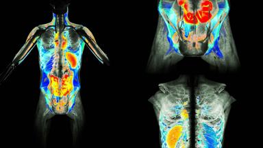
CT shows the shape of the body. PET shows what its cells are doing. One scan combines both, from head to thigh, in about 20 minutes.
A PET/CT scan, positron emission tomography with computed tomography, follows a small dose of a radioactive tracer through the body and lays the picture over a CT scan, so that active disease shows as bright spots on an exact anatomical map. The standard tracer, FDG, is a form of glucose, and most cancers take up far more glucose than normal tissue. In a randomized trial of patients about to have lung cancer surgery, adding PET to the usual tests cut operations that could not help from 41 to 21 percent (van Tinteren and colleagues, Lancet, 2002). In head and neck cancer, PET/CT after chemoradiotherapy replaced routine neck surgery, with 54 operations instead of 221 and no loss of survival (Mehanna and colleagues, New England Journal of Medicine, 2016). This page explains how PET/CT works, the tracers, what it is used for, the evidence in lung cancer, lymphoma and head and neck cancer, its limits, preparation and diabetes, the scan day, radiation, the report, and how patients from abroad arrange a PET/CT scan at Biruni Hospital in Istanbul.
21
Percent of lung cancer operations that could not help when PET was added, against 41 percent without it
54
Neck operations after PET/CT-guided follow-up, against 221 with routine surgery, with similar survival
83.7
Percent of patients with advanced Hodgkin lymphoma whose PET after two cycles was negative, allowing lighter treatment
Free
Review of your scans and reports by our nuclear medicine team
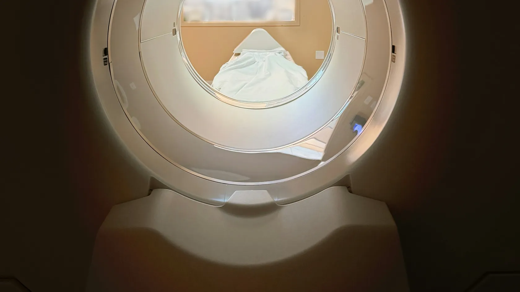
The PET part detects gamma rays given off where the tracer collects and builds a map of activity. The CT part, taken in the same session on the same table, adds a detailed picture of anatomy. Software fuses the two, so a bright spot can be placed exactly in a lymph node, a bone or the liver.
PET/CT, CT and MRI compared
Scan
What it shows
Strength
CT
Shape, size and density
Fast, detailed anatomy
MRI
Soft tissue contrast
Brain, liver, pelvis, spine
PET/CT
Metabolic activity on an anatomical map
Whole-body search for active disease
The tracer
1
FDG
Fluorodeoxyglucose, FDG, is glucose carrying radioactive fluorine-18, which has a half-life of 110 minutes. Cells take it up like sugar but cannot break it down, so it stays where it entered.
A lymph node of normal size can hold cancer, and an enlarged one can be harmless. PET/CT adds activity to size, and the combination answers questions neither answers alone.
Why sugar
Cancer cells are hungry.
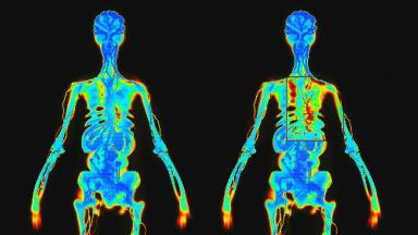
Most tumors burn glucose many times faster than the tissue around them, a pattern first described almost a century ago, and they do so even when oxygen is plentiful, because rapid growth demands the building blocks that glucose provides. FDG exploits that appetite. After injection it spreads through the blood, enters cells through the same transporters as glucose and becomes trapped inside, so an hour later the most active tissues glow brightest on the scan, whether they lie in the chest, the abdomen or a single rib.
Brain, heart and inflamed tissue burn sugar too. That shapes how scans are read.
Tracers and uses
1
Beyond FDG
Different tracers bind different targets. FDG covers most cancers, while prostate cancer, neuroendocrine tumors and some others are better seen with tracers built for them.
We have separate pages on PSMA PET/CT for prostate cancer, Ga-68 FAPI PET for cancers with dense fibrous tissue, and the thyroid and MIBG scans. This page concentrates on FDG PET/CT, the scan most people mean when they say PET.
Common PET tracers
Tracer
Target
Main use
F-18 FDG
Glucose metabolism
Most cancers, infection, heart, brain
Ga-68 or F-18 PSMA
Prostate-specific membrane antigen
Prostate cancer
Ga-68 DOTATATE
Somatostatin receptors
Neuroendocrine tumors
Ga-68 FAPI
Cancer-associated fibroblasts
Stomach, pancreas, bile duct cancer
F-18 amyloid tracers
Amyloid plaques
Memory disorders
F-18 FDG, heart protocol
Living heart muscle
Viability before bypass or stenting
PET/CT in cancer
What FDG PET/CT answers in cancer
Question
When
Effect on care
Staging, how far the cancer has spread
At diagnosis
Surgery, radiotherapy or systemic treatment
Response to treatment
During or after chemotherapy
Continue, change or stop
Recurrence
Rising markers, new symptoms
Finds the site to treat
Unknown primary
Metastasis without a known origin
Points to the source
Radiotherapy planning
Before treatment
Draws the target more precisely
A PET/CT earns its place when the answer changes treatment. Routine scans after curative treatment, without symptoms, rarely help.
Cancers that FDG shows well include lung, lymphoma, head and neck, esophageal, colorectal, melanoma, cervical and many sarcomas. Cancers that often take up little FDG include slow prostate cancer, well-differentiated neuroendocrine tumors, some kidney cancers and mucinous tumors.
1
Before surgery
Finding one unexpected distant deposit can spare a patient a major operation and send them straight to the treatment that fits.
The request from your oncologist states the question, and our nuclear medicine physician protocols the scan to answer it.
Lung cancer
1
The PLUS trial
Nine Dutch hospitals randomized 188 patients with suspected non-small cell lung cancer, all scheduled for surgery after standard tests, to go ahead or to have a PET scan first.
Addition of PET to conventional workup prevented unnecessary surgery in one out of five patients with suspected non-small-cell lung cancer (van Tinteren and colleagues, 2002).
Futile thoracotomy in the PLUS trial
Group
Futile operations
Reason counted as futile
Standard tests only
41 percent
Benign disease, unresectable tumor, relapse or death within a year
Standard tests plus PET
21 percent
Same definition
Relative reduction
51 percent
PET found spread that changed the plan
Lymphoma
Deauville five-point scale for lymphoma response
Score
Uptake in lymphoma sites
Meaning
1
None
Complete metabolic response
2
At or below the chest blood pool
Complete metabolic response
3
Above blood pool, at or below liver
Usually complete response
4
Moderately above liver
Residual disease
5
Markedly above liver or new sites
Residual or progressing disease
In the RATHL trial of 1,214 patients with advanced Hodgkin lymphoma, 83.7 percent of those scanned after two cycles of ABVD chemotherapy had a negative PET. Dropping bleomycin from the remaining cycles in that group reduced lung toxicity, with a three-year progression-free survival of 84.4 percent against 85.7 percent on full treatment, a difference that fell just short of the trial's formal margin (Johnson and colleagues, New England Journal of Medicine, 2016). Patients with a positive interim scan received stronger chemotherapy.
Head and neck cancer
1
The question
After chemoradiotherapy for head and neck cancer with large neck nodes, surgeons used to remove the neck nodes routinely in case cancer remained.
The PET-NECK trial randomized 564 patients to planned neck dissection or to PET/CT twelve weeks after treatment, with surgery only if the scan showed remaining disease. Two-year survival reached 84.9 percent with PET/CT surveillance and 81.5 percent with planned surgery, and the surveillance group needed 54 neck operations instead of 221 (Mehanna and colleagues, 2016).
2
Timing matters
PET/CT too soon after radiotherapy shows inflammation that looks like cancer. Twelve weeks gives a reliable answer.
PET-NECK trial, 564 patients
Measure
PET/CT surveillance
Planned neck dissection
Two-year survival
84.9 percent
81.5 percent
Neck dissections
54
221
Quality of life
Similar
Similar
A scan as a decision tool
Pictures do not treat anyone. Decisions do.
The three trials above share one lesson. In each, PET/CT mattered because a clear rule linked the result to an action, operate or not, add bleomycin or not, dissect the neck or not, and patients gained because they were spared treatment that could not help them.
We ask what a scan will change before we book it.
Limits and false results
Common causes of false-positive FDG uptake
Cause
Where
How we tell it apart
Infection and inflammation
Lungs, lymph nodes, joints
History, pattern, follow-up scan
Recent surgery or radiotherapy
Treated area
Timing, shape
Sarcoidosis and tuberculosis
Chest nodes, lungs
Biopsy when decisive
Brown fat
Neck, around the spine
Warm room, symmetric pattern
Bone marrow stimulation
Whole skeleton
Growth factor injections
Normal bowel and muscle activity
Gut, tense muscles
Pattern, CT correlation
A bright spot is a finding. When the result would change treatment, a biopsy makes the diagnosis.
1
Small lesions
PET resolves details down to roughly five to seven millimeters. Smaller deposits, such as tiny lung nodules, can be missed and are judged on CT.
False negatives also occur where background activity is high, in the brain, the bladder and the kidneys, and in tumors that take up little sugar.
Brain metastases are better seen on MRI.
Low-uptake tumors may need PSMA, DOTATATE or FAPI tracers.
A high blood sugar lowers tumor uptake and can hide disease.
When another scan fits better
Situation
Better choice
Why
Brain metastases
MRI with contrast
High brain background on FDG
Prostate cancer
PSMA PET/CT
Low FDG uptake
Well-differentiated neuroendocrine tumor
DOTATATE PET/CT
Receptor-based
Diabetes and blood sugar
Can I have a PET/CT scan if I have diabetes?
Yes, with some planning. High blood sugar competes with FDG and weakens the picture, and insulin drives the tracer into muscle. We book diabetic patients early in the morning, check blood sugar on arrival and give individual instructions on tablets and insulin. Short-acting insulin is not given within four hours of the injection.
If blood sugar is too high on the day, we may reschedule rather than produce a scan that cannot be trusted.
Blood sugar on the day of the scan
Blood sugar
Action
Note
Under 200 mg/dL, 11 mmol/L
Scan goes ahead
Standard
Above that level
Correct and wait, or rebook
Uptake reduced
Insulin pump
Individual plan
Discuss when booking
Preparation and the scan day
Fast for six hours before the scan. Plain water is allowed and helps. Avoid strenuous exercise for 24 hours, since working muscles take up FDG.
1
Arrival
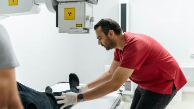
We check blood sugar, place a small cannula and inject the tracer. You then rest quietly in a warm room for about an hour, without talking much or using a phone, so that muscles stay relaxed.
The scan takes 15 to 25 minutes lying on your back with arms above the head. A CT contrast injection may be added when detailed anatomy is needed, and we ask about kidney function and allergies beforehand.
Timeline of an FDG PET/CT
Step
Duration
Note
Registration and blood sugar
15 minutes
Bring earlier scans
Injection
A few seconds
Painless
Uptake rest
About 60 minutes
Warm, quiet room
Scan
15 to 25 minutes
Breathe normally
After the scan
Leave at once
Eat and drink normally
Wear comfortable clothes without metal. Tell us if you are pregnant, breastfeeding or claustrophobic.
What to bring
Item
Why
Format
Earlier CT, MRI or PET
Comparison
Disc or secure link
Pathology report
Question to answer
Copy
Medicine list
Diabetes and contrast plan
Written list
Radiation and safety
The tracer adds a radiation dose of about 5 to 7 millisieverts for a standard adult injection, and the CT adds a further dose that depends on whether a low-dose or full diagnostic CT is used. The total is comparable to a few years of natural background radiation.
Radiation in context
Source
Typical effective dose
Note
FDG tracer
5 to 7 mSv
Depends on body weight
Low-dose CT for PET
1 to 4 mSv
Localization only
Diagnostic contrast CT
5 to 10 mSv
When detailed anatomy needed
Natural background, one year
2 to 3 mSv
Varies by place
1
After the scan
Drink plenty and empty the bladder often for the rest of the day. Keep close contact with babies and pregnant women short for a few hours, since the tracer decays quickly.
Children and pregnancy
Children receive weight-adjusted doses. In pregnancy, PET/CT is done only when the result is essential.
Reading the report
Numbers appear in every report.
The standardized uptake value, SUV, compares the tracer in a spot with an even spread through the body, and a higher value points to more active tissue, but it depends on timing, blood sugar and the scanner, so the pattern across the body and the change between scans matter more than any single figure.
Your oncologist reads the report with the whole story.
Results and next steps
Timing
1
The report
A nuclear medicine physician and a radiologist read the PET and CT together, compare them with earlier imaging and issue a report the next working day, sooner when treatment decisions are pending.
Comparison with earlier scans makes a report far more useful. Please send every previous CT, MRI and PET.
The report goes to your oncologist and, if you wish, to our tumor board, which reviews the findings with pathology and plans the next step.
Follow-up scans
Response scans use the same scanner and protocol where possible, so that values can be compared fairly.
For patients from abroad
A PET/CT needs one to two days in Istanbul, the scan on the first day and a consultation with the report on the next. You can fly the same day.
Send your pathology report, earlier imaging on disc or by secure link, a summary of treatment so far and the question your doctor wants answered. We confirm that FDG is the right tracer before you travel. After you return home, your oncologist receives the report and images, and our physicians answer follow-up questions by video call.
A typical visit
Day
Step
Note
Day 1
Consultation and scan
Fasting from the night or morning before
Day 2
Report and discussion
Tumor board if needed
Same or next day
Flight home
No restriction
One coordinator from the international patients office handles arrangements from the first message through discharge and answers the same WhatsApp number after you return. The office works in English, Arabic, French, Russian, Serbian, Romanian and Spanish and books interpreters for other languages, and it arranges the visa invitation letter, airport transfers, daily transport and accommodation near the hospital. Halal, vegetarian and diabetic meals, a prayer room on site, and a female physician on request wherever the rota allows.
1
Combined visits
Many combine the scan with a second opinion.
Cost
The price depends on the tracer, the body area covered and whether a diagnostic contrast CT is added.
We quote after reviewing your records, before you travel.
Compare quotes for the same tracer and scan range, and ask whether the report includes review by a nuclear medicine physician and comparison with your earlier scans.
What a quote covers
What a quote for PET/CT covers
Item
Included
Priced separately
Scan
FDG tracer, PET/CT from skull base to mid-thigh
Whole-body or brain protocol
Reporting
Written report with comparison
Tumor board review
Consultation
Discussion of the report
Biopsy or further imaging
References
van Tinteren H, Hoekstra OS, Smit EF, et al. Effectiveness of positron emission tomography in the preoperative assessment of patients with suspected non-small-cell lung cancer. The PLUS multicentre randomised trial. Lancet. 2002;359(9315):1388-1393.
Mehanna H, Wong WL, McConkey CC, et al. PET-CT surveillance versus neck dissection in advanced head and neck cancer. N Engl J Med. 2016;374(15):1444-1454.
Johnson P, Federico M, Kirkwood A, et al. Adapted treatment guided by interim PET-CT scan in advanced Hodgkin's lymphoma. N Engl J Med. 2016;374(25):2419-2429.
Editor's note
Written by the Biruni Hospital medical editorial team. Reviewed by Specialist Dr. Sedat OFLUOĞLU, Nuclear Medicine.