
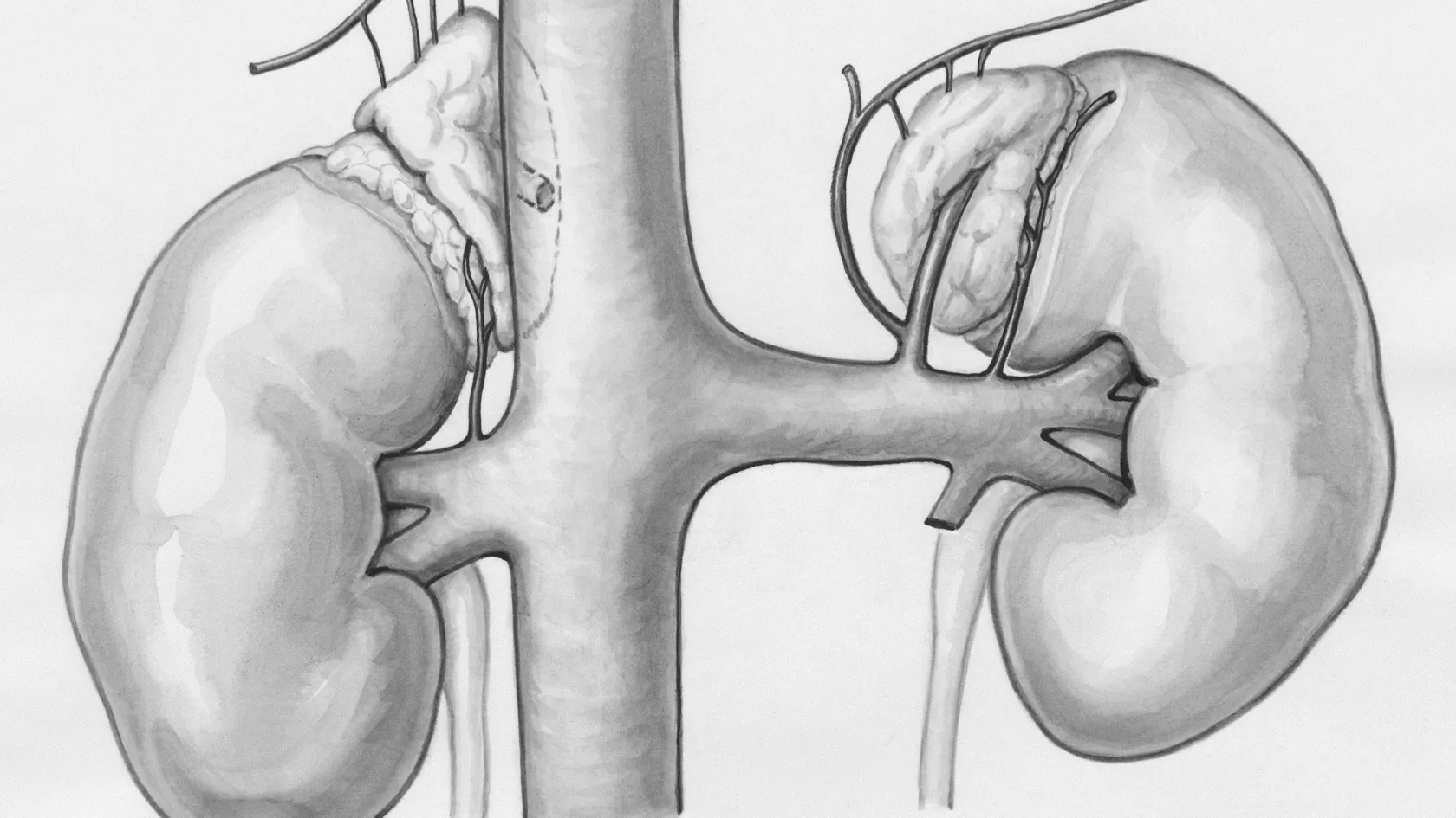
PTBD - Percutaneous Transhepatic Biliary Drainage
PTBD relieves a blocked bile duct by placing a drainage tube through the skin and the liver under ultrasound and X-ray guidance, when the endoscopic route has failed or cannot reach the blockage. It explains why bile must be drained, which cancers and benign strictures cause obstruction, how PTBD compares with ERCP and endoscopic ultrasound drainage, the procedure, living with a drain, the risks, what the studies show, and how patients from abroad arrange it in Istanbul.
About This Department
When bile cannot leave the liver, the skin turns yellow, the itch becomes unbearable and infection follows. A tube placed through the skin into a bile duct drains it within hours. Relief first. The cause afterward.
Percutaneous transhepatic biliary drainage, or PTBD, is a procedure in which an interventional radiologist passes a needle through the skin and the liver into a dilated bile duct under ultrasound and X-ray guidance, and leaves a tube that drains the bile out through the skin, into the bowel, or both. Radiologists turn to it when the ducts are blocked by a tumor, a stone or a scar and the endoscopic route through the mouth has failed or cannot be used. In a ten-year analysis of 385 patients and 2,468 drains, a drain-related complication, most often blockage, displacement or infection, affected 23 percent of drains and 40 percent of patients over the course of their treatment (Nennstiel and colleagues, Journal of Clinical Gastroenterology, 2015). A meta-analysis of nine studies and 483 patients found that endoscopic ultrasound-guided drainage, where available, had fewer adverse events and fewer repeat procedures than PTBD (Sharaiha and colleagues, Gastrointestinal Endoscopy, 2017). This page explains why bile must be drained, who needs PTBD, how it compares with the endoscopic routes, the procedure itself, life with a drain, the risks, what the studies show, and how patients from abroad arrange it at Biruni Hospital in Istanbul.
What PTBD is
The liver makes about a liter of bile a day and sends it down a tree of ducts into the intestine. Block the tree anywhere below the branches and the bile backs up, the ducts swell, and the substances bile carries, bilirubin above all, spill into the blood. PTBD finds one of the swollen branches with an ultrasound probe on the skin, punctures it with a fine needle, threads a wire down the duct, and slides a tube over the wire. Bile flows the moment the tube is in.
Why not go through the mouth first?
Why bile must be drained
Jaundice by itself harms nobody. What it signals does.
What happens if a blocked bile duct is not drained?
Who needs it
The patient with a blocked duct and no way through from below.
Which cancers block the bile ducts?
| Cause | First choice | When PTBD is used |
|---|---|---|
| Pancreatic head cancer | ERCP with a stent | ERCP fails, duodenum invaded |
| Hilar cholangiocarcinoma, Klatskin tumor | Depends on the level and the plan for surgery | High blockage, both sides need draining, or before major liver resection |
| Gallbladder cancer, hilar nodes | ERCP or PTBD | Complex anatomy at the hilum |
| Stones in the bile duct | ERCP | Stones above a stricture, or after gastric bypass |
| Benign stricture after surgery or transplant | ERCP | Bile duct rerouted to the bowel so the scope cannot reach |
| Leak from the bile duct after surgery | ERCP | Diversion through a percutaneous drain when ERCP cannot reach |
Is it only for cancer?
PTBD, ERCP or endoscopic ultrasound
Three routes into the same duct. From below, through the mouth. From the side, through the stomach wall under ultrasound, and from above, through the skin and the liver, which is the route this page describes.
The newer alternative
What is EUS-guided biliary drainage?
The procedure
Thirty to sixty minutes. Local anesthetic and sedation for most. General anesthesia for a few.
- Puncture. A fine needle through the skin between the ribs into a peripheral duct, guided by ultrasound.
- Contrast. A few milliliters of dye outline the ducts and show the blockage on X-ray.
- Wire. A guidewire passes down the duct and, where possible, through the blockage into the duodenum.
- Dilation. The track through the liver is widened over the wire with a series of dilators.
- Drain. A soft tube with side holes, 8 to 12 French in size, is placed over the wire, its tip in the bowel or above the blockage, its outer end fixed to the skin and connected to a bag.
- Check. Contrast through the drain confirms position and flow. A dressing goes on. The patient rests for four hours with blood pressure and pulse checked.
Three kinds of drain
- External drain. The tube ends above the blockage and all the bile leaves through the bag. Used when the blockage cannot be crossed, or when the bile is infected and needs to be flushed out completely for a few days.
- Internal-external drain. The tube crosses the blockage, with holes above and below, so that bile can drain into the bowel when the outside end is capped and out into the bag when it is open. The usual arrangement for the weeks after the procedure.
- Metal stent. A self-expanding mesh tube placed across the blockage through the same track, after which the external tube is removed and nothing remains outside the body. Chosen for a cancer that will not be operated on, since it stays open for months and spares the patient the drain.
Living with a drain
The bag fills. The yellow fades. Then the daily routine begins.
Occlusion, dislocation and cholangitis were the most common complications observed during PTBD therapy, and the risk factors for cholangitis and occlusion were malignant disease, prior occurrence of complications and bilateral drainage (Nennstiel and colleagues, 2015). A patient who learns to flush the drain and to recognize a blockage early avoids most of them.
- Flush. Ten milliliters of sterile saline through the drain once or twice a day, to keep it clear.
- Dressing. Changed every two to three days, and at once if wet. The skin around the tube is cleaned and dried and watched for redness.
- Watch. Fever, chills, a return of the yellow color or the itch, pain at the site, bile leaking around the tube, or a bag that has stopped filling when it should be open, each means a call the same day.
- Live. Showering with the site covered, walking, working, traveling by air with the drain capped and the supplies in hand luggage. Swimming and baths are the two things to avoid.
Risks
Higher than for most procedures done through the skin, because the liver bleeds, the bile is infected and the patients are often frail.
Later
Drain blockage and displacement, the two commonest events of long-term drainage, occur in about a quarter of drains and are handled by exchange. Tumor growth along the track is rare. Death related to the procedure itself is reported at around 1 percent in the large series, nearly always in patients who were already septic or whose liver had begun to fail before the drain was placed, which is the argument for draining early rather than late. Two habits reduce the late complications more than anything the radiologist does at the first sitting. The first is the daily flush, which keeps the side holes clear of the sludge that bile forms when it stands still. The second is the early call, because a drain that has stopped flowing is cleared in minutes on the day it blocks and becomes a septic emergency by the third day, and the patients in the Munich series who did worst were those whose blocked drains went unreported over a weekend.
What the studies show
PTBD has been done for fifty years and its results are known in detail. The studies that matter to a patient now are the ones that compare it with the alternatives.
Three studies
| Study | What it looked at | What it found |
|---|---|---|
| Munich series, 2015, 385 patients, 2,468 drains | Complications of long-term percutaneous drainage over ten years | At least one drain complication in 40 percent of patients and 23 percent of drains. Blockage, displacement and infection commonest. Cancer, previous complications and drains on both sides raised the risk |
| Meta-analysis, 2017, 9 studies, 483 patients | EUS-guided drainage against PTBD when ERCP had failed | Same technical success. EUS drainage had better clinical success, fewer adverse events and far fewer repeat procedures |
| DRAINAGE trial, 2018, 54 patients | Endoscopic against percutaneous drainage before surgery for hilar bile duct cancer | Stopped early. Mortality 41 percent after PTBD against 11 percent after endoscopic drainage, with similar drainage complications. More than half the endoscopic group later needed PTBD anyway |
The honest summary
Last resort and first resort at once.
Last, because the endoscopic routes leave nothing outside the body and carry fewer complications where they work. First, because it works when they do not, it works in the sickest patients, and it reaches the parts of the liver they cannot. A patient who has been told that PTBD is needed should ask why the other routes were not chosen, and a good answer exists in every case where the choice was right.
Coming from abroad
Few patients travel for a drain alone.
PTBD comes first for a patient who arrives jaundiced for cancer treatment, or the second step for a patient whose ERCP failed at home, and it is done in the first days so that the treatment of the cause can follow. A patient with fever and jaundice should be drained where they are, in the nearest hospital, and travel afterward.
What to send
| Item | Why | Format |
|---|---|---|
| MRI or CT of the liver and pancreas | Shows the level of the blockage and which ducts are dilated, which decides the route | Image files rather than the report alone |
| Bilirubin, liver enzymes, clotting and blood count | Decides urgency and whether blood thinners must be stopped | From the last week |
| The ERCP report, if one was attempted | Prevents a second failed attempt | With the reason it failed |
| The pathology and the treatment plan for the cause | Decides whether a temporary drain or a permanent stent is right | Reports and dates |
| The list of medicines | Bleeding is the main procedural risk | Blood thinners marked |
How long do I stay?
The week
| Day | What happens | Where |
|---|---|---|
| Day 1 | Consultation, imaging review, blood tests, antibiotics started if cholangitis is present | Clinic |
| Day 1 or 2 | PTBD under sedation, external or internal-external drain, four hours of observation | Interventional radiology, then the ward |
| Days 2 to 4 | Bile clears, bilirubin falls, drain capped, the patient or a companion learns the flushing and the dressing | Ward |
| Days 4 to 7 | Tumor board on the cause, plan for stent, surgery or chemotherapy, fitness to fly | Clinic and hotel |
| After | Exchange at two to three months here or at home, the same coordinator reachable on WhatsApp | Home |
One coordinator from the international patients office handles arrangements from the first message through discharge and answers the same WhatsApp number once you are back home. The office works in English, Arabic, French, Russian, Serbian, Romanian and Spanish and books interpreters for other languages, and it arranges the visa invitation letter, airport transfers, daily transport and accommodation near the hospital for the patient and a companion. Halal, vegetarian and diabetic meals, a prayer room on site, and a female physician on request wherever the rota allows.
Cost
One figure, after the file review.
It covers the procedure, sedation, the drain and the bag, antibiotics, the hospital nights and the teaching, and hospitals in this market quote PTBD as a procedure fee with the metal stent, if one is placed, and the later exchanges priced separately, so that a patient who goes on to a stent within the same visit should ask for the two to be quoted together rather than discovering the second on the final bill.
Confirm what the figure includes. Confirm, too, whether a second session to cross the blockage, should the first not succeed, counts as a separate procedure.
References
- Nennstiel S, Weber A, Frick G, et al. Drainage-related complications in percutaneous transhepatic biliary drainage. An analysis over 10 years. J Clin Gastroenterol. 2015;49(9):764-770.
- Sharaiha RZ, Khan MA, Kamal F, et al. Efficacy and safety of EUS-guided biliary drainage in comparison with percutaneous biliary drainage when ERCP fails. A systematic review and meta-analysis. Gastrointest Endosc. 2017;85(5):904-914.
- Coelen RJS, Roos E, Wiggers JK, et al. Endoscopic versus percutaneous biliary drainage in patients with resectable perihilar cholangiocarcinoma. A multicentre, randomised controlled trial. Lancet Gastroenterol Hepatol. 2018;3(10):681-690.
Editor's note
Written by the Biruni Hospital medical editorial team. Reviewed by Assistant Professor Yunus Emre AKPINAR, Radiology.
Medically reviewed by

Assistant Professor Yunus Emre AKPINAR
Radiology
Related Treatments
View All
Biliary Stenting - Bile Duct Stent Placement
Draining the bile before a pancreatic cancer operation doubles the complication rate, metal stents beat plastic ones by months, and both sides of a blocked liver do no better than one. Each of those findings came from trials, and each overturned what doctors assumed. This guide gives them plainly, along with the routes, the risks and the days to plan.
Bone Marrow Biopsy
Collecting the marrow takes 10 to 30 minutes, while the genetic tests on it can take three weeks, and that wait decides how long a trip to Istanbul should last.
Cryoablation - Tumor Freezing Surgery
Cryoablation destroys a tumor with a ball of ice grown around it through needles the width of a pencil lead, with no incision and one night in hospital. For small kidney tumors it matched surgery in the Mayo Clinic series, and it treats lung metastases and painful bone metastases.
CT-Guided Biopsy - Computed Tomography Guided Biopsy
A CT-guided biopsy uses the scanner to steer a needle into a lung nodule, a deep abdominal mass, a bone lesion or a lymph node and remove cores of tissue for diagnosis and molecular testing, under local anesthetic in about twenty minutes. This page explains when CT guidance is chosen, why a core of tissue matters, what the day involves, the risks of lung biopsy and of other sites with figures from large studies, what the report means, and how patients from abroad arrange it in Istanbul.
Irreversible Electroporation - Nanoknife Surgery
Irreversible electroporation, known by the brand name NanoKnife, destroys a tumor with high-voltage electrical pulses between needle electrodes while sparing the blood vessels, bile ducts and nerves around it. It covers how it works, which pancreatic, liver, prostate and kidney tumors it treats, what the registry and trial data show and what they do not, the procedure, the risks, the follow-up, and how patients from abroad arrange it in Istanbul.