The pelvis has no fascial walls to slow a tumour down, so these tumours are large by the time anyone finds them and the surgeon has to build a clear margin instead of finding one. Where the tumour sits decides nearly everything that follows.
About This Department
A tumor in the pelvis is the same disease as a tumor in the thigh, treated by an operation that bears almost no resemblance to it. The bone here is a ring rather than a column, it carries the weight of the whole upper body into both legs, and it sits against the bladder, the bowel, the iliac vessels and the nerve roots that supply the legs and control continence. Every one of those neighbors limits how much healthy tissue a surgeon can take. This article explains how pelvic tumor surgery is classified, what each type of resection removes and costs, what happens when the sacrum is involved, and what recovery and travel realistically involve.
Free consultation
Have the imaging read before you commit to anything
This review costs nothing and does not commit you to anything. Send the pelvic MRI and CT with the actual images, any staging scans, and the biopsy report if tissue has been taken. A surgeon will tell you which part of the pelvis is involved, whether the hip socket and the nerve roots can be preserved, and what a realistic operation would mean for walking afterward.
Typical theater time for a periacetabular resection with reconstruction
41 in 100
Surgical site infection rate reported in a pelvic sarcoma series
Both S3 roots
The sacral level that decides continence after a sacrectomy
A year or more
Before walking settles at its final level
Where the tumor sits changes everything
Surgeons divide the pelvis into four zones, and the classification is not academic housekeeping. It predicts the length of the operation, whether the hip joint survives it, how you will walk afterward and what the reconstruction involves. When a surgeon says a case is a type II, that single number carries most of the information.
This table scrolls sideways on a narrow screen. Swipe or drag to see every column.
The four zones of pelvic resection
Zone
What comes out
What it means for you
Type I
The ilium, meaning the broad wing of bone you feel at your waist
The hip joint stays. Walking recovers well, and many patients need no reconstruction at all
Type II
The acetabulum, the socket the thigh bone sits in
The hip joint goes with it, so something has to replace or substitute for the socket
Type III
The pubis and ischium, the front and lower part of the ring
Load-bearing is largely spared, and function afterward is usually good
Type IV
The sacrum, or the joint where the pelvis meets the spine
Nerve roots enter the discussion, and continence and sexual function may be affected
Real tumors ignore these boundaries, so the labels get combined. A type I and II resection takes the wing and the socket together, a type II and III takes the socket and the front of the ring, and a type I and IV crosses into the sacrum. Each combination costs more than either of its parts, because the surgeon has to reach further, divide more muscle and bridge a bigger defect, and because every additional zone brings another set of structures that have to be identified and protected before anything can be cut. Ask which zones your own tumor occupies. That single question extracts more useful information from a first consultation than any other, and the answer shapes everything that follows it.
The hip socket is the hard one
Everything difficult about pelvic surgery concentrates around the acetabulum. The socket carries body weight from the spine into the leg, the femoral nerve and the external iliac vessels run immediately in front of it, the sciatic nerve passes behind, and the bladder sits inside the ring a short distance away. A tumor there is in contact with all of it.
Removing the socket cleanly therefore means separating the tumor from structures that cannot themselves be sacrificed, and the margin available on some surfaces is measured in millimeters because there is simply nothing spare to take.
When the leg cannot be kept
Most pelvic tumors today are removed with the leg preserved, an operation called an internal hemipelvectomy, where part of the pelvic ring comes out and the limb stays attached. A minority cannot be managed that way. When a tumor encases the main iliac vessels and the sciatic nerve together, or has broken widely into the surrounding muscle, removing it and leaving behind a leg that has no blood supply, no nerve supply and no useful function serves nobody, and a limb preserved on paper that cannot bear weight, feel the ground or move to command is a worse outcome than the alternative for most people who end up living with it. External hemipelvectomy, sometimes called a hindquarter amputation, removes the leg with the affected part of the pelvis. Patients dread it, understandably. Within the narrow group of cases where it genuinely applies, though, it delivers both local control and a predictable recovery, where an over-ambitious limb-sparing attempt would deliver neither, and the surgeon who sets out clearly why one operation is being recommended over the other is doing the part of the job that matters most to a patient trying to decide.
Rebuilding the socket, or leaving it out
Once the acetabulum has been removed, the surgeon has to decide what fills the gap, and the honest range of options runs from an elaborate custom implant to deliberately nothing at all.
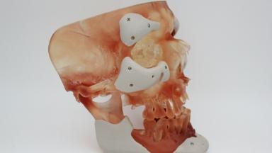
Leaving the gap unreconstructed is a real choice with a long track record. The thigh bone floats without a socket, held by scar tissue and muscle, which surgeons call a flail hip. The leg ends up shorter, the patient walks with a stick or crutches, and the hip stays unstable. In exchange no implant sits there waiting to become infected or to loosen, and in a region with this infection rate that counts as a genuine advantage rather than a consolation prize offered to patients who could not have something better. Hip transposition moves the thigh bone up against the remaining bone of the pelvis so that scar tissue forms a false joint in a controlled position, which trades some length and some stability for a construct that has no foreign material in it beyond whatever holds the bone temporarily in place. Custom implants sit at the other end of the range. Modern practice increasingly uses a 3D printed hemipelvic endoprosthesis, designed from the patient's own CT so it matches the defect exactly and carries a porous surface that bone can grow into, and according to PubMed these implants have been adopted by many specialist centers with improved outcomes, though the published evidence still comes largely from single centers with small numbers, short follow-up and considerable variation in how the implants are designed (Hu et al, 2023).
That caveat deserves weight. A custom implant is a promising technology with a thinner evidence base than a hip replacement has, and anyone being offered one should understand they are choosing the newer option rather than the better-proven one.
Sacral tumors and the nerve roots
Sacral tumors, most commonly chordomas, pose a question no other part of the skeleton poses. The nerve roots controlling the bladder, the bowel and sexual function pass directly through the bone that has to be removed, so the level of the cut determines what a patient keeps, and that trade is decided before the operation rather than discovered afterward.
The published evidence on this is unusually specific, which makes an honest conversation possible.
According to PubMed, a systematic review pooling 15 series and 244 patients found that normal ambulation was retained by 56.2 percent of patients when both S2 roots were spared and by 94.1 percent when both S3 roots were spared, while normal bladder function was present in 39.9 percent when both S2 were spared, rising to 72.7 percent with one S3 spared and 83.3 percent with both, and bowel function was normal in 94 percent of patients when both S3 roots were kept (Zoccali et al, 2016). Where even one S4 root was preserved, bladder and bowel function were normal in every reported case.
This table scrolls sideways on a narrow screen. Swipe or drag to see every column.
What the level of resection tends to leave intact
Roots preserved
Walking
Bladder and bowel
Both S2 only
Around half walk normally
Frequently impaired, with roughly two in five keeping normal bladder function
One S3
Usually preserved
Around seven in ten keep normal bladder function
Both S3
Normal in the large majority
Bowel normal in most, bladder normal in around five in six
One side only
Generally preserved
Function is retained in the great majority when the other side is intact
An older Mayo Clinic series of 53 patients points the same way, reporting normal bowel and bladder function in 87 and 89 percent of patients respectively after a one-sided resection with the opposite nerves preserved, against universally abnormal function when both S2 to S5 roots were sacrificed (Todd et al, 2002), also according to PubMed.
Two things follow from this for a patient. Ask which roots the plan sacrifices, by name, and ask what the surgeon expects that to mean for continence and sexual function specifically. Teams that hold this conversation properly before the operation have held it many times before.
The scale of the operation
Patients arriving from a consultation about a limb tumor rarely expect how much larger this is.
What a periacetabular case involves
In a published series of seven patients having a combined type II and III resection with a 3D printed hemipelvic prosthesis, operating time ran from 210 to 360 minutes, blood loss from 1300 to 2500 millilitres, and transfusion averaged around six units (Min et al, 2022), according to PubMed. Those are small numbers from one center, and they are the right order of magnitude to plan around.
Blood loss on that scale is the reason cross-matched blood is arranged in advance, why anesthetic assessment is thorough, and why intensive care for a night or two afterward is planned in advance as part of the operation. A urologist or a vascular surgeon may be scrubbed alongside the orthopedic team, and a plastic surgeon may be needed to close the wound. Several specialties in one theater is normal here.
How the admission runs
1
Before the day
Imaging is reviewed, blood is cross-matched, and the anesthesiologist assesses fitness for a long operation. Where the ureters run close to the tumor, a urologist may place stents so they can be identified and protected.
2
In theater
Several hours, often with more than one surgical team. The tumor is separated from the vessels and nerves, the bone cuts are made, the specimen comes out whole, and the reconstruction follows with fresh instruments.
3
Intensive care
A night or two, for close monitoring of blood pressure, blood counts and fluid balance after a large blood loss. Getting out of intensive care promptly matters, because a longer stay there is itself linked to infection.
4
The ward, and the weeks on it
Drains come out as output falls, the wound is inspected daily, and physiotherapy begins with sitting before it gets anywhere near standing. This is the long part of the admission.
Knowing this in advance changes how the day feels for the family waiting.
Infection is the defining complication
Every operation carries a risk of infection. In pelvic tumor surgery it stops being one item on a consent form and becomes the central problem the whole team is working against, because the wound is long, the operation is slow, the tissue planes are deep, the area sits close to the groin and the perineum, and many patients have had chemotherapy or radiotherapy first.
The number to know
In a series of 146 patients having pelvic bone or soft tissue sarcoma removed, 60 of them, or 41 percent, developed a surgical site infection. Longer operating time, more than two days in intensive care and the use of a skin flap all raised the risk, and extending preventive antibiotics from the standard couple of days out to five made no difference at all (Bensaid et al, 2023), according to PubMed.
Carry that last finding into a consultation. A longer course of antibiotics is not the answer, and a unit offering one as reassurance has misread the evidence. Three things help. A shorter operation. Careful handling of the soft tissue. Getting the patient out of intensive care as soon as that is safe.
How an infection here is treated
Infections in this region are typically polymicrobial, meaning several organisms are growing together, and antibiotics alone rarely clear them. Treatment normally means returning to theater to wash the wound out, sometimes more than once, with tissue samples taken to direct the drugs. Where an implant is present and the infection has reached it, removing that implant occasionally becomes the only route to a clean result, which is one of the arguments made for simpler reconstructions in the first place. None of this is quick. A deep infection around a pelvic reconstruction can add weeks to an admission that was already measured in weeks, and it is the single event most likely to turn a planned trip into a much longer one.
The rest of the list
Bleeding, injury to the bladder or the ureter, nerve injury affecting the leg, hernia through the surgical defect, and blood clots in the pelvic veins all belong on the consent form. None of them dominates the way infection does.
Learning to walk again
Rehabilitation after pelvic surgery is slower than after any limb operation, and expectations set against a hip replacement will be wrong by a factor of several.
Getting back on your feet
Bed rest comes first, for days rather than hours, while the wound settles and drains come out. Weight-bearing then depends entirely on what was removed and what replaced it. A type I resection with the hip joint intact allows early progress. A reconstructed socket usually means a period of protected weight-bearing with crutches or a frame while the bone grows into the implant. An unreconstructed flail hip means learning to walk on a leg that no longer has a fixed socket, which is achievable with practice and almost always needs a stick permanently.
The muscle takes longer than the bone
Muscle is the slower half of the problem. Pelvic resection divides the abductors that stabilize the hip with every step, and rebuilding that control takes months of supervised physiotherapy. In the small series quoted earlier, muscle strength on the operated side reached 68 percent of the healthy side at three months and 90 percent at twelve, which gives a realistic shape to the timeline even though the numbers come from a handful of patients with a favorable reconstruction.
Most patients are walking with support within the first few months. The final answer on how well, and with what aid, takes a year or more.
Why the surgeon's caseload matters here
Pelvic tumor resection is uncommon even inside orthopedic oncology, and it is one of the operations where how often a team does it shows up in the result.
The reasons behind that are practical rather than mystical. Units doing these regularly have an anesthesiologist who expects the blood loss, a scrub team that knows the instrument set, a urologist available to place ureteric stents before the incision, a plastic surgeon on standby for closure, a radiologist who reports these scans frequently enough to know where tumors hide, and a rehabilitation service that has taken people through this before. Each of those counts as a small advantage on its own. Stacked together, they account for most of the difference between one unit and another, and none of them shows up in a brochure.
So ask directly. How many pelvic resections does the unit perform in a year, who else scrubs in, and what happens if the wound breaks down at week three.
A team comfortable with those questions will answer them in specifics. Vagueness is itself an answer. Here the musculoskeletal tumor board reviews these cases before an operation is offered, and a patient traveling from abroad can have their scans and biopsy put to it before they commit to the trip, which is the point at which a second opinion is still worth something.
Stay, flying and the reality of the trip
Of all the operations on this site, this is the one that fits a short trip least comfortably, and saying otherwise would do a reader no favors.
Inpatient stay runs into weeks for a major pelvic resection, and longer again where intensive care or a wound complication extends it beyond what anyone planned for. Add several days at the start for imaging review, anesthetic assessment, cross-matching and the pre-operative consultation. Add a further period after discharge before anyone will clear a long flight, because the risk that matters here is a clot in a pelvic vein, in a patient who has had a long operation, a cancer diagnosis and a spell of reduced mobility all at once. Sitting brings its own problem on top of that. Somebody with a fresh pelvic wound, particularly after a sacral resection, may not tolerate six hours in an ordinary seat at all, and raising that well before the flight is booked is far better than discovering it at the airport. Bring someone with you. This is not an operation to travel for alone, because a companion needs to be present for the consent conversation, available during a long day in theater, and able to help through weeks of limited mobility afterward. The practical side of that is handled here rather than left to you. A companion bed sits in the patient rooms, and accommodation on either side of the admission is arranged through the international patients office, which matters when the stay runs into weeks rather than days.
Follow-up once you are home
Two threads run in parallel after you get home, and they need different people.
One thread covers the cancer, meaning scheduled imaging of the pelvis and the chest, starting frequent and stretching out over years, arranged near where you live. The other covers the reconstruction, watched on x-rays for loosening or failure, alongside months of rehabilitation. Where nerve roots were sacrificed, bladder and bowel management belongs to a continence specialist at home, and arranging that before departure is far easier than finding one afterward.
Travel home with the operation note describing exactly which zones and which nerve roots were taken, the full histopathology including margin status, the postoperative imaging as a baseline, and the make and reference of any implant. A surgeon revising that implant in a decade cannot easily find those details later.
Keep a line of contact to the operating team through all of it. A wound that looks wrong in the fourth week is the situation this matters for most, and the person who closed it can tell in one photograph whether it needs a local review or a flight back.
Frequently asked questions
How long will I be in hospital and in the country?
A major pelvic resection means an inpatient stay measured in weeks, with intensive care planned for the first night or two. Several days are needed before surgery for imaging review, anesthetic assessment and cross-matching, and a further period after discharge before a long flight can be cleared. Budget for weeks in the country. Do not book a return flight before the surgical team has given you a date.
Will I be able to walk normally afterward?
It depends on which zone of the pelvis was resected. A type I resection sparing the hip joint usually allows a good recovery of walking. A type II resection takes the hip socket, so walking depends on the reconstruction, and an unreconstructed flail hip generally means using a stick permanently and accepting some limb shortening. Muscle strength recovers over roughly a year, and most patients are walking with support within the first few months.
Will a sacral tumor operation affect bladder and bowel control?
That depends entirely on which sacral nerve roots have to be sacrificed. Published series report that preserving both S3 roots leaves bowel function normal in around 94 percent of patients and bladder function normal in around 83 percent, while sacrificing both S2 and everything below leaves function consistently impaired. Resection on one side only, with the opposite roots intact, preserves function in the great majority. Ask which specific roots your plan involves before consenting.
How likely is an infection after this operation?
Higher than most patients expect. One published series of 146 pelvic sarcoma resections reported surgical site infection in 41 percent of patients, with longer operating time, prolonged intensive care and the use of a skin flap increasing the risk. Extending preventive antibiotics beyond the standard short course did not reduce it. Treatment usually involves returning to theater to wash the wound out, and occasionally removing an implant if the infection is deep and persistent.
Is a 3D printed custom implant better than the alternatives?
It is the newer option and it allows an implant matched exactly to the defect, with reported outcomes that have encouraged many specialist centers to adopt it. The published evidence is still mostly single-center series with small numbers and short follow-up, and implant designs vary considerably between units. Simpler approaches, including hip transposition and leaving the defect unreconstructed, avoid having an implant that can become infected or loosen, which carries real weight in a region with this infection rate.
Can the leg always be saved?
In most cases yes, through an internal hemipelvectomy that removes part of the pelvic ring and leaves the leg attached. Where a tumor encases both the main iliac vessels and the sciatic nerve, or has spread widely through surrounding muscle, preserving the limb would leave a leg without blood supply, nerve supply or useful function. In that narrow group an external hemipelvectomy, removing the leg with the affected pelvis, gives better local control and a more predictable recovery.
References
Zoccali C, Skoch J, Patel AS, et al. Residual neurological function after sacral root resection during en-bloc sacrectomy. A systematic review. Eur Spine J. 2016;25(12):3925-3931.
Todd LT, Yaszemski MJ, Currier BL, et al. Bowel and bladder function after major sacral resection. Clin Orthop Relat Res. 2002;397:36-39.
Bensaid S, Contejean A, Morand P, et al. Surgical site infection after pelvic bone and soft tissue sarcoma resection. Risk factors, microbiology, and impact of extended postoperative antibiotic prophylaxis. J Surg Oncol. 2023;128(2):344-349.
Hu X, Lu M, Zhang Y, et al. Pelvic-girdle reconstruction with three-dimensional-printed endoprostheses after limb-salvage surgery for pelvic sarcomas. Current landscape. Br J Surg. 2023;110(12):1712-1722.
Min L, Li L, Hu X, et al. Application of modified Gibson combined with modified ilioinguinal approach in treatment of Enneking II and III pelvic malignant tumors with three-dimensional printed hemipelvic prosthesis replacement. Zhongguo Xiu Fu Chong Jian Wai Ke Za Zhi. 2022;36(7):796-803.
Editor's note
Written by the Biruni Hospital medical editorial team. Reviewed by Assoc. Prof. Dr. Suat BATAR, Orthopedics and Traumatology.