
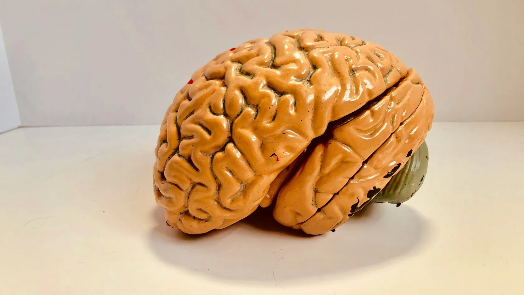
Pediatric Brain Surgery - Children's Brain Surgery
Pediatric brain surgery covers tumors, blocked spinal fluid, drug resistant epilepsy and malformations in children, and every decision is judged over decades instead of months. This page sets out what the evidence says about how much tumor to remove, why posterior fossa syndrome affects roughly one child in three, how much the choice of center matters, and what follow up a child needs for years afterward.
About This Department
A child's operation is judged in decades. Which makes the careful decision the difficult one.
Adult brain surgery is largely a question of how much can be removed. In a five year old it is also a question of what the removal costs a person who has sixty or seventy years left to live with the result. This page sets out where the evidence says to push, where it says to stop, and what a family should be told before signing anything.
What pediatric brain surgery covers
Four groups of problems bring children to a neurosurgeon. Tumors lead that list, and central nervous system tumors make up roughly a quarter of all childhood cancers, with more than half of them sitting in the posterior fossa at the back of the head. Blocked fluid comes second, whether from a tumor, from bleeding, from infection or from the way the brain formed. Epilepsy that no longer answers to medication comes third. Malformations of the skull, the spine and the blood vessels round out the list, together with the head injuries that any emergency department sees. Each of those has its own operation, its own timing and its own argument over how far to go. What they share is the thing that separates this specialty from adult neurosurgery entirely, namely that the patient is still being built. A brain that is still laying down connections responds differently to being cut, to being irradiated and to being deprived of pressure control than one that finished developing twenty years ago, and the consequences show up not in the recovery room but at school, at puberty and in the first job interview, so the operative plan has to be written with all of that in view, which is why a good pediatric neurosurgical decision often looks less impressive on the postoperative scan than an adult one would.
This page gives the third of those as much room as the first.
Why a child's brain is not a small adult brain
Start with the tumors, because they are almost entirely different animals. The commonest malignant brain tumor of adulthood barely appears in childhood, and the commonest malignant brain tumor of childhood barely appears in adults. Medulloblastoma, pilocytic astrocytoma, ependymoma and craniopharyngioma dominate the pediatric list, and they sit in different places, behave differently, respond to different treatments and carry survival figures that would be unrecognizable to anyone who has only read about adult disease. Take a pilocytic astrocytoma out completely and the child is very often cured, permanently, with nothing further required. No adult glioma behaves that way. Then the anatomy. A small child has perhaps half a liter of circulating blood, so a loss that an adult would not notice becomes a transfusion decision within minutes. Skull bone runs thinner, and in infants it has not yet closed at the sutures, which changes how pressure behaves and sometimes buys time that an adult skull would not. Posterior fossa tumors are the pediatric norm and the anterior skull base tumors of adulthood are the exception, which puts the surgical corridor in a crowded, unforgiving place next to the brainstem and the cerebellum.
Third, and most consequential, is time.
Someone of seventy treated for a brain tumor has a defined number of years in which the treatment can cause trouble. A four year old has seventy of them, and the therapies that control the disease also act on organs that have not finished forming. Radiation to a developing brain costs measurable cognitive ground, and the younger the child the steeper the cost, which is the entire reason pediatric oncology spends so much effort building protocols that delay radiotherapy, reduce its dose or avoid it in the very young altogether. Hormone axes sitting close to the surgical field can fail years after the operation. Spinal growth alters after treatment. Hearing drops. None of that is visible when the child is discharged walking and talking, and all of it belongs in the conversation before the operation is booked, because the choice between two surgical plans is frequently a choice between two different sets of late effects.
The conditions we operate on
Below runs what actually fills a pediatric neurosurgical list. Every age given is a rough one.
This table scrolls sideways on a narrow screen. Swipe or drag to see every column.
| Condition | Usual age | What surgery is for | What usually follows |
|---|---|---|---|
| Medulloblastoma | Three to nine years | Remove safely, relieve fluid blockage, obtain tissue for molecular typing | Chemotherapy, and radiotherapy in children old enough to tolerate it |
| Pilocytic astrocytoma | Five to fifteen years | Complete removal where the location permits it | Often nothing at all, with scans alone for years |
| Ependymoma | One to seven years | Maximum safe removal, which drives outcome more than in most tumors | Focal radiotherapy, sometimes a second operation first |
| Craniopharyngioma | Five to fourteen years | Protect vision and the hormone axis while controlling the tumor | Lifelong hormone replacement in most children |
| Low grade glioma of the brainstem or thalamus | Any age | Diagnosis, relief of pressure, partial removal where it is safe | Observation in more than half, chemotherapy if it progresses |
| Hydrocephalus | Birth onward | Restore fluid drainage, by endoscopy or by a shunt | Long term monitoring for blockage |
| Drug resistant focal epilepsy | Two years upward | Remove or disconnect the tissue generating the seizures | Gradual reduction of medication over years |
Notice how different the last column looks from row to row. In one line surgery is the whole treatment, in another it is only the opening move, and a family who has been told only that their child needs an operation has been given the smaller half of the information.
Getting the diagnosis right first
Almost every avoidable disaster in this field starts with an incomplete picture. A scan taken too quickly, in the wrong sequences, without contrast or without the spine included leads to an operation planned for the wrong problem, and in children the second chance is more expensive than it is in adults because the tissue around the tumor has jobs it has not learned to share yet.
So the first thing we do with any child referred from abroad is look at the imaging rather than the report. A full magnetic resonance study of the brain, with contrast, in the sequences that distinguish one tumor type from another. The whole spine imaged as well when the tumor is one that spreads through the fluid, since finding a second deposit after the operation rather than before it changes the treatment plan and wastes a general anesthetic. We use computed tomography sparingly and for specific questions, because it delivers radiation to a child who may need a great deal more of it later and the dose is cumulative across a lifetime. Molecular typing has changed this field more than any instrument has. Medulloblastoma now covers four biologically distinct diseases wearing one name, and they carry different prognoses, different treatment intensities and, as the next section describes, different answers to the question of how hard the surgeon should push. Tissue therefore has to reach a laboratory capable of that analysis, handled correctly from the moment it leaves the operating field. A center that removes a tumor beautifully and cannot type it has done part of the job.
We send back a written opinion on all of this before anyone books a flight, at no charge, and we say plainly when the right answer is an operation somewhere closer to home.
Inside the operating room
Most of what makes a pediatric operation safe happens before the first incision. Below runs the sequence, in the order the team works through it.
- Position and padding. Children lie for hours on tables built for adults, so every pressure point is padded and the neck is set at an angle that keeps the airway and the venous drainage open.
- Blood ready before the drapes go on. Cross matched and in the room, because the margin between a routine loss and a serious one is measured in tens of milliliters at this size.
- Navigation registered to the scan. The preoperative images are locked to the child's head so the surgeon knows where the tumor edge lies relative to structures that must survive.
- Monitoring of the pathways at risk. Movement, hearing, facial function and the cranial nerves of the brainstem are watched continuously, and a change in the trace is treated as an instruction rather than as information.
- Ultrasonic removal at low settings. Tumor gets emulsified and suctioned in small volumes, which protects the vessels running through it and keeps the surgeon in contact with the plane between tumor and brain.
Why the anesthetist matters as much as the surgeon
Temperature, fluid balance, blood pressure and carbon dioxide all sit with the pediatric neuroanesthetist, in a patient whose reserves are small and whose brain lies open under a microscope. Drop the blood pressure for a few minutes in a field where perfusion is already marginal and the damage will not appear until the child fails to wake properly. Allow the temperature to fall and clotting slows. Give too much fluid and the brain swells into the surgeon's view. None of this is visible in the operative report, and all of it separates a center that operates on children regularly from one that operates on adults and occasionally accepts a child.
Find out who will anesthetize your child, and how many children they put to sleep in a year. Asking costs nothing and the answer should arrive without hesitation.
How much to remove
Everything difficult about this specialty lives in one decision, and for thirty years the profession answered it the same way. Take all of it. An incomplete removal was treated as a marker of bad disease, which justified second operations to chase fragments and intensified chemotherapy and radiotherapy afterward. Then somebody checked.
What 787 children showed
The finding that changed the adviceAn international consortium pooled 787 children with medulloblastoma from 35 institutions, and for the first time sorted them by molecular subgroup before asking what surgery had contributed. Complete removal offered no overall survival advantage over near total removal, meaning a fragment smaller than one and a half square centimeters left behind. Against subtotal removal, meaning a larger piece left in place, there was a progression free survival advantage and still no overall survival advantage. Only one subgroup showed a real benefit from taking everything, and only in children whose disease had already spread. The authors concluded that removing small residual pieces is not recommended where the risk of neurological damage is high.
Read that carefully, because it does not say that surgery is unimportant. Maximum safe removal remains the standard, and the word carrying the weight is safe. What the analysis dismantled was the habit of treating the last fragment as worth any price, when a great deal of what looked like a surgical effect turned out to be the tumor's biology declaring itself through which children could be cleared easily and which could not.
Pediatric practice shows the same pattern elsewhere, arriving from a different direction. A prospective German cohort followed 116 children with low grade gliomas of the pons and medulla, treated within a defined national strategy. More extensive resection did reduce the rate of progression, clearly and statistically. It also produced a new complication in 21 of every 100 children operated on, including respiratory failure. Five year overall survival was 95 percent, and it was 95 percent whatever the surgeon had done, because these tumors are chronic conditions rather than lethal ones. Fifty nine of the 116 children received no additional treatment at all after the initial surgical step. Twenty one children with low grade tumors straddling the thalamus and the cerebral peduncle, treated in Prague, make the point at close range. Six of them had everything taken out. Every child in the series was alive at a median of six years, functioning at a level that allows normal activity with minor limitation, and the authors were explicit that excellent long term outcomes are available to children in whom complete removal is not achievable.
- We take everything when the tumor sits where it can be taken and the monitoring stays quiet throughout.
- We stop short when the remaining fragment is stuck to the brainstem, to the floor of the fourth ventricle, or to vessels that supply territory the child cannot spare.
- We say so in the operative note, we show the family the postoperative scan, and we explain what the residual means for the treatment that follows.
- We do not return for a second operation to chase a fragment unless the biology of that particular tumor says the fragment matters.
A surgeon who promises a complete removal before seeing what the tumor is attached to is promising something no honest person can promise, and what can be promised is that the decision will be made in the room, on what the tissue and the monitoring show, and that it will be explained afterward without decoration.
Posterior fossa syndrome
Here is the complication that families are almost never warned about, and it deserves a section of its own because it is the one that changes a household.
A child comes out of a posterior fossa operation awake and apparently fine. A day or two later the talking stops. Completely, in the severe form, in a child who was speaking normally before the operation and who is now mute, emotionally labile, unable to sit up straight, and sometimes moving in ways that frighten the people at the bedside. Nothing has bled. Nothing shows on the scan. The syndrome has been recognized for decades and is still not fully explained, and it arrives with a reliability that makes the silence about it hard to justify. A prospective study followed 178 children through medulloblastoma surgery and found it in 60 of them, which is 34 percent. Forty stopped speaking altogether. Twenty had markedly reduced speech. All of them had severe difficulty with balance and coordination, and among the mute group more than four in ten developed a movement disorder as well. Speech came back at a median of 2.3 months in the severe group and 0.7 months in the milder one, and walking returned at 2.1 and 1.5 months.
Those recovery figures get quoted everywhere, and they mislead. Returning and arriving describe different things. Of 27 children in the severe group followed for a year, 12 still could not walk independently at the end of it, and at a median follow up of nearly two years not a single child who had developed the syndrome had a normal neurological examination. Most families hear that speech returns, which is true, and leave with the impression that the episode closes, which is not.
Two things raised the risk in that study. Younger age, which nobody can change. And having the surgery in a center that does few of these operations, which somebody can.
Where the operation happens
The authors of that study put their conclusion in a single sentence, and the shape of that sentence deserves attention. Surgical experience emerged as the major modifiable contributor to the syndrome. Of everything that drove a child toward months of silence, the item most open to change was the choice of hospital.
That is an uncomfortable finding for a page like this one, since we are one of the centers you are choosing between, so read the three things below as questions to put to us and to everybody else on your list, with the answers compared side by side.
If any center answers the first question with a number that sounds small, that is useful information regardless of how the conversation feels.
Epilepsy surgery in children
Everything above argues for restraint. This section argues the other way, because the mistake in childhood epilepsy surgery is almost always waiting too long.
A randomized trial assigned 116 children and adolescents with epilepsy that had stopped answering to medication either to surgery with continued medical treatment or to medical treatment alone, and at twelve months 44 of 57 children in the surgical group were free of seizures, which is 77 percent. In the medically treated group the figure was 4 of 59, which is 7 percent. Seizure severity, behavior, quality of life and social maturity all moved in favor of surgery by margins that were not close. Measured intelligence did not separate significantly, which is the one result that did not deliver.
The cost side of that trial
Serious adverse events occurred in 19 of the 57 operated children, a third of them, and 15 developed weakness down one side. Those deficits were anticipated, in the sense that they followed from the region of brain that had to be removed and were discussed before consent, which is a different thing from a surgical accident but is not a small thing to a family. Consider the trade on offer. A three in four chance of a childhood without seizures, against a one in four chance of a permanent motor deficit in the specific children whose seizures come from motor territory.
What makes the delay so costly is that uncontrolled seizures are not a neutral background condition in a developing brain, since years of them cost development, schooling and independence, and those years do not come back when the seizures eventually stop. The average child reaching an epilepsy surgery program has been having seizures for a long time, usually after a succession of drugs that were never going to work, because two failed medications already predict that the third and fourth will fail too.
If your child has failed two properly chosen drugs and has a lesion on the scan, the next move is probably not another drug. Somebody should be establishing where the seizures start, and that takes a formal assessment rather than a fifth prescription.
Fluid, pressure and the growing head
Fluid made inside the brain has to leave it, and a tumor at the back of the head sits precisely where the exit runs. So a large proportion of children arriving with a posterior fossa tumor are in trouble from the blockage before the tumor itself has done anything, and the signs are recognizable once somebody names them, namely headache worst in the morning, vomiting on waking, double vision, and a head circumference climbing off the chart in an infant. These are pressure signs and they are the reason a child is sometimes operated on within hours of arriving.
What relieving the pressure involves
Removing the tumor reopens the passage in many children, and nothing further is needed. Where it does not, there are two routes. An endoscope can make a new opening in the floor of the third ventricle so fluid bypasses the blockage entirely, leaving no device behind, which suits a child with a clean obstruction and a suitable anatomy. Where that will not work, a valve and tube drain fluid into the abdomen instead. That second option works reliably and commits the child to a device that will need watching for the rest of their life, so the choice between the two is made deliberately and explained rather than defaulted.
Risks in the order families meet them
Consent conversations tend to run through a list at speed. This one runs chronologically, which is how a family actually experiences it. Nothing below is rare enough to leave out.
From the first night to the first year
Timing tells you what a complication means. The same symptom on night one and in month six points at two different problems, so the table reads down the clock.
This table scrolls sideways on a narrow screen. Swipe or drag to see every column.
| When | What | What it looks like | What is done |
|---|---|---|---|
| First 24 hours | Bleeding or swelling in the operative bed | Slow waking, a new weakness, a widening pupil | Immediate scan, return to the operating room if needed |
| Days one to five | Posterior fossa syndrome | Speech stops, balance collapses, mood swings | Therapy from day two, no operation required |
| First two weeks | Fluid leak or wound infection | Clear fluid from the wound, swelling, fever | Stitches, drainage, antibiotics, sometimes revision |
| First month | Pressure returning as fluid blocks again | Morning headache, vomiting, sleepiness | Endoscopic opening or a shunt |
| Months to years | Hormone failure, hearing loss, learning difficulty | Growth slowing, school reports changing, fatigue | Endocrine replacement, hearing aids, school support |
The bottom row is the one most likely to be skipped in a consent conversation and the one most likely to shape an adult life, since growth hormone failing quietly at nine and caught at fourteen costs height that never comes back, and the only thing standing between those two dates is somebody measuring. Somebody has to own that schedule.
Recovery, ward to classroom
The first ten days, as they usually runYour child goes to pediatric intensive care straight from the operating room and stays overnight, sometimes two nights. A scan is done within 48 hours, while what is left behind can still be told apart from healing tissue. Drips and drains come out over the next two or three days, and the child moves to the ward with a parent sleeping in the room. Sitting comes before standing, standing before walking, and physiotherapy starts whether or not the child feels like it. Speech therapy begins on day two in any child who has had a posterior fossa operation, before anyone knows whether it will be needed. Most children go home or to their hotel between day seven and day ten. Stitches or staples come out at around ten days.
Beyond those two weeks the timetable stops being a timetable. Some children, after a small tumor is taken from a quiet part of the brain, are back at school in three weeks, tired for a month, and indistinguishable from their classmates by the end of the term. Where posterior fossa syndrome developed, the scale changes entirely, with speech returning over two or three months, walking over a similar period, and a year of therapy after that to recover coordination that will probably not return all the way.
Going back to school
Return early and return partially. Half days first, with the school told what happened and what to watch for, because fatigue after brain surgery is not ordinary tiredness and a child who looks fine at nine in the morning can be finished by eleven. Attention and processing speed are usually the first things to suffer and the last to be noticed, since a quiet child who has fallen behind attracts less attention than a disruptive one. Formal neuropsychological testing at six to twelve months gives the school something concrete to work from. Ask for that report in writing. It also establishes a baseline, which matters more than the first result does, since what tells you whether a child is drifting is the comparison between that test and the one three years later.
Protect friendships as deliberately as the schoolwork. Children who spend months out of class come back to a social group that reorganized without them, and the isolation does more day to day damage than a slightly lower score on a test.
The decade nobody discusses on day one
Survival is where a family's attention sits on the day of diagnosis, understandably, and it is not where the outcome gets decided for a child who survives, as most children with the commonest pediatric brain tumors now do. What decides the outcome is whether anyone is still measuring five years later. Hormone axes sitting near the surgical field or in the radiation path fail slowly and quietly, and growth hormone usually goes first, followed by thyroid, then the adrenal and reproductive axes at puberty. Hearing drops after certain chemotherapy drugs and takes speech development with it in a young child. Vision needs testing formally and not by asking whether the child can see. Tumor surveillance runs on its own schedule, frequent at first and thinning out over years, and the interval depends on the tumor type and on what was left behind. Meanwhile the learning profile shifts as the demands of school rise, which means a child who coped in year three can struggle in year seven for reasons that have nothing to do with effort.
Who holds the file
Families who travel for surgery face a specific version of this problem, because the surgeon is in one country and the child's ordinary life is in another. So the handover has to be written rather than assumed. Every child we operate on goes home with the operative note, the postoperative imaging, the pathology including the molecular result, the treatment plan agreed with the oncology team, and a schedule saying which test is due when, in the first year and in the years after it, and that document is addressed to the doctor who will be looking after the child at home, written in English so that it travels without needing a translator at the other end. We stay reachable on the same WhatsApp number afterward, and scans taken abroad can be sent to us for a second read at any point. Remote follow up is part of the treatment and not a favor, and it costs nothing.
Bringing a child to Istanbul
Two people travel here, never one. Traveling with a sick child is harder than traveling alone, so the practical arrangements here are built around the family and not around the patient alone.
Send the scans first. Everything above is easy to organize and none of it matters if the operation is the wrong one, and that is why the free review of your child's imaging comes before any discussion of dates.
Cost, timing and travel
Plan on two to three weeks in Istanbul for a tumor operation in a child, and longer if the recovery is complicated. That covers the assessment and imaging on arrival, the operation itself, one or two nights in pediatric intensive care, seven to ten days on the ward, the early therapy, stitch removal at around ten days and a final review before you leave. Recovery complicated by posterior fossa syndrome runs longer, and we say that in advance so that nobody is booking return flights they will have to change. Fitness to fly is assessed before discharge and turns on pressure inside the head more than on the wound. A child whose fluid circulation is settled, who is eating, walking within their own capacity and scanning normally is safe on an aircraft, while one with an unresolved pressure question is not, and we will say so even when the flights are booked. Most children fly home between two and three weeks after surgery.
What moves the cost is a short list. The length of the operation and whether monitoring of the pathways is required. Time in pediatric intensive care. Where the molecular typing goes. Whether a shunt or endoscopic procedure is needed alongside the tumor operation. Whether chemotherapy or radiotherapy begins here or at home. We quote all of that in writing after reviewing the scans and we do not publish a number on this page, because a figure attached to an operation whose length nobody has established would be a guess dressed as a price.
Follow up once you are home costs nothing. Send us the scans, send us the school reports if something is worrying you, and we will read them.
Pediatric brain surgery FAQ
Families ask these questions in the first message.
Will the surgeon remove all of the tumor?
What does posterior fossa syndrome mean, and how likely is it?
Does it matter where we have the operation done?
How long will we need to stay in Istanbul?
Will my child need radiotherapy?
My child has seizures that medication cannot control. Is surgery worth considering?
Can one of us stay with our child?
References
- Thompson EM, Hielscher T, Bouffet E, Remke M, Luu B, Gururangan S, et al. Prognostic value of medulloblastoma extent of resection after accounting for molecular subgroup. A retrospective integrated clinical and molecular analysis. Lancet Oncology. 2016;17(4):484-495.
- Khan RB, Patay Z, Klimo P, Huang J, Kumar R, Boop FA, et al. Clinical features, neurologic recovery, and risk factors of postoperative posterior fossa syndrome and delayed recovery. A prospective study. Neuro-Oncology. 2021;23(9):1586-1596.
- Holzapfel J, Kandels D, Schmidt R, Pietsch T, Warmuth-Metz M, Bison B, et al. Favorable prognosis in pediatric brainstem low-grade glioma. Report from the German SIOP-LGG 2004 cohort. International Journal of Cancer. 2020;146(12):3385-3396.
- Dwivedi R, Ramanujam B, Chandra PS, Sapra S, Gulati S, Kalaivani M, et al. Surgery for drug-resistant epilepsy in children. New England Journal of Medicine. 2017;377(17):1639-1647.
- Benes V, Zapotocky M, Liby P, Taborsky J, Blazkova J, Sumerauer D, et al. Survival and functional outcomes in paediatric thalamic and thalamopeduncular low grade gliomas. Acta Neurochirurgica. 2022;164(6):1459-1472.
- Wibroe M, Cappelen J, Castor C, Clausen N, Grillner P, Gudrunardottir T, et al. Cerebellar mutism syndrome in children with brain tumours of the posterior fossa. A multicentre prospective study. BMC Cancer. 2017;17(1):439.
Editor's note
Written by the Biruni Hospital medical editorial team. Reviewed by Assistant Professor Özcan ÇIKLATEKERLİO, Neurosurgery.
Medically reviewed by
Assistant Professor Özcan ÇIKLATEKERLİO
Neurosurgery
Related Treatments
View All
ALIF - Anterior Lumbar Interbody Fusion Surgery
Coming in from the front restores the curve of the lower back better than any posterior route and leaves the back muscles untouched. It also carries one risk most pages will not name, and this page names it.

Brain AVM Surgery - Arteriovenous Malformation Surgery
An unruptured brain AVM bleeds at roughly 2.2 percent a year, and surgery spends its whole risk on a single morning. This page sets those two shapes of risk side by side, explains what the ARUBA trial does and does not say about your case, and shows how a Spetzler-Martin grade changes the answer.
Brain Tumor Surgery - Brain Tumor Removal
Brain surgery is one trade repeated all day, how much tumor comes out against what the brain around it still does. This page puts the published numbers on both sides of that trade.
Cervical Microdiscectomy - Minimally Invasive Neck Disc Surgery
Neck disc surgery is done from the front, through the space between your windpipe and your carotid artery, and almost everything that goes wrong that is not the disc happens in that corridor. What has been measured in it.

Chiari Malformation Surgery
A Chiari malformation is measured in millimeters and treated according to symptoms, and those two things come apart more often than patients are told. Most people with the finding never need an operation. This page sets out who does, what posterior fossa decompression removes, why surgeons still argue over opening the dura, and what the pooled studies say about both sides of that argument.