Most osteosarcoma pages stop at the leg. The harder question is what happens once the disease has reached the lungs, and whether cutting it out there still ends in a cure. For a proportion of patients it does.
About This Department
It started as an ache above the knee that got worse at night and did not settle with rest, and somebody called it growing pains or a football knock. Osteosarcoma announces itself that way in most teenagers, and the interval between that first symptom and the diagnosis therefore runs into months. This page explains what removing an osteosarcoma actually means, why the operation on the bone is only part of it, and how the whole thing is arranged when you are travelling for treatment.
Free consultation
Send the biopsy report and the chest scan together
A sarcoma team reads the pathology, the MRI of the whole affected bone and the chest CT in one sitting, then tells you what removing this tumour would involve, whether anything in the lungs changes the plan, and where the chemotherapy either side of surgery would be given. The review costs nothing and carries no obligation.
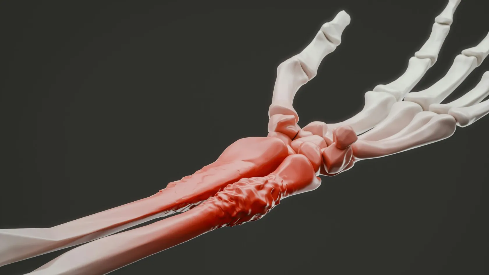
Osteosarcoma is a cancer of the bone-forming cells, so the tumour it produces makes bone, badly and in the wrong place. That single fact explains most of what follows. It explains the appearance on an X ray, where new bone is being laid down in disorganised streaks and the periosteum is being lifted off the shaft. It explains the hardness of the mass, the way it destroys the cortex from the inside, and why radiotherapy has so little to offer against it compared with the drugs and the knife. Three quarters of these tumours arise around the knee, in the lower end of the femur or the upper end of the tibia, because that is where bone grows fastest and longest. Two age groups get it. The larger peak sits in adolescence and the early twenties, tracking the growth spurt so closely that tall teenagers are over-represented, and a second, smaller peak appears after sixty, often in bone already altered by Paget's disease or by radiotherapy given decades earlier for something else. Those two populations tolerate treatment very differently, and a plan written for a fifteen year old does not transfer to a seventy year old.
Osteosarcoma travels to the lungs.
It reaches them through the bloodstream and not through lymph nodes, so a chest CT belongs to the first assessment and not to the follow up. That also explains why one in five patients already has visible lung deposits on the day of diagnosis. The disease also seeds within the same bone, producing separate nodules above or below the main tumour that are called skip metastases and that carry their own weight in the prognosis. Skip metastasis at diagnosis raised the hazard of death by a factor of 2.52 in the National Cancer Database series described below, close to the effect of lung disease itself.
How it announces itself
Pain comes first, and it has a character worth recognising, because it sits deep, it builds over weeks instead of arriving with an injury, it wakes the patient at night, and it refuses to follow activity in the way a strain does. Swelling arrives later. Sometimes much later, and by then the lump is firm, fixed to the bone underneath, and large enough that the family remembers noticing it weeks before anyone measured it. A limp, a reduced range at the nearest joint, or a bone that breaks under trivial force can all be the presenting event. Fever and weight loss are uncommon enough that their absence reassures nobody. The delay between that first ache and the diagnosis is longer than families expect, and it is mostly biology doing that. A retrospective cohort of 1,227 patients with high grade bone sarcomas found a median diagnostic interval of four months, with tumour characteristics predicting who waited longest and social ones barely registering. Reassuringly, the length of that interval did not change the amputation rate or survival. It did change one thing, because good necrosis after pre-operative chemotherapy was less common in the patients who had waited, at 25 percent against 34 percent.
Plain X rays of the painful area find most osteosarcomas, and they cost almost nothing. For a parent whose child has been sore for six weeks, that sentence matters more than anything else here.
Everything after the X ray belongs in a sarcoma unit. The MRI has to cover the whole bone and not just the painful segment, because a scan cropped to the knee misses a skip lesion at the hip and the operation then gets planned around incomplete information. The surgeon who will perform the resection plans the biopsy, and the track goes where that surgeon can take it out later in one piece with the tumour.
What removal has to include
Removing an osteosarcoma means removing every deposit of it that can be reached. Every is the word that gets missed. Three targets have to be accounted for before an operating date means anything.
The primary tumour in the bone, taken inside a continuous cuff of healthy tissue that is never breached during the dissection.
Any skip lesion in the same bone, so the MRI runs from joint to joint and the resection sometimes has to be far longer than the visible mass suggests.
Deposits in the lungs, taken by a thoracic surgeon in the same overall plan, sometimes at more than one sitting and sometimes years apart.
The staging investigations exist to define those three, and each one answers a specific question rather than adding reassurance.
This table scrolls sideways on a narrow screen. Swipe or drag to see every column.
What each staging investigation is looking for
Investigation
The question it answers
What changes if it is positive
MRI, whole bone
Where the tumour ends inside the marrow, whether it has crossed the growth plate, and how it sits against the artery, vein and nerve.
Sets the length of bone removed and decides whether the limb can be reconstructed at all.
Chest CT
Whether there are lung deposits, how many, and which side they sit on.
Adds a thoracic operation to the plan. It does not remove the bone surgery from it.
Bone scan or PET
Whether any other bone is involved, and how metabolically active the primary is before treatment starts. The baseline matters as much as the finding.
A second bone site changes the intent of the whole plan and is discussed openly before anything begins.
Planned biopsy
The exact histological subtype and grade, from tissue taken along a track the resection can later remove.
Confirms the diagnosis and selects the chemotherapy regimen. A badly placed track can cost the limb.
Heart and kidney tests
Whether the patient can tolerate doxorubicin, cisplatin and high dose methotrexate safely.
Doses and drug choices are adjusted, which matters most in the older group.
The order it happens in
Treatment runs in a fixed order and the order is doing real work. Chemotherapy first shrinks the tumour and hardens its edge so that less bone has to come out, it treats the microscopic disease already circulating on the day of diagnosis, and it produces the specimen from which the pathologist reads how well the drugs are working, which is a measurement no scan can supply and no clinician can guess. Reverse the sequence and all three are lost.
1
Diagnosis and staging
Whole bone MRI, chest CT, bone scan or PET, planned biopsy, heart and kidney function. One to two weeks, and the multidisciplinary meeting sits at the end of it.
2
Chemotherapy before surgery
Methotrexate, doxorubicin and cisplatin, given as cycles over about ten weeks through a central line. Hair loss, mouth ulcers, nausea and low counts all come with it, and every one of them is managed rather than endured, which is worth saying to a teenager who has read the internet.
3
The resection
The tumour, its biopsy track and a cuff of normal tissue come out in one piece. The bone defect is reconstructed in the same anaesthetic. Blood counts have to have recovered before the date is fixed.
4
The pathology report
Two to three weeks later it gives the margin status and the percentage of tumour killed. Those two lines shape the year ahead. They matter more than anything said in the consulting room beforehand.
5
Chemotherapy after surgery
Restarted once the wound has closed and continued for up to a year in total. Lung surgery, where it is needed, is fitted into gaps between cycles by agreement between the two surgical teams.
Delays inside that sequence cost more than delays before it. A wound that will not heal keeps the drugs out of the patient for weeks, which is one of the strongest arguments for having plastic surgical cover planned into the resection instead of improvised afterwards.
The number that predicts the rest
When the resected specimen reaches the laboratory, the pathologist maps it and counts what proportion of the tumour cells the chemotherapy has killed. That proportion is the necrosis rate. Nothing else on the report competes with it.
Ninety percent marks the line, and somebody tested it instead of assuming it
Researchers working through the National Cancer Database took 1,580 patients treated between 2010 and 2015 and went looking for a better cutoff. They did not find one. Five year overall survival was 74 percent for specimens showing 95 to 100 percent necrosis and 70 percent for 90 to 94 percent, a difference that was not statistically meaningful. Below 90 percent the hazard of death doubled, at a hazard ratio of 2.00. Among tumours that responded poorly, no further gradation predicted anything, so 82 percent necrosis and 40 percent necrosis carried the same message.
So what does the number change? Not the drugs. Trials that intensified treatment for poor responders have not shown a survival gain, and the same study found metastasis at diagnosis a stronger predictor than the necrosis rate anyway, at a hazard ratio of 2.85. The score is information for planning and for setting the intensity of surveillance, and a family should hear it in those terms and not as a verdict delivered on a child. One finding in that paper deserves saying out loud. Patients without insurance, and patients from the lowest income groups, were significantly less likely to reach 90 percent necrosis, which almost certainly reflects delayed presentation and interrupted treatment, and not biology. Access to uninterrupted chemotherapy counts as part of the treatment itself, and for a family travelling for care that is a practical question about where the cycles will be given.
Taking out the bone tumour
Limb sparing surgery is the standard operation and Cancer Research UK puts it at around 85 in every 100 osteosarcomas. The surgeon removes the affected segment of bone with its cuff of healthy tissue and rebuilds the gap in the same sitting, usually with a modular metal endoprosthesis that replaces the bone and the joint at its end. Amputation is reserved for tumours that encase the main nerve or the main vessels, that have broken through the skin, or that would leave too little muscle behind to move and cover a reconstruction.
Three quarters of this work happens at the knee.
Endoprosthetic replacement wins on speed. The construct bears weight on the day it goes in, the patient starts walking within days, and chemotherapy restarts on schedule instead of waiting for bone to knit under the influence of drugs that suppress healing and leave the graft with nothing to work with. Biological reconstructions matter where the resection is intercalary, meaning a length of shaft is removed while both neighbouring joints stay intact. One approach removes the affected segment, irradiates it outside the body with a sterilising dose, and screws the patient's own bone back in as a graft. A tertiary centre series of 96 osteotomies in 48 patients, mean age 15.8 years, reported non-union in 11.4 percent, graft failure in 2.1 percent, infection in 12.5 percent and soft tissue local recurrence in 14.5 percent, and found that a V-shaped diaphyseal osteotomy and an additional small plate at that osteotomy both shortened the time to union. Those are the details that separate a graft that unites from one that does not, and they are decided in theatre rather than in the clinic.
Vascularised fibula transfer is the other biological option. The patient's own fibula moves with its artery and vein, a microsurgeon plumbs it into the recipient site, and it thickens over years as it takes load.
Removing disease from the lungs
Osteosarcoma parts company with almost every other cancer a family will have read about at exactly this point, and it is the section most pages leave out. In most solid tumours, disease in the lungs means the intent of treatment changes and surgery stops being curative. In osteosarcoma it does not. Deposits in the lungs are cut out, and cutting them out cures a proportion of patients outright. The strongest evidence comes from a consecutive series of 463 children, adolescents and young adults treated with lung metastasectomy at three Italian centres between 1973 and 2014, followed for a median of 18.6 years. Overall survival was 34.0 percent at five years and 29.7 percent at twenty, and the flatness of that curve between five and twenty years is the point, because it means the patients alive at five years were largely cured, and not merely held in check. Of the 138 patients still alive, 42 had recurred and been cured by further metastasectomies, some of them repeatedly. Three factors predicted who benefited, being the length of the disease free interval before the lungs were involved, the number of deposits, and whether the resection removed all of them.
Complete clearance is the whole game.
A partial clearance carries the operative risk without the survival benefit, which is why the thoracic surgeon inspects and palpates every lobe rather than treating only what the scan showed, and why an open approach is still chosen over a keyhole one in many units for exactly that reason. Hands find what CT misses. Small deposits below the resolution of the scan turn up under the fingers more often than a family is led to expect. Where both sides are involved, the two operations are staged a few weeks apart, so that one lung has recovered its volume before the other is opened, and so that the patient is not asked to breathe on two freshly operated lungs at once in the middle of a chemotherapy cycle. Recurrence in the lungs after a clear interval is therefore not the end of treatment. It is a reason to scan, to count, and to operate again if the deposits are countable and the patient is fit, and the Italian series is what allows that to be said plainly. Families who understand this in advance cope far better with the first abnormal surveillance scan.
Two practical consequences follow for anyone arranging treatment across a border. The first is that the thoracic surgeon has to be part of the team from the beginning and not summoned once a scan turns abnormal, because the timing of lung surgery is negotiated against the chemotherapy calendar and against the bone operation, and a service that treats the chest as somebody else's problem will lose weeks arranging what should already have been planned. The second is that surveillance imaging has to be readable by the people who would operate. A chest CT reported in another country and described over the phone is not the same as the images themselves sitting in front of the surgeon who would take the deposits out, and the difference between those two things has decided whether an operation happened at all.
Ask early who the thoracic surgeon is.
Children who are still growing
Resecting the lower femur in an eight year old removes the growth plate that supplies most of the leg's remaining length, so a fixed implant leaves that child finishing growth with one leg several centimetres shorter than the other. Non-invasive expandable prostheses solve it by lengthening under an external magnetic field, in an outpatient visit with no incision.
Growth is the whole problem in a small child.
The technology has improved sharply within one generation of implants, and the difference is documented. A Paris paediatric series followed 40 children with a mean age of 8.8 years at surgery, 36 of them with osteosarcoma around the knee, comparing an older expandable design against a current one. Every surviving implant in the older group eventually came out again, mechanical failure being the usual reason. In the current group implant survival was 100 percent at a mean follow up of just over six years, the mean function score was 87.6 percent, mean knee flexion was 112 degrees, and 84 percent of those children finished with legs of equal length. Infection across the whole cohort ran at 7.5 percent. Rotationplasty remains the other serious option for a young child with a tumour around the knee, and it is the one families find hardest to hear described. The knee region is removed, the lower leg is turned through 180 degrees and reattached, and the ankle then works as a knee inside a prosthesis. Function and durability are excellent, there is no implant to fail, and children who have it run and play sport. The appearance is confronting. Families need time and proper counselling, and nobody should be rushed into the decision or away from it by a surgeon who finds the conversation uncomfortable.
Fertility preservation belongs in the conversation before the first cycle, because alkylating agents and cisplatin both threaten it and the window for storing sperm or ova closes the moment treatment starts. Ask for that conversation if nobody has raised it.
What can go wrong
Three sources of harm stack on top of each other here, being a long operation, a large implant and a year of cytotoxic drugs, in a patient immunosuppressed for much of that time. Naming them properly is what lets a family recognise trouble early.
Deep infection of the reconstruction. The most feared surgical complication, running to roughly one in ten in published endoprosthesis series and to 12.5 percent in the irradiated autograft series quoted earlier. Fever, a wound that reopens or discharges, and pain increasing in a limb that had been settling all mean same day assessment. Treatment runs from washout and antibiotics to a staged implant exchange.
Fever is the emergency, every time.
Local recurrence. The tumour returning at the operated site, driven by a close margin or a poor chemotherapy response and seldom by bad luck. A new lump, new pain or new swelling gets an MRI rather than a period of watching.
Heart and kidney damage from the drugs. Doxorubicin is toxic to heart muscle in a dose dependent way and cisplatin damages the kidneys and the hearing, so cardiac function, renal function and audiometry are checked before and during treatment, and the total anthracycline dose is tracked across the whole year.
Non-union and mechanical failure. Biological reconstructions can fail to knit, at 11.4 percent in the series above, and metal ones loosen, fracture or wear out over years. Revision is planned surgery and every reconstruction is designed to be revisable.
Neutropenic sepsis. The one complication that kills quickly and the one every family must be drilled on. A temperature above 38 degrees at any point in the chemotherapy year means intravenous antibiotics within an hour, at the nearest hospital, without waiting for a call back from anyone.
Second cancers many years later are a real if uncommon consequence of anthracyclines and alkylating agents, and they keep survivorship follow up running long after the sarcoma clinic has stopped worrying about the sarcoma.
Recovery and the year around it
Physiotherapy begins the day after the resection. Weight bearing through a cemented endoprosthesis in the femur or the tibia starts within the first few days, because the construct is stable from the moment it goes in, whereas an uncemented stem or a graft reconstruction is protected for six to twelve weeks and the surgeon sets that limit from what was actually done in theatre. The ward stay for a limb resection runs five to ten nights. A lung metastasectomy adds a shorter admission of its own, typically with a chest drain for the first few days.
Sutures come out at two to three weeks and chemotherapy restarts once the wound is closed.
Beyond that the calendar belongs to the drugs rather than to the limb, and this is the part families are least prepared for. Strength returns in the gaps between cycles and then goes backwards during each one, quadriceps power after a distal femoral replacement rebuilds slowly enough to be genuinely demoralising, and the patient is neither ill enough to stay in hospital nor well enough to go back to school or work for most of the year. Most people walk without aids by three months, an upper limb reconstruction reaches useful function around the same point although overhead reach after a proximal humeral replacement stays limited for good, and the reconstruction stops feeling like a foreign object somewhere between six and twelve months. Strength comes back in fits and starts. Plan the year around the chemotherapy calendar and it becomes survivable at home. Planning it around the surgery does not work.
Running, jumping and contact sport come off the list permanently with a joint replacing endoprosthesis. Swimming, cycling, walking and machine based gym work are all encouraged, and a rotationplasty or a fibular reconstruction in a young patient permits considerably more than that, which is one of the reasons families who take the time to understand rotationplasty properly sometimes choose it over an implant for a nine year old who wants to play football again.
Staying, flying and bringing someone
Treatment here occupies about a year, which is nothing like the surgical trip most medical travel is built around, so the arrangement has to be built for a year from the outset or it comes apart halfway through.
Diagnosis and staging take one to two weeks on the ground. The surgical phase needs four to six weeks in the country, and that block covers the pre-operative workup, the resection itself, five to ten nights on the ward, the wound review at two to three weeks, the first physiotherapy and the conversation about what the pathology report turned out to say. Where lung surgery is part of the plan it is scheduled into a chemotherapy gap and adds roughly another week.
Chemotherapy decides everything else about the arrangement. Given entirely abroad it means the better part of a year away from home, which few families can do. Given at home under a shared written protocol, with the sarcoma team here setting the regimen and reviewing the counts and the imaging between cycles, it becomes possible, and this is the single arrangement to settle first. Book nothing until it is agreed in writing. Flying after the bone resection is cleared at around three to four weeks, subject to a healed wound, no fever and an agreed plan for clot prevention. After a thoracotomy or a thoracoscopic metastasectomy the constraint is different, because the lung has to be fully re-expanded and any air leak resolved on a chest X ray before a pressurised cabin is safe, and two to three weeks is the usual wait. Book an aisle seat, walk hourly, wear the stocking and carry the prescribed injections in hand luggage.
Bring somebody with you. For a child that means a parent for the whole stay, and the invitation letter for the visa can name both parents if you ask when the appointment is made.
What drives the cost
Published figures for osteosarcoma are close to meaningless because the treatment is a year long programme whose shape is not known when the first quote is written. Six things move the total, and knowing them lets you read any quote you are sent.
Start with the site and length of the resection, since a distal femoral replacement, a total femoral replacement and a pelvic resection sit at three different scales. The reconstruction, because a modular endoprosthesis, a custom implant made to the patient's own CT, an extracorporeally irradiated autograft and a free vascularised fibula carry very different device, theatre and lead time costs, and an expandable paediatric implant adds years of lengthening visits on top. The number of chemotherapy cycles, which the response determines and nobody fixes in advance. Whether lung surgery is needed at all, and if so on one side or both, at one sitting or across several separated by weeks of drugs. Whether a plastic surgical flap is required for wound cover, which brings in a second surgical team. And how much inpatient rehabilitation the case turns out to need.
Age, kidney and heart function, previous surgery at the same site and any existing condition that changes drug dosing all move it further.
Published packages in this market tend to wrap up the airport transfers, the scans and blood tests done before admission, the surgeon and the anaesthetist, the device itself, an agreed allowance of nights on the ward, ward physiotherapy, interpreting, a fixed block of hotel nights for the companion and the appointments before you leave. Left outside it, almost always, are the flights, any travel insurance, nights beyond the allowance, intensive care nobody planned for and whatever a complication costs to treat. Chemotherapy sits outside the surgical package far more often than inside it, and in osteosarcoma that one boundary explains most of the gap between two quotes that appear to describe the same treatment. Six questions turn a headline into something comparable. What length of resection does this assume, and what happens if the MRI shows more. Is the implant named, and is it modular, custom or expandable. How many chemotherapy cycles sit inside the figure. Is lung surgery included or excluded. How many ward nights are allowed before extra nights are charged. And where the financial responsibility falls if a complication sends the patient back to theatre.
No figure means anything until a surgeon and an oncologist have read this patient's own reports and imaging. That reading costs nothing. The contact details at the top of this page are where it starts.
Watching the lungs afterwards
Surveillance after osteosarcoma is aimed mainly at the chest, and it is intensive for the first two years because that is when most recurrences appear. Chest imaging and examination of the operated limb happen every three months for two years, every four to six months to five years, then yearly. Implant radiographs run alongside on the same visits.
Skipped scans are how a curable recurrence becomes an incurable one.
Cardiac function gets rechecked at intervals for years afterwards because of the anthracycline dose.
That schedule exists for countability. A recurrence found as three small nodules is a resectable problem with a documented chance of cure behind it, as the Italian series shows. Count them. Do not despair of them. The same recurrence found eighteen months later, because a scan was skipped, may not be.
Take home the operative note, the pathology report including the necrosis percentage, the implant card with catalogue and serial numbers, the full chemotherapy record with cumulative doxorubicin dose, and the discharge imaging, in a language your own doctors read. That cumulative dose matters for the rest of the patient's life and no local cardiologist can guess it.
Once you are home the team stays reachable between scans by message and video call, with imaging sent as files. Send the images themselves rather than a description of them.
Frequently asked questions about osteosarcoma removal
The cancer has already spread to the lungs. Is surgery still worth doing?
Yes, in osteosarcoma lung deposits are removed surgically and doing so cures a proportion of patients. In a consecutive series of 463 children and young adults followed for a median of 18.6 years, overall survival after lung metastasectomy was 34.0 percent at five years and 29.7 percent at twenty, and 42 of the surviving patients had recurred and been cured by further operations. What matters is whether every deposit can be taken out, how many there are, and how long the disease free interval was.
Why is chemotherapy given before the operation rather than after it?
It does three jobs that cannot be done in the other order. It shrinks the tumour so less bone has to be removed, it treats microscopic disease that is already circulating at diagnosis, and it produces the specimen from which the pathologist measures how well the drugs are working. Necrosis above 90 percent counts as a good response. That threshold is associated with markedly better survival and has been tested against alternatives without a better one being found.
Can my child have the chemotherapy at home and only travel for the surgery?
Frequently yes, and it is the arrangement that makes treatment abroad practical for most families. It needs a written shared protocol, a local oncology service willing to deliver the cycles, and agreement in advance on who reviews the blood counts and the imaging between cycles. Settle that before booking anything, because the surgery cannot be scheduled around a chemotherapy plan that does not yet exist.
How long do we need to be in Turkey, and when can we fly home?
Diagnosis and staging take one to two weeks. The surgical phase needs four to six weeks, with about another week if lung surgery is part of the plan. Flying after a bone resection is cleared at around three to four weeks, given a healed wound, no fever and an agreed plan for clot prevention. After lung surgery the wait runs to two or three weeks and turns on the lung being fully re-expanded with no air leak on a chest X ray.
Will my child's operated leg end up shorter than the other one?
Not with a modern expandable implant, which lengthens under an external magnetic field without an incision. In a paediatric series of 40 children with a mean age of 8.8 years at surgery, 84 percent of those given a current generation expandable prosthesis finished with legs of equal length, mean knee flexion was 112 degrees and no implant had needed removal at a mean follow up of just over six years.
What happens if there is a problem after we get home?
A temperature above 38 degrees during the chemotherapy year is an emergency and needs intravenous antibiotics at the nearest hospital within an hour, without waiting to contact anyone here. Everything else, including implant problems, a wound that reopens or an abnormal surveillance scan, is handled by sending the images and reports to the operating team, who can then advise the local doctors or arrange a return.
References
Pastorino U, Palmerini E, Porcu L, et al. Lung metastasectomy for osteosarcoma in children, adolescents, and young adults, proof of permanent cure. Tumori. 2023;109(1):79-85.
Richardson SM, Wurtz LD, Collier CD. Ninety percent or greater tumor necrosis is associated with survival and social determinants of health in patients with osteosarcoma in the National Cancer Database. Clinical Orthopaedics and Related Research. 2023;481(3):512-522.
Sasi A, Ganguly S, Biswas B, et al. Determinants and impact of diagnostic interval in bone sarcomas, a retrospective cohort study. Pediatric Blood and Cancer. 2023;70(3):e30135.
Kapoor L, Singh H, Sahoo B, et al. Factors affecting the incorporation of extracorporeally irradiated autograft for the treatment of bone tumours, a retrospective analysis from a tertiary referral centre. International Orthopaedics. 2023;47(5):1361-1372.
Dukan R, Mascard E, Langlais T, et al. Long-term outcomes of non-invasive expandable endoprostheses for primary malignant tumors around the knee in skeletally-immature patients. Archives of Orthopaedic and Trauma Surgery. 2022;142(6):927-936.
Editor's note
Written by the Biruni Hospital medical editorial team. Reviewed by Assoc. Prof. Dr. Suat BATAR, Orthopedics and Traumatology.