
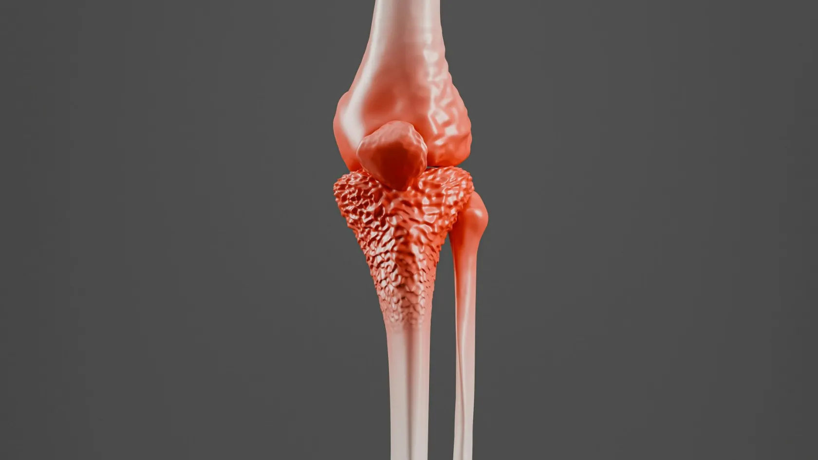
Orthopedic Oncology Surgery
Where the first biopsy is taken can decide whether a limb is saved, which is why a specialist should see the imaging before anyone reaches for a blade.
About This Department
A tumor found in a bone is not the same thing as bone cancer, and the two get confused constantly. Many of the growths an orthopedic oncology unit removes are benign. Many others are deposits from a cancer that started somewhere else entirely, in the breast, the lung, the kidney or the prostate. Only a small share are primary bone sarcomas. This article explains what each of those situations involves surgically, how the decision between saving a limb and removing it is actually made, and what a patient traveling from another country needs to think about before booking anything.
Free consultation
Have the scans and the pathology read together
This review costs nothing and does not commit you to anything. Send the MRI of the affected area, any staging scans already done, and the biopsy or pathology report if a biopsy has been taken. An orthopedic oncology surgeon will tell you what the imaging suggests, whether the tissue diagnosis is complete enough to plan from, and whether an operation is the right next step or a later one.
Three different operations under one name
Orthopedic oncology is the branch of surgery that treats tumors of bone and soft tissue, meaning muscle, fat, nerve sheath and connective tissue. The specialty covers benign bone lesions such as giant cell tumors, bone cysts and osteoid osteomas, primary bone cancers including osteosarcoma, Ewing sarcoma and chondrosarcoma, soft tissue sarcomas of the arm, leg, trunk or pelvis, and metastatic bone disease, which is cancer that spread to bone from an organ elsewhere in the body.
Those four groups need genuinely different operations, and a page that blurs them together is no use to a reader trying to work out which one applies.
Surgeons treat a benign tumor by scraping the lesion out of the bone and filling the cavity, an operation called curettage, sometimes with cement or graft material packed into the defect. Some benign lesions need no operation at all and get watched with repeat imaging instead. A primary sarcoma differs in kind. It requires the tumor to be removed as one intact block with a cuff of healthy tissue around it, so that no cancer cells are left at the cut edge, and that resection usually leaves a hole in the skeleton that has to be reconstructed in the same operation. Metastatic bone disease sits somewhere else again, because the aim there is rarely to cure. A bone weakened by a deposit is stabilized with a rod, a plate or a joint replacement so it does not break, or fixed after it has already broken, and the point of the surgery is to keep the patient walking and out of pain while the systemic treatment does the oncological work.
Naming which of these four you are dealing with is the first thing a specialist opinion should give you. It changes the size of the operation, the length of the stay, the reconstruction, the rehabilitation and the entire timeline, and no useful planning happens before it is settled.
The biopsy decides more than people expect
Most patients think of the biopsy as a test. Surgically it is the first step of the operation, and a badly planned one narrows every choice that comes afterward.
Mechanics explain it. A biopsy needle or incision contaminates the tissue it passes through with tumor cells, so that track has to be removed along with the tumor at the definitive operation. Placed correctly, along the line of the planned surgical approach, the track comes out with the specimen and costs the patient nothing. Placed across the limb, through a different muscle compartment, or through the wrong side of a joint, it forces the surgeon to remove far more tissue than the tumor itself required, and in some cases it turns a limb that could have been saved into a limb that cannot be.
If a lump has not been biopsied yet, stop and ask one question
Ask whether the person about to take the biopsy is the person who would perform the cancer operation, or is working directly with them. A deep lump larger than about five centimeters, or any lump that is growing, deserves an MRI and a specialist opinion before a needle or a blade goes anywhere near it.
The published data on this is uncomfortable. According to PubMed, a multicentre Japanese study of 97 patients referred to sarcoma centers after an unplanned excision, meaning a soft tissue sarcoma removed by a surgeon who did not know it was a sarcoma, found that 45 of them had been operated on with no prior MRI at all, 42 had received a transverse skin incision that complicates any later reconstruction, and 82 of the 97 needed a second, wider operation to clear tissue the first one had left behind (Nakamura et al, 2024). Of those re-excised patients, 59 then required soft tissue reconstruction to close the resulting defect, and the average gap between the first operation and the corrective one ran to 96 days. A separate American series of 124 similar referrals, also reported on PubMed, found that the initial unplanned removal came most often from general surgery at 44 percent and orthopedic surgery at 28 percent, and that preoperative imaging of any kind had been obtained in only 59 percent of cases (Wise et al, 2025).
None of this means those surgeons were careless. It means soft tissue sarcoma is rare enough that a competent general surgeon may see one or two in a career, and it looks exactly like the harmless lipoma they have removed hundreds of times.
The practical consequence for a reader deciding where to be treated is simple. Send the imaging for review before anyone operates, and if a biopsy has already been done elsewhere, send the pathology report and ask whether the slides themselves should be reviewed again, since sarcoma subtyping is difficult and a second reading by a specialist pathologist sometimes changes the diagnosis and therefore the whole plan.
Will I lose the limb?
For most patients with a bone or soft tissue tumor in an arm or a leg, the limb can be preserved. Limb salvage, also called limb-sparing surgery, has been the standard approach for decades now, and amputation has become the exception rather than the expected outcome.
Anatomy decides this, and courage or preference do not come into it. A tumor can be removed with the limb intact when the surgeon can achieve a clear margin all the way around it and still leave behind a limb that has blood supply, nerve supply, skin cover and enough working muscle to be useful. When a tumor has grown around the main artery and nerve of the limb, or has broken through into surrounding tissue so extensively that clearing it would leave a numb, pulseless, functionless leg, amputation is the operation that gives the better result. Saying so plainly is part of an honest consultation.
Function afterward lands closer between the two groups than most people assume. Comparative studies consistently find that limb salvage patients score higher on formal functional scales while amputees report more physical activity and less anxiety about damaging the limb, and both groups describe a mild reduction in quality of life against people who never had either operation. For a tumor low down in the leg, near the ankle or the foot, a well-fitted below-knee prosthesis can outperform a salvaged limb outright.
What the survival figures do and do not show
Published survival data comparing the two operations needs reading carefully, because the comparison is not a fair one. According to PubMed, a 2024 systematic review and meta-analysis pooling 8412 patients with high-grade osteosarcoma reported a five-year overall survival of 64 percent across the whole group, with 69 percent among patients treated by limb salvage against 47 percent among those who had an amputation (Papakonstantinou et al, 2024).
That gap is real, and it is mostly not caused by the operation.
Patients offered limb salvage are the ones whose tumors were smaller, better contained and further from critical structures in the first place, which is exactly the group that was always going to do better. The same analysis found what genuinely predicts survival, namely whether the disease had already spread at diagnosis, which raised the risk of death more than threefold, and how well the tumor responded to chemotherapy before surgery, where good responders reached 81 percent five-year survival against 56 percent for poor responders. Anyone who tells you that choosing limb salvage improves your survival is misreading the same numbers.
How the bone is rebuilt
Removing a segment of femur or tibia leaves a gap that has to be bridged in the same operation. Four approaches cover most reconstructions, and they behave very differently over a lifetime, which matters enormously for a patient in their teens or twenties who may live with the result for fifty years.
This table scrolls sideways on a narrow screen. Swipe or drag to see every column.
| Method | What is used | Trade-off to understand |
|---|---|---|
| Endoprosthesis | A modular metal implant replacing the removed bone and the neighboring joint | Stable immediately and allows early weight-bearing, but wears and loosens over the years and will need revision |
| Bone allograft | Size-matched donor bone from a tissue bank, fixed to the patient's remaining bone | Muscle and ligament reattach to real bone, but it takes months to unite and carries a higher infection risk during chemotherapy |
| Allograft-prosthetic composite | Donor bone combined with a prosthetic joint surface | Aims to combine the soft tissue attachment of allograft with the reliability of an implant, at the cost of a longer, more complex operation |
| Expandable prosthesis | A lengthening implant for a child whose skeleton is still growing | Modern designs lengthen without opening the limb again, though the mechanism itself adds failure modes over time |
Rotationplasty deserves a separate mention, because it is the option most often dismissed by families on first hearing and most often praised by the patients who have had it. In a rotationplasty the knee and the tumor are removed, the lower leg is rotated through 180 degrees and reattached to the thigh bone, and the ankle then works as a knee joint inside a below-knee prosthesis. It looks unusual. Mechanically it excels, lasting decades without revision surgery, and it suits young children whose growth would defeat most implants.
Implant survival gets the least attention before surgery and the most afterward. Implant failure is a recognized and common complication of endoprosthetic reconstruction, according to PubMed research developing prediction models for exactly this problem, which found the length of bone resected to be the strongest predictor of early implant survival, with patient age becoming the dominant factor at two and three years (Goker et al, 2025). Find out before surgery what happens when the implant reaches the end of its life, because for a young patient that conversation is a certainty.
Surgery is one part of a longer plan
For a high-grade sarcoma, the operation sits in the middle of the treatment rather than at the end of it.
Chemotherapy usually comes first, over a period of months, to shrink the tumor and to treat cells that may already have traveled elsewhere. The surgery follows. More chemotherapy then follows the surgery, guided partly by how much of the tumor was found dead when the pathologist examined the removed specimen, which is the response measurement that the survival figures earlier in this article turn on. Radiotherapy joins the sequence for many soft tissue sarcomas, before or after the resection depending on the case.
For an international patient this matters more than almost anything else on this page, and hospital websites tend to leave it out. A sarcoma pathway is not a procedure you fly in for. Think of it as a course of treatment measured in months, coordinated by one team, in which the surgeon, the medical oncologist, the radiation oncologist, the radiologist and the pathologist all discuss the same case together in a formal meeting.
Benign lesions and metastatic bone stabilization are different. Those genuinely are discrete operations with a defined start and finish, and they fit a planned trip abroad far more comfortably than a newly diagnosed sarcoma does. For the sarcomas, one structural question matters more than any other. Whether a musculoskeletal tumor board reviews the case before an operation is offered, and which specialties sit at it. Radiology, pathology, medical oncology, radiation oncology and surgery in one room is the arrangement that produces a plan, and a single surgeon deciding alone is not. That board meets here, and a patient traveling from abroad can have their imaging and biopsy presented to it before they get on a plane.
Complications, and when they show up
Tumor resection and reconstruction take hours in theater, frequently on patients whose immune systems chemotherapy has already suppressed, and the complication rates reflect that honestly. Reported figures for megaprosthetic reconstruction around the knee reach as high as 55 percent when every complication over the full follow-up period is counted, and success rates for osteoarticular allograft reconstruction of the distal femur have been reported across a range as wide as 27 to 73 percent depending on the series and the length of follow-up.
Those numbers frighten people, and context changes what they mean. They count everything from a wound that took longer to heal through to an implant that needed replacing after fifteen years, and they cover the entire life of the reconstruction instead of the weeks immediately after surgery.
Early, in the first weeks
Wound healing problems and infection dominate this period, and both are more likely when chemotherapy has recently suppressed the bone marrow or is due to restart soon. Deep infection around a large implant is the complication surgeons fear most, because clearing it can mean removing the reconstruction, treating the infection for weeks and rebuilding afterward. Bleeding, blood clots in the leg and nerve injury from the retraction needed to reach the tumor also belong to this window.
Later, over years
Aseptic loosening, where the implant gradually separates from the bone without any infection, and mechanical wear of the moving parts are the ordinary consequences of a metal joint being used for a long time. Periprosthetic fracture, meaning a break in the bone next to the implant, and failure of an allograft to unite with the host bone belong here too. So does local recurrence of the tumor, and surveillance imaging continues for years because of it, as the section further down this page explains.
Young patients having a knee endoprosthesis today should expect further surgery at some point in life. Plan for it from the start.
Recovery and rehabilitation
Hospital stay after a resection with reconstruction commonly runs one to two weeks, varying with the size of the operation, the reconstruction used and the patient's general condition going into it.
What happens next depends heavily on which reconstruction was chosen, and you want to understand it before the operation. An endoprosthesis is mechanically stable the day it goes in, so partial weight-bearing with crutches or a frame starts within days. A biological reconstruction using donor bone has to unite with the patient's own bone before it can carry load, which delays full weight-bearing by weeks or months and means a longer period of restricted walking.
Rehabilitation takes the longest. Getting back to ordinary daily activity takes several months, and full functional recovery runs six to twelve months or longer, particularly where chemotherapy continues after the operation and repeatedly knocks back the patient's energy for training. Published guidance on limb salvage after major bone resection puts the average time to relearn walking at around a year. Physiotherapy does the real work throughout that period, and a structured, supervised program produces measurably better function than a sheet of exercises handed over at discharge, which is the practical argument for arranging your rehabilitation at home before you leave hospital instead of scrambling for it once you land.
Which cases suit treatment abroad
Some of them do, comfortably. Others do not, and a hospital that tells you so serves you better than one that says yes to everything.
Four situations share the same convenient shape. A benign bone tumor needing curettage. A solitary bone metastasis needing stabilization. A low-grade lesion that needs a clean resection and nothing else. A planned revision of a reconstruction done years ago. Each of them has a defined operation, a predictable hospital stay of one to two weeks, a recovery that can continue at home, and no dependence on months of coordinated systemic treatment delivered in the same building. Those cases travel well.
A newly diagnosed high-grade sarcoma poses a harder question, and the answer splits in two. Traveling for the whole pathway, meaning diagnosis, chemotherapy, surgery and the chemotherapy that follows it, under one team, works and is done regularly. Traveling for the surgery alone while chemotherapy is delivered by a different team in a different country is where patients get hurt, because the timing between the last chemotherapy cycle and the operation is a clinical decision that needs one group of people holding the whole picture, and because the response assessment on the removed specimen has to reach whoever is prescribing the treatment afterward.
So settle first which of those two you are actually asking for. That question matters more than any question about the hospital.
On flying, the constraint after major limb surgery is less about the wound than about the blood clot risk that comes with a long operation, reduced mobility and a cancer diagnosis all at once, since cancer itself raises clotting risk before any surgery is added to it. Clearance depends on wound healing, on mobility, and on whether the patient is on anticoagulation, so it is settled at a review before departure. No honest team promises you a fixed number of days in advance. Ask for a fitness-to-fly letter before you leave the hospital, since airlines sometimes request one after major surgery. One point matters more than the flight rules for anyone with a sarcoma. Radiotherapy and chemotherapy are both delivered on this site, alongside the surgery, so the phases of a sarcoma pathway do not have to be split between institutions or between countries. The international patients team works in English, Arabic, French, Russian, Serbian, Romanian and Spanish, with other languages arranged on request, and a companion bed in the room means a parent stays overnight with a child rather than leaving at visiting time.
What moves the cost
Quotes for orthopedic oncology surgery vary more widely than quotes for almost any other orthopedic operation, and the spread is not padding. It reflects how differently these cases are built.
The reconstruction drives the largest share of it. A custom or modular endoprosthesis bridging a long femoral resection sits in a different class of expense from a curettage filled with cement, and an expandable pediatric implant with a lengthening mechanism sits higher again. How much bone comes out drives implant length, which drives cost directly. Whether the tumor sits in a limb or in the pelvis changes the operation from a long one into a very long one, and it frequently needs a second surgical team. Soft tissue coverage forks the cost again, since a defect that cannot be closed directly needs a plastic surgical flap and the theater time that goes with it. Then comes everything around the operation, meaning the chemotherapy cycles, the radiotherapy, the staging scans, the pathology work on the resected specimen, intensive care where the case warrants it, and any inpatient rehabilitation, all of which belong to the treatment and all of which appear differently on different quotes.
Patient factors move it too. Age, nutritional state, kidney and heart function, previous surgery at the same site, and whether chemotherapy has left the patient's counts low enough to delay the operation all change how the case runs.
Packages published in this market for musculoskeletal tumor surgery generally cover the surgeon and anesthesiologist fees, theater and hospital stay, pre-operative imaging and blood work, the histopathology, transfers and interpreting. Flights, accommodation beyond the stated nights, systemic treatment given outside the package period, treatment of a complication and any second operation sit outside them. Compare two quotes on those lists first, because a lower number that excludes the implant is not a lower number.
Five things need pinning down before you accept any figure. Which reconstruction does it assume, and does it name the implant. What happens financially if the plan changes once the surgeon is operating and finds something the scans did not show. Whether the staging scans and the histopathology sit inside the figure or arrive as separate invoices afterward. How many nights it counts, and what the nightly rate becomes past that point. And whether any chemotherapy or radiotherapy is included at all, since a surgical figure quoted beside a sarcoma pathway can look far lower than the treatment it belongs to. You will only get answers that mean anything from a surgeon who has read your own imaging, and that review costs nothing.
Follow-up once you are home
Sarcoma follow-up runs for years, and it has two separate purposes that are easy to confuse.
One is oncological surveillance, looking for local recurrence at the operated site and for spread to the lungs, which is where bone and soft tissue sarcomas most commonly go. That means scheduled imaging of the operated limb and of the chest at intervals that start frequent and stretch out over time. The other is mechanical, watching the reconstruction itself for loosening, wear and fracture, which is a different set of x-rays read by a different eye.
Both have to happen somewhere near where you live. Nobody flies back every three months for a chest scan.
Practically, that means leaving the hospital with the operative note, the full histopathology report including margin status and the chemotherapy response assessment, a copy of the imaging, and the exact make and reference of any implant, since a surgeon revising it years later needs that information and cannot easily obtain it afterward. A written surveillance schedule matters just as much, so that a doctor at home knows what to arrange, and when, without having to work it out from scratch. Keep a line of contact to the operating team as well, because when a scan at home shows something ambiguous, the surgeon who performed the resection can say whether it looks like the tissue they deliberately left behind or like something new, and that judgment comes hard to anyone who was not in the operating room. That line stays open here after discharge. The coordinator who handled your case keeps the same WhatsApp number once you are home, and imaging done abroad is reviewed at no charge, which is the arrangement a surveillance schedule needs behind it to be worth anything.
Symptoms that need attention this week
Most of this article describes decisions taken over weeks, with time to gather opinions and compare them properly. A few situations compress that timeline.
Deep bone pain that wakes you at night and does not settle with ordinary painkillers deserves imaging, particularly in someone with a previous cancer diagnosis, and reassurance alone is the wrong response to it. A lump that is enlarging, sits deeper than the layer of fat under the skin, or measures more than about five centimeters should be imaged before it is removed. Pain in a bone known to contain a metastatic deposit, especially in the thigh or the hip, can mean the bone is close to breaking, and stabilizing it before it fractures is a much better operation than fixing it afterward.
Two situations need assessment the same day, in the nearest hospital that can provide it. A bone that has already fractured through a tumor. New weakness or numbness suggesting a deposit is pressing on the spinal cord. Nobody plans a trip around those.
Frequently asked questions
Can I come for the surgery only and have chemotherapy at home?
How long will I be in hospital, and how long in the country?
I have already had the lump removed elsewhere. Does that change anything?
Will my child need more operations as they grow?
What should I send for a remote opinion?
References
- Papakonstantinou E, Athanasiadou KI, Markozannes G, et al. Prognostic factors in high-grade pediatric osteosarcoma among children and young adults. Greek Nationwide Registry data along with a systematic review and meta-analysis. Cancer Epidemiol. 2024;90:102551.
- Nakamura T, Ogura K, Hayakawa K, et al. Real-world Referral Pattern of Unplanned Excision in Patients With Soft-tissue Sarcoma. A Multicenter Study by the Bone and Soft-tissue Tumor Study Group of the Japan Clinical Oncology Group. In Vivo. 2024;38(6):2712-2717.
- Wise CE, Le C, Pham NS, et al. Prevention in non-oncologic intent sarcoma surgery. Surg Oncol. 2025;62:102273.
- Goker B, Brook A, Zhang R, et al. Development of Machine Learning Models to Predict Tumor Endoprosthesis Survival. J Surg Oncol. 2025;132(1):226-234.
- Jamshidi K, Ammar W, Kargar Shooroki K, Mirzaei A. Outcomes of Megaprosthesis Reconstruction for the Salvage of Failed Osteoarticular Allograft Around the Knee Implanted before Skeletal Maturity in Primary Bone Sarcoma. A Case-Series. Arch Bone Jt Surg. 2024;12(3):211-218.
Editor's note
Written by the Biruni Hospital medical editorial team. Reviewed by Assoc. Prof. Dr. Suat BATAR, Orthopedics and Traumatology.
Medically reviewed by
Assoc. Prof. Dr. Suat BATAR
Orthopedics and Traumatology
Related Treatments
View All-
Achilles Tendon Repair
Achilles tendon repair stitches a torn tendon back together and cuts the risk of a second rupture, while a boot with early movement gives the same function at one year in trials. This guide covers the signs of a tear, surgery or a boot, open and minimally invasive repair, rehabilitation week by week and the stay.
-
Bone Tumor Resection
Most people arrive expecting to lose the limb. For the large majority of bone tumours today the limb is saved, the bone is rebuilt with an implant or a graft, and survival is no worse for it. What decides the answer is the margin, and the margin is planned from an MRI of the whole bone before anything is cut.
-

Chondrosarcoma Surgery and Bone Tumor Resection
Chemotherapy and radiation barely touch this tumor, which sounds like the worst news on the page and is the reason a single operation cures it outright. For a grade 1 chondrosarcoma in an arm or a leg the bone is scraped clean from the inside and the natural joint stays where it is.
-
Complex Knee Revision Surgery
Infection now causes more failed knee replacements than any other single reason, 21.6 percent across registries in seven countries, and it can turn a revision into two operations with weeks of antibiotics between them. This page explains how the cause is found before surgery, when cones or a hinge are needed, and how a two-stage revision fits around travel.
-
Complex Trauma Reconstruction
A fracture that has not healed after a year, healed crooked or became infected can still be repaired, and the treatment often runs for months in an external frame. This page explains nonunion, malunion and bone loss repair, the choice between bone transport and the Masquelet technique, and how a long treatment fits around travel to Istanbul.