
Neurosurgical ICU (Neurological Intensive Care Unit)
Brain swelling peaks days after the injury, seldom on the day of it. That is what this unit is built to catch, and the hourly examination catches it earlier than any monitor in the room.
About This Department
The monitor bolted into the skull did not beat the bedside examination. The nurse waking you every hour is the monitor.
In the only large randomized comparison, managing severe head injury by an intracranial pressure monitor gave no better survival and no better recovery than managing it by the clinical examination and repeated scans. Six month mortality came out at 39 percent against 41 percent. Everything a neurosurgical intensive care unit does runs off that examination.
What makes this unit different from any other
A neurosurgical intensive care unit treats one organ that behaves unlike every other organ in the body. The brain sits inside a rigid box of fixed volume, so anything taking up extra room inside the skull, whether that is blood from a bleed, swollen tissue around an injury, or fluid that has stopped draining, has to push something else out of the way, and what gets pushed out is blood flow to tissue that dies within minutes of losing it. The whole unit exists to catch that happening before it finishes.
Why the staffing looks the way it does
General intensive care units are built around the heart and the lungs, and their alarms fire on blood pressure, oxygen and rhythm. Here the decisive change stays silent on every machine in the room. A pupil that was equal an hour ago and is now larger on one side, an arm that pushed your hand away this morning and drifts today, a patient who opened his eyes to his name at noon and needs a shout at four. None of that trips an alarm. Not one. A nurse trained to look for them finds them, and the difference between finding it at twenty minutes and finding it at two hours decides how much brain is still there afterwards, which is a sentence worth reading twice before comparing hospitals on anything else. That is the whole argument for a separate unit with separately trained staff, and it also makes the nursing ratio the first number to ask for, ahead of anything on the equipment list.
The examination that outranks the machines
Intracranial pressure monitoring has been standard practice in severe head injury for decades, and in 2012 somebody finally tested it properly.
Chesnut and colleagues, writing in the New England Journal of Medicine, randomized 324 patients with severe traumatic brain injury to management guided by an intraparenchymal pressure monitor or to management guided by the clinical examination and repeated imaging. The composite outcome, built from 21 measures of function and cognition at three and six months, came out at 56 against 53 with no significant difference. Six month mortality was 39 percent in the monitored group and 41 percent in the examination group, and intensive care stay ran 12 days against 9. One clear difference did emerge, and it ran opposite to what most people expect, because the group managed without a monitor received more brain-directed treatment than the monitored group did, at 4.8 treatment days against 3.4. Read that last figure carefully, because it explains what the monitor is actually for. Rather than finding trouble the examination misses, it tells a team when to stop treating, which spares a patient hyperosmolar fluids and spells of hyperventilation nobody needed to give.
Why monitors still go in
The reasons are good ones.
A patient deep under sedation cannot be examined at all, a patient in a scanner is out of reach for half an hour, and a pressure trend running upward overnight shows itself before a pupil does. What the trial settled is the hierarchy. The examination leads and the numbers support it, and a unit that treats the screen instead of the patient has the order backwards.
What the numbers on the screen mean
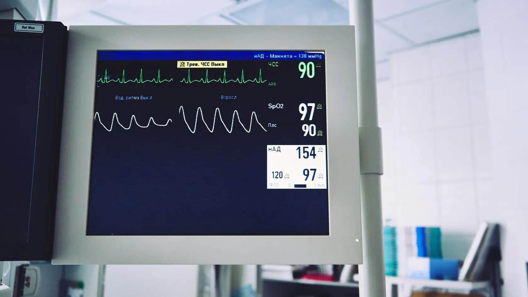
Families spend hours watching these figures without being told which ones matter, and the screen makes no distinction between a number the team is acting on and a number it is simply recording, which is how a relative ends up frightened by a heart rate while the figure that actually changed the plan scrolled past unremarked an hour earlier. Below is the working set, in the order a neurosurgical team reads it, with what moves each one. Learn these six and the room stops being noise.
Narrow screens scroll this table sideways. Swipe or drag to reach every column.
| Measurement | What it tells the team | What makes it move |
|---|---|---|
| Glasgow Coma Scale | Eye opening, speech and movement scored out of 15, repeated by every shift so a drop of even one point is visible | Rising pressure, a new bleed, a seizure, or simply the sedation still wearing off |
| Pupil size and reaction | The earliest sign that pressure is pushing the brain against the nerves running underneath it | A clot enlarging, swelling peaking, or a drain that has stopped working |
| Intracranial pressure | Pressure inside the skull, read from a probe in the brain or a drain in the fluid spaces, with treatment thresholds set by international guidelines around 20 to 22 mmHg | Position of the head, coughing, fever, seizures, low blood sodium, and the injury itself |
| Cerebral perfusion pressure | What is left of the blood pressure once the pressure inside the skull has been subtracted, which is the number that decides whether tissue is being fed | Blood pressure drugs, fluid balance, and every change in intracranial pressure |
| Blood sodium | Low sodium pulls water into brain tissue and makes swelling worse, so it gets checked several times a day instead of once | Salt losing states that follow brain injury, fluids given, and hyperosmolar treatment |
| Blood glucose | Both extremes damage injured brain, and the safe corridor is narrower here than on a general ward | Stress hormones after injury, steroids, feeding, and insulin |
| Continuous EEG where it is used | Finds seizures that produce no movement at all, which are invisible on every other monitor in the room | The injury, fever, low sodium, and certain drugs |
Single readings mean very little. Direction over hours is what the team acts on, so a ward round here spends longer looking at the trend line than at the number currently on the screen, and a relative who asks what the pressure is right now will get an answer covering the last six hours instead.
Who ends up in a neurosurgical intensive care unit
Five broad groups arrive here, and the reason for admission differs sharply between them, so a single answer to how long the stay lasts does not exist. The first two account for most of the beds.
Those two groups look identical from the corridor while being entirely different situations, so ask your surgeon which one applies to your case, because the answer changes what the first forty eight hours mean and it changes what a quiet night is evidence of. Put the question before the operation.
Two further groups turn up regularly. Patients whose fluid spaces have blocked and who need a drain, and patients admitted after a seizure that will not stop. A fifth group is admitted purely for the monitoring that no ward can provide, usually after an aneurysm bleed, where the danger arrives on a schedule that the first days give no hint of.
Sedation, and the problem it creates
Every drug that keeps a ventilated patient comfortable also switches off the examination the unit depends on, and that tension runs through every single day of a neurosurgical intensive care stay without any clean solution to it, only a compromise that gets managed shift by shift. Nobody pretends otherwise.
Sedation holds, and what they are for
Sedation is kept as light as the situation tolerates, and it is lifted deliberately.
Sedation holds work like this. The drugs get stopped or reduced at a planned moment, in the morning with the full team present, so the patient can be examined properly. The team wants to see eyes opening, a hand squeezing on request, both sides of the body moving equally, and the ventilator being fought rather than accepted. Then the sedation goes back on before pressure or agitation becomes a problem. Where the pressure inside the skull is unstable, the hold gets postponed, because rousing a patient raises that pressure for several minutes and the price is not worth paying that morning.
Families watch this moment and read it as the patient waking up, when it is a test of the examination and nothing more, so ask the team directly which of the two they believe they are looking at. They will say.
When the pressure will not come down
Treatment for raised pressure runs as a staircase. Each step carries more risk than the step below it, so nobody skips ahead, and most patients never leave the first two. The upper steps are where the decisions get hard, and where a family needs to understand what is being traded before anyone asks them to agree to it.
- Simple measures first. Head of the bed raised, the neck straight so the veins draining the head are not kinked, fever treated aggressively because every degree raises the brain demand for blood, and pain and coughing controlled.
- Deeper sedation, and drainage of cerebrospinal fluid through a drain if one is already in place. Removing a few milliliters of fluid from a rigid box drops the pressure immediately.
- Hyperosmolar treatment, meaning concentrated salt solution or mannitol given through a central line, which pulls water out of brain tissue for a period of hours and buys time for something definitive.
- Brief controlled hyperventilation, which shrinks the blood vessels and the volume of blood inside the skull within minutes. It is a rescue measure for a deteriorating patient on the way to a scanner, because holding it for long starves the tissue it is meant to protect.
- Barbiturate coma, which shuts brain activity down to its lowest metabolic setting. The price is steep, because it drops blood pressure, suppresses immunity and removes the examination completely for days on end.
- Removing part of the skull. Swelling tissue then has somewhere to go.
That last step needs its own paragraph. The evidence behind it is the most sobering in this specialty.
Hutchinson and colleagues randomized 408 patients with head injury and pressure above 25 mmHg that had resisted everything else, reporting in the New England Journal of Medicine in 2016. At twelve months, death had occurred in 30.4 percent of the surgical group against 52.0 percent of the medical group, so the operation saved roughly one life in five. The survivors did not arrive evenly across the outcome scale. Vegetative state ran at 6.2 percent against 1.7 percent, and severe disability with dependence on others for care ran at 18.0 percent against 14.0 percent. Good recovery came out at 9.8 percent against 8.4 percent, which is the same within chance.
Removing part of the skull converts deaths into survivors, and most of those survivors live with severe disability. Surgeons put that in front of families before the operation rather than after it.
Two treatments that sounded right and were not
Neurocritical care has a particular history of ideas that made perfect physiological sense and then failed when somebody tested them. Both examples below changed practice by being negative.
Cooling the brain
Lowering body temperature reduces the brain demand for oxygen and reliably brings intracranial pressure down, so cooling looked like an obvious treatment for years. Andrews and colleagues enrolled 387 patients across 47 centers in 18 countries, randomizing those whose pressure stayed above 20 mmHg despite basic measures to cooling plus standard care or to standard care alone. Recruitment was suspended on safety grounds before the planned number was reached. Cooling did what it was supposed to do, reducing the need for the harsher later steps from 54 percent to 44 percent, and the patients did worse anyway. A favorable outcome at six months occurred in 26 percent of the cooled group against 37 percent of the others.
Why that result mattered beyond cooling
Cooling hit its physiological target and missed the only target that counts. Controlling a number and helping a patient turned out to be two separate achievements, and the trial is now the standard example used to explain why those two get confused.
Driving the blood sugar down
Tight glucose control was adopted widely in intensive care before the neurological evidence existed. A 2018 meta-analysis in Critical Care pooled ten randomized trials covering 1,066 head injury patients and found no mortality difference between tight and conventional targets, a borderline advantage for tight control on neurological outcome, and a fivefold higher rate of severe hypoglycemia with it. An injured brain tolerates a low sugar worse than almost any other tissue, so most units now run a moderate target and check the level several times a day. Chasing a number into a range the brain cannot survive is the same mistake the cooling trial made, in a different unit of measurement.
Neither result should make you distrust intensive care. Both are reasons to ask what a unit does and why it does it, because a unit still cooling head injuries to bring a pressure number down in 2026 is working from a 2009 understanding of the evidence and has not read the trial that closed the question.
The cheap tablet that genuinely works
After an aneurysm bleeds into the space around the brain, the arteries there can narrow days later and starve territory that survived the original bleed. This delayed injury, rather than the bleed itself, is what takes a good proportion of patients who reach hospital alive.
An oral calcium channel blocker reduces it. A Cochrane review pooled 16 trials covering 3,361 patients and found that oral nimodipine cut the risk of a poor outcome by roughly a third, with a relative risk of 0.67 and a confidence interval running from 0.55 to 0.81. Given by vein it did not show the same benefit, and other drugs in the same class did not either, which leaves an old, inexpensive and thoroughly unglamorous tablet standing as one of the very few treatments in this field with a clear positive trial behind it.
How it is given
Three weeks of it, by mouth or by tube, starting as soon as the diagnosis is made.
The question to ask on day one
Ask whether it has started. No question a family asks on the first day after an aneurysm bleed is more useful.
What families see, and what it actually means
Almost everything alarming in this room has an explanation. Most of them sound far less frightening once somebody says them out loud, and the list below covers what relatives ask first, in roughly the order they notice things.
Columns run past the edge on a small screen. Slide the table across to read them.
| What you see | What it usually means |
|---|---|
| The eyes are open but nobody is there | Common in the first days and rarely permanent. Eye opening returns before awareness does, because they are driven by different parts of the brain |
| An arm or leg stiffens into an odd posture | A reflex movement that appears when the injured brain loses control over the spinal cord below it. The team grades it carefully because the pattern carries real information |
| A nurse shines a light in the eyes every hour, all night | The single most informative test in the unit. Sleep is being sacrificed deliberately, and the team knows it |
| A tube through the mouth becomes a tube in the neck | A tracheostomy, done when the airway will be needed for more than a week or two. It lets sedation come down, which usually makes the patient more awake rather than sleepier |
| A bandaged hollow where part of the skull was | The bone was removed to give swelling somewhere to go, and it is stored and replaced at a second operation once the swelling has settled |
| A bag of fluid draining from the head | An external drain removing cerebrospinal fluid and, on the same line, measuring the pressure. It is usually temporary |
| The patient is moved onto a scanner again | Routine, and rarely ominous. Repeat imaging is how a change in the examination gets explained, and most repeat scans show nothing new |
Get the nurse to explain anything you have been staring at. Nobody here minds.
Why nobody will give you a number in the first week
Families want a percentage. An early percentage would be worse than no answer at all.
Two reasons, and they compound each other. The first is biological. Swelling has not finished, sedation is still in the system, and the brain has a slow recovery curve that does not begin to show itself for weeks, so an examination on day three describes day three and very little else. The second is a trap the specialty has documented at length. A pessimistic prediction made early tends to change the treatment that follows it, treatment that has been reduced produces a worse outcome, and the worse outcome then appears to confirm the prediction that caused it.
How good units guard against it
Deliberate safeguards exist, and good units write them down.
They set a period during which full treatment continues whatever anyone suspects, they involve more than one senior clinician in any conversation on limiting treatment, and they keep the examination going across enough days for a trend to appear.
What a prognosis conversation should contain
When that conversation does happen, it should reference the imaging, the examination and its trajectory, and any electrical testing that has been done, and it should come with an explicit statement of how certain the team is. Any team that answers a percentage question on day two is guessing.
Leaving the unit, and the road after it
Moving to a neurosurgical ward happens once the examination has held steady for a day or more, pressure control no longer needs drugs, the airway is safe and any drain has come out. Alarms go quiet. One to one nursing stops, and most families find the first ward day harder than they expected it to be, because the constant watching they had learned to read as danger had also become the thing reassuring them, and its removal registers as abandonment before it registers as progress.
Recovery here runs on a different clock. Useful improvement continues for months, and in younger patients for a year or more, which is the encouraging part and the exhausting part at the same time, because a family that expects the curve to finish in six weeks will read a normal recovery as a failure and give up on therapy that was still working. Early gains come quickly and then the curve flattens into slow work, and the things that come back last are attention, memory, word finding and the ability to hold a plan in mind, none of which show up on a scan and all of which decide whether somebody returns to their old life. Physiotherapy, occupational therapy and speech and language therapy carry that stage, and starting them while the patient is still an inpatient matters more than which building they happen in.
Putting the skull back
Where part of the skull was removed, it goes back.
Surgeons store the bone and replace it at a second operation called a cranioplasty, once the swelling has fully settled, which takes weeks to months. Until then a protective helmet is worn for anything active, and your team gives you the timing in writing so the second operation can be planned around travel instead of the other way round.
Planning the trip, the stay and the flight home
Neurosurgical cases divide into two travel situations, and they need different planning.
A planned operation with a booked bed is straightforward to arrange. Send the imaging first, get the written opinion, then travel with the dates fixed. An emergency admission during a trip is a different problem, and the thing to understand in advance is that flying is the constrained part. Air travel after a brain injury or intracranial surgery carries specific issues that have nothing to do with how well the patient looks. Air trapped inside the skull after an operation expands as the cabin pressure drops, the lower oxygen at cabin altitude is tolerated poorly by injured brain, seizure risk is highest in the early weeks, and a patient who still needs oxygen or suction needs an airline that will carry them, and an escort as well. Your surgeon sets the date, it is a separate clearance from discharge, and for intracranial surgery it usually runs several weeks rather than several days.
Book a changeable return ticket. Buy insurance covering an extended stay and a medical repatriation, and read that policy properly before you buy on price, because repatriation for a neurosurgical patient means a stretcher, an escort and sometimes an air ambulance, which is the expensive kind and the kind cheap policies quietly exclude.
Send what you already have before you commit to anything. For a brain or spine problem that means the CT and MRI images themselves and not only the report, any angiogram, the operation note if surgery has already happened, the current medicine list with doses, and a plain description of what the patient can do today compared with a week ago. A neurosurgeon and an intensive care physician read them together and tell you what the plan would look like, whether a reserved bed forms part of it, and where they would advise against moving the patient at all. That review is free. It commits you to nothing. For the paperwork, our international patients office puts your appointment confirmation together with an invitation letter carrying the hospital name and the name of the treating doctor, which is the document most consulates ask for with a medical visa application, and it goes out ten days or so ahead of travel.
After you get back home
Follow up for a neurosurgical patient happens mostly where you live, and it gets arranged before you leave, never improvised afterwards. You go home with the discharge summary, the operation note, the pathology result where there is one, and the imaging on disc, which together let a neurologist or neurosurgeon at home take over without requesting records from another country. Your coordinator stays reachable on the same WhatsApp number, so a question about a wound, a drug dose or a scan result reaches somebody who already knows the case. Remote check-ins follow intervals that match the operation, and you send the local scan reports in as they are done so the surgeon here looks at the same pictures your own doctor has. Two things send you straight to a local emergency department first, and they are a seizure, and any new or worsening weakness, confusion or severe headache, none of which should wait for a message to reach another country before somebody puts a scanner over the patient. Tell us afterwards. Tell us early.
Conversations here are harder than on almost any other page of this site, so language matters more. Seven languages are covered directly by our international patients team, namely English, Arabic, French, Russian, Serbian, Romanian and Spanish, with interpreting in anything else arranged on request. One coordinator takes your case from the first message, sits in on the conversations that matter, follows it through discharge and keeps the same WhatsApp number live once you have flown home. Visiting inside the unit follows the unit rules, which your coordinator sends you in writing before you arrive, and once the patient moves to a normal room one person stays overnight because the rooms have a companion bed. Halal, vegetarian and diabetic diets come from the hospital kitchen, there is a prayer room on site, and a request for a female physician goes to the department and is accommodated wherever the rota allows.
What moves the cost of a neurosurgical intensive care stay
No published figure will apply to your case, because the cost of this stay follows what the stay turns into. Learn which items move it instead.
Drag the table sideways where a phone cuts off the last column.
| What moves the total | Which direction, and why |
|---|---|
| Nights in the unit | The dominant item, and the one every other item eventually works through, because almost everything that goes wrong here adds nights |
| Monitoring hardware | A pressure probe, a drain or continuous electrical monitoring each add a device, an insertion procedure and the staff time to run them |
| Time on the ventilator, and any tracheostomy | Days of ventilation bring their own nursing and drugs, and a tracheostomy adds a procedure while often shortening the unit stay |
| A second operation | Returning to theater for a clot, a drain revision or a decompression restarts the sequence. Cranioplasty later is a separate admission and is quoted separately |
| Complications that carry their own treatment | Dialysis, resistant infection and its isolation, or repeated blood products each add a cost line of their own |
| Rehabilitation before discharge | Inpatient physiotherapy, occupational therapy and speech therapy are counted by session, and a neurological patient needs many more of them than a general surgical patient does |
Health the patient arrives with moves the figure before anything happens. Age, diabetes, heart and lung disease, blood thinning medication and a previous operation at the same site all raise the chance of an extra night and an extra intervention.
Packages published by hospitals and medical travel agencies in this market typically cover the surgeon and anesthesia fees, the planned ward nights, pre-operative tests, any implant named in the quote, an interpreter, transfers and a set number of hotel nights. Flights, travel insurance, extra hospital nights and the treatment of a complication sit outside almost every one of them, and so does intensive care beyond the nights the quote assumed. On a neurosurgical case that last exclusion is the one that matters.
Five questions to put in writing before you commit
How many intensive care nights does this figure assume, and what happens financially if the count goes up. Does it include monitoring hardware and any tracheostomy. Would a return to theater be covered, or billed separately afterwards as a fresh admission with its own nights attached. Is the cranioplasty quoted at all, and if it is, when does that quote expire and what happens to the price if the second operation has to wait six months for the swelling to settle.
Any number that means something comes from a clinician who has read the imaging. That reading costs you nothing.
Neurosurgical ICU FAQ
How long does a stay in a neurosurgical intensive care unit last?
After a planned brain or spine operation, one night is the commonest answer and the patient moves to a ward the following morning once the examination has been stable overnight. After a severe head injury, a large bleed or an aneurysm rupture, the stay runs from several days to several weeks, because the danger period for swelling and for delayed narrowing of the arteries extends well beyond the first forty eight hours and the unit keeps the patient until that window has closed.
Can the patient hear us?
Assume yes and speak accordingly. Hearing survives when nothing else responds, sedation dulls recall without necessarily removing awareness, and patients who recover from long stays frequently describe remembering voices. Familiar voices saying ordinary things, where the patient is, what day it is, who is in the room, are useful as well as kind.
Why does the nurse keep waking the patient through the night?
The neurological examination is the main monitor in this unit, and it only works when it is repeated often enough to catch a change while the change is still reversible. Twenty minutes and three hours are different worlds here. Sleep is being traded for that margin on purpose, and the intervals lengthen as the patient stabilizes.
Will anyone there speak our language?
Seven languages are covered directly, namely English, Arabic, French, Russian, Serbian, Romanian and Spanish, with interpreting in anything else arranged on request. One coordinator takes the case from your first message and follows it through discharge, which counts for most in the conversations about what the imaging shows and which options are genuinely open.
Can one of us stay with the patient overnight?
Inside the intensive care unit that follows the unit rules, and your coordinator sends you those rules in writing before you arrive so nobody finds out at the door. Once the patient moves to a normal room, one person stays overnight for the rest of the admission because the rooms have a companion bed, and accommodation for the nights either side of the admission is arranged by the international patients office.
When can a patient fly home after neurosurgery or a brain injury?
Later than most people expect, and the date belongs to the surgeon. Air trapped inside the skull after an operation expands as the aircraft climbs, cabin oxygen sits below ground level, and seizure risk concentrates into the early weeks, which together push the clearance for intracranial surgery out to several weeks rather than several days. Ask for it in writing at discharge and book a changeable ticket.
What happens if there is a problem once we are home?
Your coordinator stays reachable on the same WhatsApp number, so the first message goes to somebody who already knows the case. You leave with a written discharge summary, the operation note and the imaging on disc, which together are what a neurologist or neurosurgeon at home needs in order to act on the same day. Anything urgent goes to your nearest emergency department first and to us second.
References
- Chesnut RM, Temkin N, Carney N, Dikmen S, Rondina C, Videtta W, et al. A trial of intracranial-pressure monitoring in traumatic brain injury. New England Journal of Medicine. 2012;367(26):2471-2481.
- Hutchinson PJ, Kolias AG, Timofeev IS, Corteen EA, Czosnyka M, Timothy J, et al. Trial of decompressive craniectomy for traumatic intracranial hypertension. New England Journal of Medicine. 2016;375(12):1119-1130.
- Andrews PJD, Sinclair HL, Rodriguez A, Harris BA, Battison CG, Rhodes JKJ, et al. Hypothermia for intracranial hypertension after traumatic brain injury. New England Journal of Medicine. 2015;373(25):2403-2412.
- Dorhout Mees SM, Rinkel GJE, Feigin VL, Algra A, van den Bergh WM, Vermeulen M, et al. Calcium antagonists for aneurysmal subarachnoid haemorrhage. Cochrane Database of Systematic Reviews. 2007;2007(3):CD000277.
- Hermanides J, Plummer MP, Finnis M, Deane AM, Coles JP, Menon DK. Glycaemic control targets after traumatic brain injury. A systematic review and meta-analysis. Critical Care. 2018;22(1):11.
Editor's note
Written by the Biruni Hospital medical editorial team. Reviewed by Prof. Dr. Mehmet İlke BÜGET, Anesthesia and Reanimation.
Medically reviewed by

Prof. Dr. Mehmet İlke BÜGET
Anesthesia and Reanimation
Related Treatments
View All
Bone Marrow Transplant Unit (BMT Unit)
Between the mid 1990s and the mid 2000s, death within 200 days of an allogeneic transplant that was not caused by relapse fell by 60 percent, and almost none of that came from a new drug. This page is about the part nobody photographs, meaning the filtered room, the rules that have evidence behind them and the ones that do not, the laboratory chain behind the cells, and what a family can actually do.
Burn ICU (Burn Intensive Care Unit)
Age, burn size and smoke inhalation are the three numbers that set the outlook after a major burn. This page walks through fluids, early surgery and grafting, the long healing phase, and when a burn patient can be flown abroad.

Burn Unit (Burn Center)
Burn care runs on one figure, the percentage of the body surface burned, and a systematic review of 28 studies found that referring hospitals overestimate it in half or more of cases. Fluid volume, the transfer decision and the survival estimate are all calculated from that number, which is why this page starts with how it is measured and who should be measuring it.

Cancer Pain Management Unit
The Cancer Pain Management Unit at Biruni Hospital in Istanbul treats pain caused by cancer and by its treatment. Two in three people with advanced cancer report pain, and reviews show nearly one in three receive medication too weak for it. The unit uses stepwise medicines, radiotherapy for bone pain, nerve blocks and spinal pumps, and plans follow-up for patients who return home abroad. The pain team reviews reports and current medicines free of charge.
Cancer Patient ICU Care
Cancer once counted as a reason not to offer an intensive care bed at all. Pooled data from 7,354 critically ill cancer patients retired that rule, and the argument now is about which patient, for what, and how long full support runs before anybody formally reassesses it.