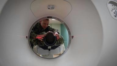
Ultrasound shows the prostate and rarely the tumor, so blind biopsies missed cancers for decades. An MRI-guided biopsy aims the needle at the lesion the scan revealed, finds significant prostate cancer in 38 percent of men against 26 percent with standard biopsy, and spares one man in four the needle entirely. In the breast, it samples lesions that only MRI can see.
About This Department
Radiology and Urology
A needle aimed at what the MRI showed finds more cancer with fewer samples. In the prostate it also leaves one man in four without a biopsy at all.
An MRI-guided biopsy takes tissue from a spot that magnetic resonance imaging has shown and that ultrasound or mammography cannot see well enough to target. Urologists use it most, matching the MRI picture to live ultrasound or working inside the scanner, and breast radiologists use it for lesions that only MRI shows. In the PRECISION trial, the MRI route found clinically significant prostate cancer in 38 percent of men against 26 percent with the standard biopsy, diagnosed fewer harmless cancers, and spared 28 percent of men any biopsy because their MRI was clear.
38% vs 26%
Significant prostate cancer found, MRI route against standard biopsy, NEJM 2018
28 in 100
Men whose clear MRI meant no biopsy, NEJM 2018
93% vs 48%
Sensitivity of MRI against standard biopsy for significant cancer, Lancet 2017
Free
Radiologist review of your MRI
Free consultation
Send the MRI report and, if you can, the images on a disc or a link, together with the PSA history and any earlier biopsy results for the prostate, or the mammogram, ultrasound and MRI reports for the breast. A radiologist and a urologist or breast specialist say whether the lesion needs sampling, which form of guidance suits it, and whether the scan should be repeated first. The opinion is free and commits you to nothing.
An MRI-guided biopsy is a diagnostic test. This page treats it as one and explains where the MRI comes in, how the needle reaches the spot, what the trials measured, and how a patient from abroad has it done in Istanbul in a few days. Most of the page concerns the prostate, where the method has changed practice, and one section covers the breast.
What an MRI-guided biopsy is
A biopsy proves or rules out cancer. It takes a piece of tissue for a pathologist. Where the needle goes decides what it finds, and for decades it went blind. Ten to twelve cores were taken from the prostate in a fixed pattern under ultrasound, which shows the gland and rarely the tumor. Multiparametric MRI shows the tumor. It combines three kinds of image, anatomy, water movement and blood flow, into one examination, and a radiologist grades each suspicious area from one to five on the PI-RADS scale, where five means cancer is highly likely. Doctors sample areas scored four or five and leave areas scored one or two alone. The MRI-guided biopsy is the step that connects the picture to the needle.
Two organs, two problems. In the prostate the difficulty is that ultrasound cannot see most tumors, so the MRI has to be brought into the ultrasound room. In the breast the difficulty is that some lesions appear only on MRI, so the biopsy has to be done inside the MRI scanner itself, with a needle that works in a magnetic field.
Three ways to aim at a prostate lesion
Once the MRI has marked a target, the needle gets guided in three ways. Open each one.
Fusion biopsy, the most common
The MRI images are loaded into an ultrasound machine before the biopsy. Software overlays them on the live ultrasound picture and tracks the probe, so the lesion the radiologist outlined appears as a target on the screen and the urologist steers the needle into it. Two to four cores are taken from each target, usually with a set of systematic cores as well. The procedure is done in an outpatient room under local anesthetic or light sedation and takes 15 to 30 minutes.
In-bore biopsy, inside the scanner
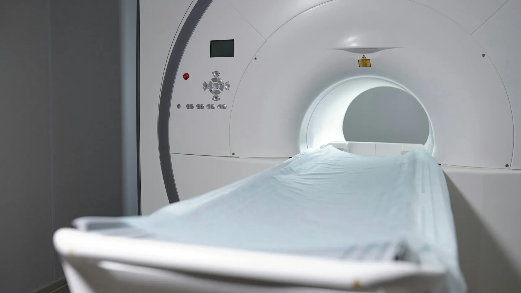
The patient lies in the MRI scanner and the needle is advanced through a guide, with scans repeated to confirm its tip lies in the lesion before each core. Nothing is estimated, since the target and the needle are seen on the same image. It takes longer, 45 to 60 minutes, needs a scanner slot, and is kept for lesions that fusion cannot reach or that an earlier fusion biopsy missed.
Cognitive fusion, in the operator's head
The urologist studies the MRI, then aims the ultrasound-guided needle at where the lesion should be. No software is involved. It works for large, obvious lesions in experienced hands and is less reliable for small or awkwardly placed ones. Ask which of the three is planned for you, and why.
Who needs one, prostate
Guidelines in Europe and the United States now put the MRI before the first biopsy, and the biopsy is targeted at what the MRI shows. Four situations bring most men to it.
A raised or rising PSA with no previous biopsy. The MRI decides whether a biopsy is needed at all. A clear scan with a low PSA density lets many men avoid the needle and return to PSA checks.
A negative standard biopsy with a PSA that keeps rising. The old blind biopsy missed something. The MRI shows where, and a targeted biopsy reaches it, which the Cochrane review found detected 44 percent more significant cancers than repeating the systematic biopsy.
Active surveillance for a low-risk cancer. Men who are watching a small cancer rather than treating it have an MRI and a targeted biopsy at intervals to confirm that nothing has grown or changed grade.
A lesion in the front of the gland. Anterior tumors sit beyond the reach of a standard transrectal biopsy and are found almost only by MRI.
Two groups sit between the lines.
Men with a suspicious MRI and a normal PSA, and men whose MRI is clear while their PSA density is high, are the difficult cases. They need a urologist to weigh the two, and a targeted biopsy alone is sometimes too little for them.
MRI-guided breast biopsy
Doctors use breast MRI to screen women at high genetic risk, to measure the extent of a known cancer before surgery, and to look for a primary tumor when cancer has appeared in a lymph node. It finds lesions that mammography and ultrasound miss. The first step after such a finding is a second-look ultrasound, aimed at the spot the MRI marked, because an ultrasound-guided biopsy is quicker and cheaper. Spick and Baltzer pooled 17 studies with 2,201 MRI-detected lesions and found that second-look ultrasound located 57.5 percent of them, more often when the lesion was a mass and when it was malignant. The rest, more than four in ten, have to be sampled inside the scanner.
The MRI-guided breast biopsy is done lying face down on a table with the breast in a grid. Contrast is injected, the lesion is located, the skin is numbed, and a vacuum-assisted needle removes several samples through one small opening. A tiny titanium marker is left at the site so that the spot can be found again on mammography if surgery is needed. It takes 30 to 60 minutes, and the marker is safe for future scans. A lesion that has faded by the time of the biopsy, which happens with hormonal changes, is not sampled, and a repeat MRI in six months takes its place.
What the studies found
The randomized trial. 38% vs 26%, and 28 percent spared a biopsy
In PRECISION, 500 men with a raised PSA and no previous biopsy were randomized to MRI with targeted biopsy only if the scan was suspicious, or to standard transrectal biopsy. Clinically significant cancer was found in 38 percent on the MRI route and 26 percent on the standard route, harmless low-grade cancer was diagnosed 13 percentage points less often, and 71 of 252 men on the MRI route needed no biopsy at all. Kasivisvanathan and colleagues, N Engl J Med, 2018.
The accuracy study. 93% vs 48%
Sensitivity of multiparametric MRI and of standard biopsy for clinically significant cancer in PROMIS, where 576 men had both tests and a saturation template biopsy as the reference. MRI was less specific, at 41 percent. The authors calculated that using MRI as a triage test would let 27 percent of men avoid a biopsy, and 5.9 percent of participants had a serious adverse event from the biopsies, including eight cases of sepsis. Ahmed and colleagues, Lancet, 2017.
Why systematic cores are often kept. 5.2 percent missed without systematic cores, 7.6 percent without targeted cores
In MRI-FIRST, 251 men had both a targeted and a systematic biopsy in the same session, with different operators blind to each other. Each method found significant cancers the other missed. The authors concluded that MRI improves detection but does not yet remove the need for systematic sampling in men having a first biopsy. Rouviere and colleagues, Lancet Oncol, 2019.
The review. Detection ratio 1.44 after a negative biopsy, 1.05 before any biopsy
The Cochrane review by Drost and colleagues pooled 43 studies. Against systematic biopsy, the MRI route found 44 percent more significant cancers in men whose earlier biopsy was negative and about the same number in men having a first biopsy, while diagnosing fewer insignificant ones. The certainty of the evidence was rated low because of the way patients were selected. Cochrane Database Syst Rev, 2019.
The day of the biopsy
Both procedures fit into a morning.
Prostate, from arrival to leaving
An antibiotic tablet before the procedure, or none if the transperineal route through the skin behind the scrotum is used, which carries far less infection risk and is now preferred by many centers. Blood thinners paused on the schedule your doctor set. Local anesthetic, or light sedation on request. The probe is placed, the MRI overlay is aligned, and the cores are taken in 15 to 30 minutes. Some blood in the urine, stool or semen for one to three weeks is normal. Most men are back at the hotel within two hours and can fly the next day.
Breast, from arrival to leaving
No fasting unless sedation is planned. Blood thinners paused as instructed. Face down in the scanner, contrast through a vein, local anesthetic, the vacuum samples, the marker, then pressure on the site and a small dressing. Bruising is common and settles in a week or two. Most women leave within an hour and can fly the next day.
Pathology takes three to five working days.
Risks and side effects
See a doctor the same day for a fever or shaking chills after a prostate biopsy, being unable to pass urine, heavy bleeding from the rectum or in the urine, or after a breast biopsy, a breast that swells rapidly or a wound that reddens and leaks. Fever after a transrectal biopsy is treated as sepsis until proven otherwise.
The targeting itself adds no risk. What remains comes with any biopsy of that organ, and for the prostate it depends heavily on the route. A transrectal biopsy passes the needle through the bowel wall, and infection follows in a few percent of men, with the serious cases seen in PROMIS. A transperineal biopsy passes through skin and brings the infection rate close to zero. It costs a little more discomfort. Bleeding in the urine, stool and semen, temporary difficulty passing urine and a brief effect on erections are common to both routes and settle within weeks. For the breast the risks are bruising, a small hematoma, infection at the site and a marker that shifts a few millimeters, none of them frequent, and the contrast agent used for MRI causes a reaction in a very small number of people and is avoided in severe kidney disease.
Days in Istanbul, cost and the trip
How long, and when to fly, and what to send
Three working days cover the length of stay. Day one holds the consultation and, if the MRI has to be repeated or done here, the scan. Day two holds the biopsy. Pathology takes three to five working days, so the results are either given on a fourth or fifth day or explained by video after you have flown home, with the written report and the slides available for a second opinion. Flying the day after the biopsy is fine for both organs, and the visit fits inside any short visa unless you stay on for treatment. Send these before you book.
MRI report and images, PSA history with dates, and any earlier biopsy report
For the breast, the mammogram, ultrasound and MRI reports, and the date of the last period if relevant to timing
A list of medication, with blood thinners marked
Allergies, kidney disease and any pacemaker or metal implant, which decide whether MRI and contrast are possible
Whether you want sedation, and whether a companion will travel with you
Cost comes next. The MRI itself, if it has to be done or repeated in Istanbul, is the largest item apart from the biopsy. A fusion biopsy costs more than a cognitive one because of the software and the planning, an in-bore biopsy more than a fusion biopsy because of the scanner time, and a breast biopsy inside the scanner more than one under ultrasound. Sedation, the number of targets, pathology with any immunohistochemistry, and a second opinion on outside slides are separate items. Published packages in Istanbul cover the consultation, the biopsy, pathology, an interpreter and transfers, and leave out the hotel, flights and the MRI unless it is named. One coordinator handles the case in English, Arabic, French, Russian, Serbian, Romanian or Spanish, or through an interpreter, sends the appointment confirmation and an invitation letter naming the hospital and the treating doctor ten days before travel, and books the hotel and the transfers. A female radiologist or breast specialist can be requested when you first write, and the department arranges it wherever the rota allows. A quote follows the doctor's review of the MRI, which costs nothing.
Results, and after you fly home
The pathology report leads to one of a small number of next steps. The table gives them for the prostate, where the grading system needs a word of explanation, since pathologists grade the cancer from 1 to 5 in the system of the International Society of Urological Pathology and grade group 1 marks the cancer that guidelines advise watching instead of treating.
This table scrolls sideways on a narrow screen. Swipe or drag to see every column.
What the prostate result usually leads to
Result
What it means
Usual next step
No cancer, MRI lesion sampled well
The lesion was inflammation or benign tissue
PSA checks, repeat MRI in one to two years if PSA rises
Grade group 1
Low-grade cancer that rarely spreads
Active surveillance with PSA, MRI and repeat biopsy at intervals
Grade group 2
Intermediate cancer, the commonest finding
A conversation about surgery, radiotherapy or surveillance in selected men
Grade group 3 to 5
Higher-grade cancer
Staging scans, then surgery or radiotherapy with hormone treatment
No cancer, but the target was missed
The cores did not reach the lesion
Repeat with in-bore guidance or a second fusion biopsy
Breast results, and once you are back home
A benign result that matches what the MRI showed ends the matter. Some women have a repeat MRI at six months. A high-risk lesion, such as atypical hyperplasia, leads to surgical removal because a minority hide cancer nearby. A cancer result leads to staging and a treatment plan with the breast surgeon and the oncologist, and the marker left at the biopsy site guides the surgery months later. Radiotherapy and chemotherapy are both delivered at Biruni Hospital on the same site if the treatment is planned here.
Once you are back home, the discharge file speaks for you. It holds the MRI report and images, the biopsy report with the grade and the number of positive cores, and a letter in English for your urologist or breast team, and the slides can be sent to a pathologist at home for a second reading. The coordinator stays reachable on the same WhatsApp number. Clinical questions go to the doctor who did the biopsy.
MRI-Guided Biopsy FAQ
Is an MRI-guided biopsy better than a standard prostate biopsy?
For finding significant cancer, yes. In the PRECISION trial the MRI route found it in 38 percent of men against 26 percent with standard biopsy, diagnosed fewer harmless cancers, and spared 28 percent of men a biopsy because their scan was clear. Many centers still add systematic cores at a first biopsy, because each method finds some cancers the other misses.
What is the difference between fusion and in-bore biopsy?
A fusion biopsy overlays the MRI on live ultrasound in an outpatient room and takes 15 to 30 minutes. An in-bore biopsy is done inside the MRI scanner with the needle checked on repeat scans, takes 45 to 60 minutes, and is kept for lesions that fusion cannot reach or missed.
Can I avoid a biopsy if my prostate MRI is clear?
Often. In PRECISION, 28 percent of men had a clear MRI and no biopsy. A clear scan with a high PSA density still needs a urologist's judgment, since MRI misses some cancers.
Why do I need an MRI-guided breast biopsy instead of an ultrasound one?
Because some lesions only appear on MRI. A second-look ultrasound finds about 57 percent of MRI-detected lesions, and the rest can only be sampled inside the scanner with a vacuum needle.
How long do I need to stay in Istanbul?
Three working days for the consultation, any repeat MRI and the biopsy. Pathology takes three to five working days and can be explained by video after you fly home, or in person if you stay.
Is the biopsy painful, and can I fly the next day?
Local anesthetic keeps the biopsy itself to pressure and brief discomfort, and sedation is available. Both prostate and breast biopsies allow a flight the next day. Blood in the urine or semen for a few weeks, or breast bruising for a week or two, is expected.
References
Kasivisvanathan V, Rannikko AS, Borghi M, et al. MRI-Targeted or Standard Biopsy for Prostate-Cancer Diagnosis. N Engl J Med. 2018;378(19):1767-1777.
Ahmed HU, El-Shater Bosaily A, Brown LC, et al. Diagnostic accuracy of multi-parametric MRI and TRUS biopsy in prostate cancer (PROMIS): a paired validating confirmatory study. Lancet. 2017;389(10071):815-822.
Rouviere O, Puech P, Renard-Penna R, et al. Use of prostate systematic and targeted biopsy on the basis of multiparametric MRI in biopsy-naive patients (MRI-FIRST): a prospective, multicentre, paired diagnostic study. Lancet Oncol. 2019;20(1):100-109.
Drost FH, Osses DF, Nieboer D, et al. Prostate MRI, with or without MRI-targeted biopsy, and systematic biopsy for detecting prostate cancer. Cochrane Database Syst Rev. 2019;4(4):CD012663.
Spick C, Baltzer PA. Diagnostic utility of second-look US for breast lesions identified at MR imaging: systematic review and meta-analysis. Radiology. 2014;273(2):401-9.
Editor's note
Written by the Biruni Hospital medical editorial team. Reviewed by Prof. Dr. Bülent ACUNAŞ, Radiology.