
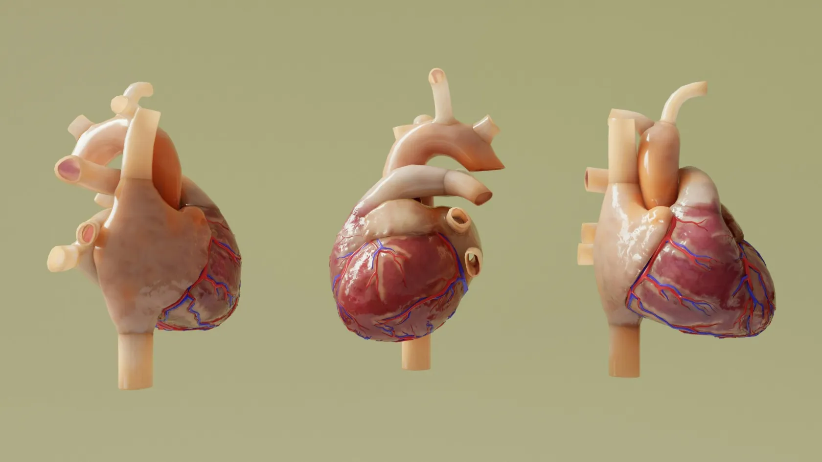
Mitral Valve Repair
A repaired mitral valve is still your own valve, and that turns out to matter for how long you live. This page covers when the operation should be done, which incision suits which valve, and how soon a patient who traveled for it can fly home.
About This Department
Fifteen years after mitral valve repair, 53.3 percent of patients in a 1,493-patient Canadian registry were still alive, against 46.0 percent of those whose valve was replaced instead. That gap held at every age. That difference explains why surgeons work hard to preserve a leaking mitral valve instead of exchanging it for a prosthesis, and it makes the single most useful fact to carry before you decide where to have this operation.
Free consultation
Send your echocardiogram and find out whether your valve can be repaired
Reports alone are rarely enough, so send the moving images from the echo as well, on a disc or as a file, together with any cardiology letter and your medication list. A cardiac surgeon reads them and tells you whether this valve looks repairable, whether it needs doing soon, and which incision your anatomy allows. The review costs nothing, commits you to nothing, and a coordinator writes back in your own language.
What goes wrong with the valve
Your mitral valve sits between the left atrium and the left ventricle, the two chambers on the side of the heart that pushes blood out to the body. It has two leaflets, a ring of tissue they hang from called the annulus, and a set of fine cords tethering their free edges to muscles in the ventricle wall, and when the ventricle contracts those cords hold the leaflets shut so the whole stroke of blood goes forward into the aorta with none of it washing back toward the lungs. Stretch the annulus, thin the leaflets or break a cord, and blood leaks backward. Doctors call that leak mitral regurgitation.
Degenerative leak, meaning the valve itself has worn out
Leaflet tissue thickens and stretches over decades, most often on the posterior leaflet, until a segment billows back into the atrium during contraction, and sometimes a cord snaps outright and a whole segment flails free with nothing holding its edge down. Doctors call this degenerative or primary mitral regurgitation, or Barlow's disease and fibroelastic deficiency depending on the pattern, and it is the group where repair works best. A skilled surgeon can rebuild most of these valves.
Secondary leak, meaning the valve is healthy and the heart is not
Here the leaflets are normal.
Enlargement of the ventricle after a heart attack or from a cardiomyopathy, which drags the papillary muscles apart and pulls the leaflets away from each other so they no longer meet in the middle. The valve suffers for the ventricle behind it. This distinction matters enormously for what your treatment should be, because an operation that fixes the leaflets does not fix the ventricle behind them, and the evidence for surgery in this group is far weaker than it is in degenerative disease.
Narrowing rather than leaking
Mitral stenosis presents the opposite problem, with the valve opening scarred down and blood struggling to get through it into the ventricle.
Rheumatic fever in childhood causes most of it. That is why mitral stenosis remains common across the Middle East, the Balkans, Central Asia and Africa while it has all but vanished from Western Europe, and it is why a surgeon here sees far more of these valves than a surgeon in London does. Selected rheumatic valves can still be repaired. Attempting it in a young patient is the right instinct, since a repair spares them a prosthesis for some years, though scarred rheumatic tissue keeps changing after the operation and a rheumatic repair therefore carries a higher chance of needing a second operation later than a degenerative one does.
Why repair rather than replace
Repair keeps your own tissue and the cord apparatus underneath it, which continues to hold the shape of the left ventricle after the operation the way a set of guy ropes holds a tent. Cut that apparatus out and put a prosthesis in its place, and the ventricle loses part of its scaffolding.
That scaffolding explains the survival difference, and the difference runs deep.
Researchers in British Columbia followed 1,493 patients across five hospitals who had isolated mitral surgery for degenerative disease between 2000 and 2017, matched 415 pairs by propensity score, and reported survival at one, five, ten and fifteen years of 95.7, 88.8, 71.4 and 53.3 percent after repair against 93.0, 81.6, 61.3 and 46.0 percent after replacement. Thirty-day mortality ran at 2.4 percent for repair and 3.6 percent for replacement. On multivariable analysis replacement came out as an independent risk factor for both early and late death, and the hazard ratio stayed above 1.0 across every age band, which is the finding that dismantles the old argument that older patients may as well have a replacement.
What a replacement commits you to
Mechanical valves last a lifetime and demand warfarin for a lifetime in exchange, with regular blood tests, a diet that has to account for vitamin K, and a bleeding risk that never goes away. Tissue valves avoid the warfarin and wear out instead, faster in younger patients, so a woman of 55 accepting a tissue valve is signing up for a second operation in her seventies. Your own repaired valve needs no blood thinner beyond the first few weeks in most cases and carries a lower long-term risk of infection settling on it. Repair asks something in return, which is a surgeon who does enough of these to be good at them, and that is a fair trade, but it is also the reason repair rates differ so sharply between two hospitals doing the same operation on the same kind of valve. Ask about that difference.
Ask any surgeon you consult what proportion of degenerative valves they repair and what proportion they replace, and ask it before you ask about anything else.
One useful benchmark. A unit doing this work properly repairs the great majority of degenerative valves it operates on, and a surgeon comfortable with the whole toolkit will quote you a figure without hesitating. Vagueness in reply to that question is itself an answer. It matters more here than for most operations, because the alternative to a good repair is a prosthesis with everything a prosthesis brings, and that substitution happens in the operating room on a decision you are not awake for and cannot revisit afterwards. Settling it in the consultation, in writing, is the only leverage a patient has. The second question concerns durability. A repair that leaks again in three years has bought you nothing but an extra operation, and durability turns out to depend on the technique and the tissue together rather than on either alone, so a ring plus neochords on a single prolapsing posterior segment in otherwise good tissue tends to last decades while a valve with widely redundant tissue, heavy annular calcium or rheumatic scarring is a much harder proposition. Your surgeon should tell you which of those you have. Ask before you agree to anything.
One thing repair does not require is a simple valve. A meta-analysis of ten studies comparing repair of the anterior or both leaflets against repair of the posterior leaflet alone found no significant difference in long-term survival, in freedom from moderate or severe recurrent leak, or in freedom from reoperation. Anterior leaflet disease is harder to repair, so it is more often sent for replacement, and the outcome data do not support that reflex.
When is the right time to operate
Most patients arrive at this page having been told to wait and watch. The waiting frightens them, and it follows a real logic, and it also has a point past which it starts costing them something permanent, which is the part nobody explains.
Severe leaks mean the ventricle is pumping a large volume of blood twice over, forward into the aorta and backward into the atrium. It copes by enlarging, and for years it copes well enough that you feel nothing. Then the muscle begins to fail, and the tragedy of mitral regurgitation is that the failure starts silently and is only partly reversible once it has started. Repairing the valve after the ventricle has been damaged fixes the leak and leaves you with a weakened heart. So the triggers for surgery in the 2020 ACC and AHA valvular heart disease guideline are not symptoms alone. The guideline recommends surgery for severe primary mitral regurgitation once symptoms appear, and also in patients with no symptoms at all when the left ventricle starts to show measurable strain, meaning the ejection fraction has fallen to 60 percent or below or the end-systolic diameter has reached 40 millimeters. The guideline goes further for patients without symptoms and with normal ventricular function, where surgery becomes reasonable when the valve is very likely to be repairable and the operation is being done in a center that repairs reliably.
Read that last clause again, because it is the clause that decides where you go. Early surgery on a normal heart is only justified if the valve is genuinely going to be repaired, since a replacement in that situation trades a symptomless leak for a prosthesis and its lifelong obligations. The threshold for operating early and the quality of the surgery are the same question.
Two other findings move the timing forward regardless of symptoms. New atrial fibrillation, and a pulmonary artery pressure that has climbed above 50 millimeters of mercury at rest, both indicate the heart has started to pay for the leak.
How the valve is actually repaired
Repair covers several operations.
The surgeon combines techniques according to what the valve in front of them needs, and knowing the names helps you follow your own operation note afterwards.
The annuloplasty ring
Surgeons stitch a ring or a partial band of fabric-covered metal around the annulus to bring it back to the size and shape it had before it stretched, which pulls the leaflets toward each other so they meet properly again. Almost every mitral repair includes one. It also protects the rest of the repair from the slow re-stretching that would otherwise undo it over years, and it is the reason a repaired valve holds up instead of leaking again in a decade.
Artificial cords
Where a cord has ruptured and a segment flails, the surgeon anchors expanded polytetrafluoroethylene sutures to the papillary muscle and to the free edge of the leaflet, then sets their length so the segment sits level with its neighbors again. Surgeons call them neochords. Preserving the whole leaflet is what has pushed this technique ahead of cutting tissue away, and it suits a small incision better than resection does. Setting the length is the whole art of it, since a neochord a millimeter too long leaves the segment prolapsing and a millimeter too short tethers it down, and the surgeon judges that against the rest of the leaflet on a heart that is not beating.
Removing excess leaflet
Billowing posterior segments can instead be cut out and the gap closed, an approach known as quadrangular or triangular resection. Older, durable, still right for some valves. A surgeon who uses both, choosing by anatomy, will repair more valves than one who only has one method.
The check before you leave the operating room
Intraoperative echo is the step patients rarely hear about and the one that decides whether the repair holds. Once the repair is finished and the heart is beating and pumping again, an ultrasound probe positioned in the esophagus looks directly at the valve while it works under real pressure. The surgeon sees what leak remains and how the leaflets move. At Biruni Hospital that check is done in every mitral case, and if the result is not good enough the patient goes back on the bypass machine and the valve is corrected in the same operation.
Nobody leaves the theater with a repair that was assumed to be adequate.
Start to finish, three to five hours. Most of it is the careful part. Your heart stops and a heart-lung machine takes over the circulation for roughly one to two hours of that, and the machine is one of the reasons this operation needs a proper cardiac unit around it and not just a good surgeon.
Which incision, and what it changes
Three routes reach the mitral valve and all three are offered here. The classical one divides the breastbone down the middle. A minimally invasive approach goes through a small incision between the ribs on the right side and reaches the valve with long instruments and a camera, leaving the breastbone whole. A robotic approach uses the same right-side ports, with the surgeon working from a console.
The trade runs deeper than the marketing around it suggests.
Pooling two randomized trials and nineteen propensity-matched studies found the minimally invasive approach reduced permanent pacemaker implantation, surgical site infection and transfusion compared with sternotomy, and it also found something rarely quoted, which is that after mitral repair specifically, reoperation on the valve came more frequently after the minimally invasive route than after sternotomy, with a hazard ratio of 7.33. The robotic approach showed no such difference against sternotomy, and carried more returns to theater for bleeding. A separate meta-analysis of twelve studies and 4,514 patients having repeat mitral surgery found the right-side approach gave shorter hospital stays, lower thirty-day mortality and less new kidney failure than opening an old sternotomy again. Read that practically. The incision should follow the valve, and never the other way round. A straightforward posterior leaflet prolapse in a patient with suitable groin vessels is an excellent small-incision case, while a complex bileaflet repair, a valve needing several techniques, or an operation combined with bypass grafting or a tricuspid repair belongs to the sternotomy every time. Choosing a keyhole approach and accepting a less complete repair, or a replacement instead of a repair, is a bad trade in every dataset there is.
This table scrolls sideways on a narrow screen. Swipe or drag to see every column.
| Route | Suits | What you live with |
|---|---|---|
| Full sternotomy | Complex or bileaflet disease, a second procedure on the same trip, heavy calcification, poor leg vessels. | A vertical scar down the chest and a breastbone that takes six to eight weeks to knit, which sets the driving and lifting rules. |
| Right mini-thoracotomy | Isolated posterior or single-segment prolapse. Redo surgery, where the right chest offers untouched tissue instead of an old sternotomy to reopen. Anyone wanting a faster return to normal. | A scar under the right breast and an intact breastbone. Less transfusion and fewer wound infections. Rib pain that outlasts the wound itself. |
| Robotic | The same anatomy as the mini-thoracotomy, where fine suturing deep in the chest is easier with wristed instruments. | Small port scars and durability that matched sternotomy in the pooled data, against a higher rate of returning to theater for bleeding. |
What can go wrong
Mitral valve repair is open heart surgery, and pretending otherwise would be dishonest.
In an isolated degenerative valve operated on before the ventricle fails, it also ranks among the safer major operations in medicine, with thirty-day mortality of 2.4 percent in the matched registry above and lower still in younger patients without other disease. What the numbers sit alongside follows.
Bleeding and a return to theater
Chest drains measure the loss hour by hour on the intensive care unit, and if the volume stays high the patient goes back to theater so the source can be found and stopped, which is a short procedure and a far better outcome than a slow accumulation nobody acted on. It happens in a small minority. Dealing with it that night beats watching it. You stop blood thinners and aspirin on a schedule set before you travel.
Atrial fibrillation
An irregular heart rhythm in the days after cardiac surgery is common enough to count as expected recovery, and the enlarged left atrium behind a leaking mitral valve makes it likelier still. Medication controls it, sometimes with an electrical shock under sedation, most cases settle, and a few need blood thinning for several weeks afterwards.
Stroke
Stroke is the most feared complication and an uncommon one.
Air or debris entering the circulation while the heart is open is the mechanism, and modern practice controls it with carbon dioxide flooding of the field, careful de-airing before the heart restarts, and imaging of the aorta beforehand to find calcification the surgeon should avoid. Weakness on one side, a facial droop or slurred speech is an emergency.
A pacemaker
Electrical conduction runs close to the mitral annulus, so stitches placed there can bruise it and leave the rhythm too slow. Most such disturbances recover within days on temporary wires. A minority persist. Those patients need a permanent pacemaker before discharge, and the network meta-analysis found this happened less often after the minimally invasive approach than after sternotomy.
The repair failing later
Repaired valves can leak again, usually because a stitch or a cord gives way or because the underlying disease progresses in tissue that was already abnormal.
Most recurrences appear within the first year or two and show up on the annual echocardiogram before you feel anything, and that alone justifies attending the scan. A second operation is possible. Surgeons usually go through the right chest for it.
Wound problems and infection
Sternal wound infection is the one that matters, because the breastbone is bone and infected bone is a long problem.
Diabetes, obesity, smoking and chronic lung disease raise the risk, and each of those repays attention in the weeks before surgery, while there is still time for it to count. Fever, a wound that weeps or opens, or a breastbone that clicks and moves when you cough all need to be seen quickly.
Fluid around the lung and the heart
Fluid collecting in the space around the left lung is common after mitral surgery, particularly after a right-side incision, and it shows up as breathlessness and a dry cough in the second or third week when everything else is improving. A chest X-ray finds it and a needle drains it under local anesthetic if it is large enough to matter. Fluid gathering around the heart itself is less common and more serious, and it declares itself through breathlessness with a low blood pressure and a fast pulse. Any of those three appearing together in the weeks after discharge earns an echocardiogram the same day. Attend the echo and the chest film before you fly.
Who should think twice
Surgery has the least to offer in severe secondary mitral regurgitation with a badly weakened ventricle, and a catheter-based clip or medical therapy may serve that patient better. Advanced lung disease, a recent stroke, active infection elsewhere in the body, severe kidney failure and frailty all change the calculation, sometimes decisively, and none of them is a reason to hide the answer from you. Being told the operation is not for you is a real answer. It travels better by message than after a flight.
Recovery, and when you can fly
The days in hospital
You wake on the intensive care unit with a breathing tube still in place, and it comes out within a few hours once you are warm, awake and breathing well. Published packages describe one to two days there and a total ward stay of five to seven. Drains and pacing wires come out over the first two or three days, each removal taking a minute and feeling odd more than painful, and physiotherapy starts on day one, because sitting still after heart surgery is what fills the bases of the lungs and causes half the fevers on any cardiac ward. Move early.
The first month
Tiredness dominates and it surprises people who expected pain to be the problem.
Sleep breaks up for two or three weeks, appetite takes a while to return, and mood can dip around the second week in a way that is well described and passes. Walking treats most of this, starting with ten minutes and adding a little every day. If your breastbone was divided, you keep your arms close to your body when pushing up out of a chair, avoid lifting anything heavier than a full kettle, and do not drive until six weeks have passed, because an emergency stop puts the seatbelt across a bone that has not knitted.
After a right-side incision those restrictions are shorter, since no bone was cut. Rib pain and a numb patch under the breast are the trade. Both fade.
Months two and three
Energy returns properly somewhere in this window, with most people back at desk work by week six and at physical work between weeks ten and twelve, though the exact date depends as much on what your job asks of your chest and arms as on the calendar. Cardiac rehabilitation, meaning a supervised exercise program, measurably improves how far and how confidently you get back, so join one wherever you live. The first check echo happens before discharge, then at three months, then yearly.
The flight home
Flying home decides your return ticket, and the answer runs like this. Ten to fourteen days after a minimally invasive or robotic repair is the usual clearance for a long flight, and two to three weeks after a sternotomy, provided the wound is dry, the rhythm is stable, any fluid around the lungs has settled and you are off strong painkillers. Cabin pressure itself is not the problem. Sitting immobile for hours with a fresh operation behind you is, so the advice is an aisle seat, a walk every hour, plenty of water and compression stockings. Somebody examines you and writes the date on your discharge summary, and it moves if the wound or the rhythm gives a reason.
If the valve cannot be repaired
Sometimes the valve turns out to be worse than the echo suggested. Heavy calcification of the annulus, destruction from infection, rheumatic tissue that has lost all its pliability, or a repair that will not hold on the intraoperative check all lead to the same conversation, and it is one you want to have before the operation, while you can still take part in it.
Ask which prosthesis would be used if it came to that, and why. The answer should be personal to you.
Under roughly 60 to 65 a mechanical valve is often preferred because it will outlast you, and the price is warfarin for life. Over that age a tissue valve usually makes more sense, because its expected durability begins to match your own life expectancy and you avoid the anticoagulation. Pregnancy plans, occupation, bleeding history, whether you can attend regular blood tests where you live, and your own preference all belong in that decision, so settle it in the consultation and written into the consent form as a plan instead of being left to the surgeon's judgment on the day. One route avoids surgery altogether. A clip delivered on a catheter through a vein in the groin grips the two leaflets together at the point of the leak, and for patients with heart failure and secondary mitral regurgitation who remain symptomatic on full medical therapy it changed the outlook. The COAPT trial randomized 614 such patients and found heart failure hospitalizations fell from 67.9 to 35.8 per hundred patient-years and death within two years from 46.1 to 29.1 percent. At Biruni Hospital the choice between an operation and a catheter-based option is worked out case by case with cardiology, and where the catheter route is the better answer for you, that is what you are told and that is where you are directed.
A hospital that only offers what it does is not assessing you.
What moves the price of a quote
No figure appears on this page. A number quoted before anybody has watched your valve move is a number for a different patient, and the moment it exists you end up negotiating around it instead of describing your own heart. Learning to read what you are sent serves you better. Six things move the total in mitral surgery specifically. Whether the operation is a repair or a replacement, and if a replacement, whether the valve is mechanical or tissue. Which incision the surgeon plans, since the minimally invasive and robotic routes carry their own equipment costs. Whether anything else is being done on the same bypass run, meaning a tricuspid repair, a maze procedure for atrial fibrillation, closure of a hole between the atria or coronary grafting, each of which lengthens the operation. How many nights on intensive care the quote assumes rather than hopes for. Whether the pre-operative work-up, which for a heart operation means a coronary angiogram in most adults over 40, a carotid scan and a dental check, sits inside the figure. And whether a second operation for bleeding, if it were needed, is covered.
Your own condition moves it as much as the surgery does. Poor kidney function, diabetes, chronic lung disease, obesity, a previous cardiac operation, a weak ventricle and pulmonary hypertension each raise the expected time on intensive care, and intensive care is the expensive part of any cardiac quote.
Packages published by Turkish hospitals and medical travel agencies for cardiac surgery typically cover the airport transfer, the pre-operative tests, the surgeon, anesthesia and perfusion fees, the theater, the planned intensive care and ward nights, an interpreter, and the follow-up appointments before departure. They typically exclude flights, travel insurance, unplanned extra intensive care, the treatment of a complication and any prosthesis upgrade. Read what you are sent against that list.
A figure means something only after a surgeon has watched your valve move. That review costs nothing here and carries no obligation.
Coming to Istanbul and going home
Plan on three to four weeks in the city.
That covers the consultation and the tests that have to be repeated here, the operation, one to two days on intensive care, five to seven days on the ward, the recovery time in a hotel before you are fit to fly, and the wound and echo check before departure.
Bring the moving echo images, and not only the report. A cardiologist's summary tells a surgeon the leak is severe, and the loops tell them which segment is prolapsing and whether the valve can be rebuilt, which is a different question and the one your trip depends on.
The hospital assigns you a coordinator when you first write, and she stays with you until discharge. The international patients team works in English, Arabic, French, Russian, Serbian, Romanian and Spanish, with interpreting in other languages arranged on request, and the two conversations where that matters most are the consent discussion before surgery and the ward round on the morning the drains come out. Patient rooms have a companion bed, so one person stays overnight with you for the whole admission apart from the intensive care days, where visiting is limited by unit rules. Accommodation for you and your companion for the nights on either side of the admission is arranged by the international patients office, along with airport transfers and the transport between hotel and hospital. The hospital kitchen handles halal, vegetarian and diabetic diets. A prayer room is on site. Requests for a female physician go to the department and are met wherever the rota allows.
For the visa, the international patients office issues an appointment confirmation and an invitation letter naming the hospital and your treating doctor, the document most consulates ask for with a medical visa application, and it goes out roughly ten days before travel.
After you land at home
Your coordinator stays reachable on the same WhatsApp number, so a photograph of a wound that looks wrong reaches somebody who already knows your case. You leave with the operation note naming every technique used and the size and model of any ring, the intraoperative echo findings, the discharge echo, your medication list with a stop date for anything temporary, and a letter to your own cardiologist. That package lets a doctor at home take over without repeating anything. Long-term follow-up means an echocardiogram and a cardiology review at three months and then once a year, wherever you live. Tell any dentist and any surgeon that you have had valve surgery, because antibiotic cover before certain procedures is standard after a repair with a ring. Breathlessness that returns, ankles that swell, a heartbeat that turns irregular, or a fever with no obvious cause are the four things to report promptly, without waiting for the next appointment.
Mitral valve repair FAQ
How long do I need to stay in Turkey for mitral valve repair?
When can I fly home after heart valve surgery?
Will I need blood thinners for the rest of my life?
How long does a mitral valve repair last?
Can my husband or wife stay in the room with me?
Can I have keyhole surgery instead of having my chest opened?
What happens if something goes wrong once I am back home?
References
- Kakuta T, Peng D, Yong MS, Skarsgard P, Cook R, Ye J. Long-term outcome of isolated mitral valve repair versus replacement for degenerative mitral regurgitation in propensity-matched patients. JTCVS Open. 2023;17:84-97.
- Otto CM, Nishimura RA, Bonow RO, Carabello BA, Erwin JP, Gentile F, Jneid H, Krieger EV, Mack M, McLeod C, O'Gara PT, Rigolin VH, Sundt TM, Thompson A, Toly C. 2020 ACC/AHA guideline for the management of patients with valvular heart disease. Journal of the American College of Cardiology. 2021;77(4):e25-e197.
- Yokoyama Y, Kuno T, Takagi H, Briasoulis A, Ota T. Conventional sternotomy versus right mini-thoracotomy versus robotic approach for mitral valve replacement or repair. Insights from a network meta-analysis. The Journal of Cardiovascular Surgery. 2022;63(4):492-497.
- Stone GW, Lindenfeld J, Abraham WT, Kar S, Lim DS, Mishell JM, Whisenant B, Grayburn PA, Rinaldi M, Kapadia SR, Rajagopal V, Sarembock IJ, Brieke A, Marx SO, Cohen DJ, Weissman NJ, Mack MJ. Transcatheter mitral-valve repair in patients with heart failure. The New England Journal of Medicine. 2018;379(24):2307-2318.
- Iqbal K, Haque IU, Shaikh VF, Rathore SS, Yasmin F, Iqbal A, Shariff M, Kumar A, Stulak JM. Mitral valve repair for anterior or bi-leaflet versus posterior leaflet degenerative mitral valve disease. A systematic review and meta-analysis. Current Problems in Cardiology. 2022;47(12):101355.
Editor's note
Written by the Biruni Hospital medical editorial team. Reviewed by Op. Dr. Mohammed N.I. SKAIK, Cardiovascular Surgery.
Medically reviewed by
Op. Dr. Mohammed N.I. SKAIK
Cardiovascular Surgery
Related Treatments
View All-
Aortic Aneurysm Repair - Aortic Aneurysm Surgery
Aortic aneurysm repair prevents rupture, which killed 37 percent of patients in emergency surgery against 6 percent in planned repair in a large screening trial. This guide explains when to watch and when to repair, size thresholds for men and women, EVAR against open surgery, long-term trial results, recovery and the stay in Istanbul.
-

Aortic Valve Repair
Being told the aortic valve leaks is usually followed by a conversation about which prosthesis to have. For a large group of patients there is a third answer, which is keeping the valve they were born with, and this page explains who qualifies.
-

Aortic Valve Replacement
The catheter valve wins the first year and the five-year data show the two routes ending up level. This page sets out what that means for a patient of 60 as opposed to a patient of 85, and which prosthesis belongs in each of them.
-
Coronary Angioplasty
In 5,179 patients with stable coronary disease, opening the narrowings did not reduce heart attacks or deaths. In somebody having a heart attack, the same procedure saves lives. Almost every argument about stents comes from confusing those two situations.
-
Endovascular Aneurysm Repair - EVAR
Three randomized trials compared keyhole aneurysm repair with open surgery and disagreed about long-term survival. They agreed on something else, and it is the part that decides how you will spend the next twenty years.