A thin antenna placed inside a tumor under ultrasound or CT heats it above 60 degrees, and the tissue dies within minutes with no incision and one night in hospital. Microwave ablation treats small liver, lung, kidney and thyroid lesions in patients who cannot have surgery, and in a randomized liver cancer trial it matched radiofrequency ablation on survival with faster, larger ablation zones.
About This Department
Interventional Radiology
A needle heats a tumor to over 60 degrees and it dies in minutes. No incision, one night in hospital, and the same control as radiofrequency in a randomized trial.
Microwave ablation destroys a tumor from the inside with heat. An antenna the width of a knitting needle is guided into the lesion under ultrasound or CT. Microwaves agitate the water molecules around its tip. The tissue cooks to a set size within a few minutes. It treats liver, lung, kidney and thyroid lesions that are small enough to fit inside that zone, in patients who cannot or should not have surgery. In a randomized trial of liver cancer, 6 percent of lesions treated with microwaves had regrown at two years against 12 percent after radiofrequency, and no patient died of either treatment.
6% vs 12%
Regrowth at 2 years, microwave against radiofrequency, Lancet Gastroenterol Hepatol 2018
73 in 100
Lung metastases fully ablated, 90 in 100 when under 3 cm, Radiology 2011
40% lower
Distant recurrence with microwaves, 7 randomized trials, Cancers 2020
Free
Interventional radiologist review of your scans
Free consultation
Send the CT or MRI images with their reports, the pathology if a biopsy has been done, liver or kidney function tests, blood clotting results and your medication list. An interventional radiologist tells you whether the lesion can be ablated in one session, whether surgery or radiotherapy would serve you better, and how many days to plan. The opinion costs nothing and asks nothing of you in return.
Radiologists write MWA. Patients search for microwave ablation. This page carries both in its title, Microwave Ablation - MWA. The method belongs to a family of treatments called thermal ablation, and a good part of what follows compares the members of that family, because the choice among them matters more than the brand of the machine.
How microwave ablation works
Microwaves at 915 or 2450 megahertz, the same frequencies as a kitchen oven and a wireless router, make water molecules spin. Spinning molecules generate heat. In microwave ablation the source of the microwaves is the tip of a thin antenna placed inside the tumor, so the heat forms in the tissue itself instead of being conducted into it from a hot electrode, and that single difference from the older technique, radiofrequency ablation, gives microwaves three advantages. They heat a larger volume in less time. Charring does not stop them, whereas it stops an electrical current. Blood flowing through nearby vessels cools them less, so lesions next to large veins are treated more reliably. Tissue above 60 degrees dies at once. The radiologist plans the ablation zone to cover the tumor plus a margin of at least five millimeters of normal tissue, and the margin decides whether the treatment holds.
At Biruni Hospital microwave ablation is done by the interventional radiology team under ultrasound or CT guidance, with an anesthesiologist present, and the patient stays one night for observation. Surgery, stereotactic radiotherapy on the Elekta Versa HD and medical oncology are on the same site, so the ablation is chosen at a tumor board that has the other options in front of it.
Organ by organ
Four organs account for nearly every procedure.
Liver
Ablation began in the liver, and the liver holds the strongest evidence. Guidelines for hepatocellular carcinoma recommend it as a curative treatment for tumors under three centimeters in patients who are not candidates for surgery or transplant, and as an alternative to surgery for the smallest ones. Metastases from bowel cancer get the same treatment when they are few and small, often together with chemotherapy or after a liver resection has removed the larger ones. The needle goes through the skin below the ribs under ultrasound. One session reaches lesions of three to four centimeters.
Lung
Lung metastases, and small primary lung cancers in patients who cannot have surgery, are treated under CT guidance with the patient lying on the table and the antenna passed between the ribs. Air changes the physics. It conducts heat poorly, so the lung insulates the ablation zone well and carries microwaves into it badly, and the results therefore depend on size and position more than in any other organ.
Kidney
Small kidney tumors under four centimeters, found by chance on a scan, are increasingly ablated and left in place, above all in older patients and in those with one kidney or poor kidney function, where preserving every part of the organ counts. A biopsy comes first. A quarter of such tumors turn out to be benign.
Thyroid
Benign thyroid nodules that press on the windpipe or show in the neck can be shrunk with a thin antenna under ultrasound in an outpatient session, and the nodule loses most of its volume over a year. Nothing else on this page treats a benign condition. Surgery, the alternative, removes half the gland.
What the studies found
Each study below is open. Close the ones you have read.
Microwave against radiofrequency in liver cancer, the randomized trial
Vietti Violi and colleagues randomized 152 patients with chronic liver disease and up to three hepatocellular carcinomas of four centimeters or less at four university centers in France and Switzerland. At two years, six of 98 lesions treated with microwaves had regrown at the edge of the ablation zone, 6 percent, against 12 of 104 treated with radiofrequency, 12 percent. The difference did not reach statistical significance. Two grade 4 complications, both arterial bleeding controlled by embolization, occurred in the microwave group, and three grade 3 complications in the radiofrequency group. No patient died of either treatment. Lancet Gastroenterology and Hepatology, 2018.
All the randomized trials together
Facciorusso and colleagues pooled seven randomized trials with 921 patients. Complete ablation was the same with both techniques, survival was the same at every year up to five, local recurrence did not differ, and adverse events were as frequent with one as with the other. Distant recurrence was 40 percent lower with microwaves, and disease-free survival at five years favored them, findings the authors said need confirmation. Cancers, 2020.
Lung metastases, size and position decide
Vogl and colleagues treated 130 lung metastases in 80 patients with CT-guided microwave ablation. Complete ablation was achieved in 73 percent of lesions overall, in 90 of 110 lesions of three centimeters or smaller, and in 5 of 20 larger ones. Peripheral lesions succeeded in 80 percent and central ones in 50 percent. Survival was 91 percent at one year and 75 percent at two. A collapsed lung followed 8.5 percent of sessions and needed a drain once, and no patient died within 60 days. Radiology, 2011.
Benign thyroid nodules over three years and more
Cho and colleagues pooled studies with at least three years of follow-up after thermal ablation of benign thyroid nodules. Volume fell fast in the first year, held steady from one to three years and fell further after that. Complications occurred in 3.8 percent, and no unexpected late problems appeared. Most of the pooled data came from radiofrequency and laser devices, and microwave results are assumed to follow the same pattern. Endocrinology and Metabolism, 2020.
Who it suits
The consensus document on tumor ablation by Ahmed and colleagues defines technical success as an ablation zone that covers the tumor and its margin on the first scan afterward. Patient selection serves that definition. Four things have to be true.
The lesion is small enough. Three centimeters or less gives the most reliable result. Between three and five centimeters the zone may need two antennae or two sessions. Above five, ablation rarely clears the margin and other treatments come first.
There are few lesions. Up to three in the liver, and a handful in the lung, can be treated in one or two sessions. Beyond that, drugs or radiotherapy treat the organ as a whole.
The lesion can be reached and seen. A needle path that avoids bowel, gallbladder, large vessels and the diaphragm, and a lesion that shows on ultrasound or CT during the procedure. Lesions at the dome of the liver or beside the heart are the hardest.
Surgery is not the better choice. A fit patient with a single resectable liver tumor and no cirrhosis is offered surgery, which removes more tissue with a wider margin. Ablation is for patients who cannot have surgery, do not want it, or would lose too much organ to it.
Some patients wait. Blood thinners that cannot be stopped, an uncontrolled clotting disorder, a lesion less than a centimeter from a large bile duct or the bowel, and an active infection are reasons to postpone or to choose something else, and the radiologist says which.
The procedure
Before the day
A CT or MRI within four to six weeks, blood tests for clotting, liver and kidney function, and a tumor marker as a baseline. Blood thinners are paused on the schedule your own doctor sets. Fasting from midnight. An anesthesia review the day before, since most ablations are done under deep sedation or general anesthesia so that breathing can be held still while the antenna is placed.
1
Positioning and planning
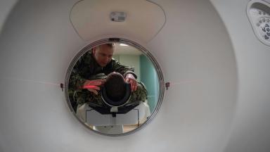
You lie on the CT table or beside the ultrasound machine in the position that gives the straightest path. The radiologist marks the entry point on the skin and measures the depth and angle from the images.
2
Placing the antenna
After local anesthetic and a nick in the skin, the antenna is advanced under real-time imaging until its tip sits in the far side of the tumor, with a scan or ultrasound sweep to confirm before any energy is delivered. Large lesions receive a second antenna.
3
Ablation
The generator runs for three to ten minutes at a set power. Gas bubbles form in the heated tissue and show on ultrasound as a growing cloud, and on CT the zone appears as a dark area. The radiologist stops when the zone covers the tumor and the planned margin.
4
Track ablation and withdrawal
The antenna is heated as it is withdrawn, which seals the path and keeps tumor cells from being carried out along it. Pressure on the skin and a small dressing follow.
5
The first 24 hours
A CT or contrast ultrasound within a day confirms the ablation zone. Pain at the site and a low fever for two or three days are expected and come from the dead tissue. Most patients go home the next morning.
Risks and complications
Every organ has its own list.
This table scrolls sideways on a narrow screen. Swipe or drag to see every column.
Complications of microwave ablation by organ
Organ
Main risks
How common, and what is done
Liver
Bleeding, abscess in the ablation zone, injury to the bile ducts or the bowel, a burn on the skin or the diaphragm
Serious events in a few percent. Bleeding is embolized, an abscess drained, and antibiotics are given when the bile ducts have been operated on before
Lung
Collapsed lung, bleeding into the airway, fluid around the lung, infection of the ablated tissue
A collapsed lung in 8.5 percent of sessions in the Frankfurt series, most needing no drain. Coughing up blood for a day or two is common
Kidney
Bleeding around the kidney, injury to the ureter or bowel, a burn on the skin
Uncommon. Fluid can be injected to push the bowel away before ablation
Thyroid
Voice change from heat near the nerve, bleeding, a burn on the skin
Around 3.8 percent overall. Voice change is usually temporary
All sites
Post-ablation syndrome, which is fever, tiredness and aching for two to five days
Very common and self-limiting. Paracetamol and fluids
All sites
Tumor seeding along the needle track
Rare, and the reason the track is heated on withdrawal
How it compares with the alternatives
A sentence on radiofrequency. Radiofrequency ablation has thirty years of results, is the technique most guidelines still name, and in the randomized trials it matched microwaves on survival and complications. Microwaves win on speed, on the size of the zone and on lesions beside vessels. A center that offers only one of the two is not wrong, and a patient should ask about the operator's experience before the choice of energy.
This table scrolls sideways on a narrow screen. Swipe or drag to see every column.
Treatments for a small liver or lung tumor
Treatment
What it involves
Where it fits best
Microwave ablation
A needle through the skin, sedation, one night
Lesions under 3 cm, up to three, in patients who cannot have surgery
Radiofrequency ablation
The same approach with an electrode and a slower, smaller zone
Lesions under 3 cm away from large vessels
Cryoablation
Freezing through a probe, visible as an ice ball on CT
Kidney and some lung lesions, and tumors near nerves where cold is safer than heat
Surgical resection
Removal of the tumor with a wide margin under general anesthesia
Fit patients with a single resectable tumor and enough organ to spare
Stereotactic body radiotherapy
Three to five sessions of focused high-dose radiation, no needle
Lesions that cannot be reached by needle, patients who cannot have sedation, and central lung lesions
Chemotherapy, targeted drugs, immunotherapy
Treatment of the whole body
Many lesions, or disease outside the organ
Days in Istanbul, cost and the trip
Four to six days cover the length of stay. Day one holds the consultation, the imaging review and the anesthesia check. Day two is the ablation and the night in hospital. The next-day scan and discharge follow, and the radiologist sees you once more two or three days later, when the fever has settled and the skin site is checked, before you fly. Flying after a lung ablation waits for a chest X-ray that shows no air around the lung.
CT or MRI images with reports, and the pathology if a biopsy was done
Liver or kidney function, clotting tests and a recent blood count
The full medication list with blood thinners marked, and any allergy to contrast or sedation
The history of the cancer, with dates of surgery, chemotherapy and earlier ablations
Whether a companion will travel with you, and any request for a female doctor
Cost follows the number of lesions and sessions, the organ, the type of anesthesia, the imaging before and after, the antenna, which is a single-use device, and the night in hospital. Two antennae or two sessions for a large lesion double the device cost, more or less. Published packages in Istanbul cover the consultation, the ablation, anesthesia, one night, the follow-up scan, an interpreter and transfers. Hotel, flights, a PET-CT and any chemotherapy fall outside them, and your own health enters through the anesthesia plan and through liver or kidney function. A figure follows the radiologist's review of the images, which costs nothing. One coordinator handles the case in English, Arabic, French, Russian, Serbian, Romanian or Spanish, or through an interpreter, sends the appointment confirmation and an invitation letter naming the hospital and the treating doctor ten days before travel, and books the hotel and the transfers. The room has a companion bed. Halal, vegetarian and diabetic meals are available, a prayer room is on site, and a female doctor can be requested when you first write.
Results, and after you fly home
The first scan decides, and the second confirms
Complete ablation is judged on the CT or MRI taken within a day or a month, where the zone should cover the tumor with a margin. A scan at three months then separates a shrinking zone from regrowth at its edge, and scans continue every three to four months for two years, then every six. Regrowth at the edge is treated with a second ablation in most cases, and new lesions elsewhere in the organ are treated when found small. The consensus document by Ahmed and colleagues sets out this reporting and follow-up standard.
Once you are back home
The discharge file holds the images before and after, the ablation report with the power, the time and the zone size, the pathology if any, a tumor marker baseline, and a letter in English for your oncologist setting out the scan schedule. Fever above 38.5 degrees after the first week, worsening pain, jaundice, breathlessness or coughing up more than a streak of blood need a local doctor the same day. The coordinator stays reachable on the same WhatsApp number, and clinical questions go to the radiologist who did the ablation.
Microwave Ablation - MWA FAQ
Is microwave ablation better than radiofrequency ablation?
In the randomized trial of liver cancer, 6 percent of lesions regrew at two years after microwave ablation against 12 percent after radiofrequency, a difference that did not reach statistical significance. Across seven trials, survival and complications were the same. Microwaves are faster, treat larger zones and cope better with lesions beside vessels.
How big can a tumor be for microwave ablation?
Three centimeters or less gives the most reliable result. In a lung series, complete ablation succeeded in 90 of 110 lesions under 3 cm and in 5 of 20 larger ones. Lesions of three to five centimeters may need two antennae or two sessions, and larger ones are treated another way.
Is the procedure painful, and how long do I stay in hospital?
Nothing is felt during it, because the ablation is done under sedation or general anesthesia. Pain at the site and a low fever for two to five days afterward are expected. Most patients stay one night and fly home within four to six days, once the radiologist has checked the site and, after a lung ablation, a chest X-ray.
Can ablation be repeated if the tumor comes back?
Yes. Regrowth at the edge of the ablation zone is usually treated with a second ablation, and new lesions elsewhere in the organ are treated when found small, which is why scans continue every three to four months for two years.
Is microwave ablation a cure?
For a single small liver tumor it can be, with results close to surgery in patients who cannot be operated on. For metastases it controls the treated lesions as part of a plan that includes drugs and follow-up scans, and the outcome depends on the cancer of origin more than on the ablation.
What is the difference between ablation and radiotherapy for a small lung lesion?
Ablation needs a needle path and sedation and gives a result on the next day's scan. Stereotactic radiotherapy needs no needle and suits central lesions and patients who cannot have sedation, at the cost of not knowing what the lesion was and of waiting months to judge the response. The tumor board chooses between them from the same scans.
References
Vietti Violi N, Duran R, Guiu B, et al. Efficacy of microwave ablation versus radiofrequency ablation for the treatment of hepatocellular carcinoma in patients with chronic liver disease: a randomised controlled phase 2 trial. Lancet Gastroenterol Hepatol. 2018;3(5):317-325.
Facciorusso A, Abd El Aziz MA, Tartaglia N, et al. Microwave Ablation Versus Radiofrequency Ablation for Treatment of Hepatocellular Carcinoma: A Meta-Analysis of Randomized Controlled Trials. Cancers (Basel). 2020;12(12):3796.
Vogl TJ, Naguib NN, Gruber-Rouh T, Koitka K, Lehnert T, Nour-Eldin NE. Microwave ablation therapy: clinical utility in treatment of pulmonary metastases. Radiology. 2011;261(2):643-51.
Cho SJ, Baek JH, Chung SR, Choi YJ, Lee JH. Long-Term Results of Thermal Ablation of Benign Thyroid Nodules: A Systematic Review and Meta-Analysis. Endocrinol Metab (Seoul). 2020;35(2):339-350.
Ahmed M, Solbiati L, Brace CL, et al. Image-guided tumor ablation: standardization of terminology and reporting criteria, a 10-year update. J Vasc Interv Radiol. 2014;25(11):1691-705.
Editor's note
Written by the Biruni Hospital medical editorial team. Reviewed by Assoc. Prof. Dr. Utku Mahir YILDIRIM, Radiology.