
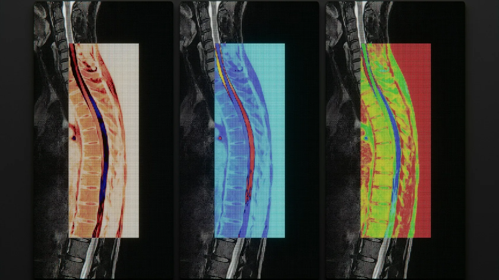
Microdiscectomy - Minimally Invasive Disc Surgery
A tube instead of a retractor changes the scar. The operation underneath it stays the same, and what minimally invasive buys turns out to be smaller than the brochures suggest.
About This Department
Somebody has recommended a microdiscectomy and used the phrase minimally invasive, and you now want to know what that actually buys. This page takes the operation apart. How it is done, what the tube and the endoscope change and what they leave alone, what the Cochrane evidence says when the techniques are compared head to head, how the day after feels, and what an international patient needs to arrange around it.
Free consultation
Send the MRI and get a technique recommendation with a reason attached
The review costs nothing and commits you to nothing. Send the lumbar MRI images and the radiologist's report, the level and side of the pain, how long it has been going on, and any weakness or numbness you have noticed. A spine surgeon reads the films and answers the question this page is about. Which technique fits this herniation, at this level, in this patient, and why that one instead of the others.
What the phrase covers
Minimally invasive describes the route to the disc. Nothing more than that.
Underneath every version of this operation the surgeon does the identical thing, which is to reach the nerve root, lift it gently to one side, and take out the piece of disc pressing on it. Identical fragment, identical decompression. What changes between an open microdiscectomy, a tubular one and a full-endoscopic one is how much muscle gets disturbed on the way in, how big the scar ends up, and which optical device the surgeon looks through, and those three things matter to the first fortnight far more than they matter to the result at one year. Anybody selling the technique as a different operation with a different outcome has skipped past the evidence, which this page comes to shortly.
Three routes are in common use. An open microdiscectomy goes through a midline incision of fifteen to twenty-five millimetres with the muscle held aside by a retractor and the work done under an operating microscope, which gives the surgeon the widest view of the three and remains the technique every newer method is measured against in the trials. A tubular microdiscectomy passes a series of dilators through the muscle fibres and leaves a working tube of sixteen to twenty-two millimetres in place, so the muscle is spread instead of stripped. A full-endoscopic discectomy uses a tube of around eight millimetres with a camera and light at its tip, entering either between the laminae or through the foramen at the side, frequently with the patient awake and sedated.
Scar sizes differ by a centimetre or so, which is the true scale of the visible difference between a technique described as minimally invasive and one that is not.
Two terms cause confusion and are worth separating now. A discectomy removes the herniated fragment and leaves the rest of the disc doing its job as a spacer between the vertebrae. A fusion removes the whole disc and joins the two vertebrae with cages and screws so that segment stops moving. Patients frequently arrive having read about the second while being offered the first, and the two sit at opposite ends of the spinal surgery spectrum in every way that matters, in operating time, in recovery, in what the spine can do afterwards and in cost. A discectomy takes an hour or so and sends somebody home the next morning with a segment that still bends. A single-level fusion takes three or four hours, keeps a patient in for several days, commits that segment permanently, and transfers load onto the discs above and below it for the rest of their life. Surgeons reserve fusion for instability, for repeated recurrences at the same level, or for a slipped vertebra sitting on top of the herniation, and a patient offered fusion for a straightforward first-time disc prolapse should ask, politely and directly, what specifically about their spine takes them out of the discectomy group.
The operation, step by step
Sixty to ninety minutes covers most of these, from the moment the patient is positioned to the moment the dressing goes on.
The five stages inside the theatre
Positioning and locating the level
The patient lies face down with the abdomen free, which drops venous pressure around the spine and reduces bleeding. An X-ray image confirms the level before anything is opened, and that single picture is what stands between a routine operation and the classic never event in spinal surgery, which is a technically flawless discectomy performed one level away from the herniation that brought the patient in.
Reaching the spine
A small incision opens over the level. Muscle is retracted, or dilated apart if a tube is being used, and the ligament between the two laminae comes into view. No bone has been touched yet.
Opening the window
Removing the ligamentum flavum comes first, followed in many cases by a few millimetres of bone from the edge of the lamina. This is the window through which everything else happens, and it is sized to the surgeon's need to see the nerve safely.
Retracting the root and removing the fragment
The nerve root is eased medially with a blunt retractor. Underneath it sits the herniation, which comes out in one piece where the outer ring has held it contained, or in several where it has burst through and migrated up or down behind the vertebral body, and retrieving those scattered pieces is the part of the operation that separates a fifty minute case from a ninety minute one. The surgeon then feels along the root to check nothing remains behind it.
Checking and closing
With the fragment out, the root should move freely across the disc space, and that free movement is the endpoint the surgeon is working towards. The wound closes in layers, frequently with a dissolving stitch under the skin and glue on top, so there is nothing to remove later.
One detail patients rarely hear, and it explains a great deal about how the next few weeks feel. The hole in the outer wall of the disc, the one the fragment came through, is left open. Surgeons do not stitch it, because the tissue does not hold a stitch, and the disc seals it with scar over roughly six weeks. Every restriction in this article traces back to that unclosed hole, and so does the whole idea of a recurrence, and so does the reason a patient who feels perfectly well at three weeks still cannot lift a suitcase.
Tube, endoscope or microscope
Herniations sit in different places along and around the nerve root, and that position on the scan drives the choice of technique far more than fashion, marketing or whatever equipment a hospital happens to have bought in the last three years. A standard paramedian herniation at L4 to L5 or L5 to S1 suits any of the three. A far lateral herniation, sitting outside the canal beside the exiting nerve, is reached more directly through the foramen with an endoscope than by going through the midline. A large sequestered fragment that has migrated two centimetres up behind the vertebral body is easier to retrieve through a wider working channel. Recurrent herniations, surrounded by scar from a previous operation, favour whichever technique gives the surgeon the best view of tissue planes that no longer look the way the textbook says. Two anatomical details also constrain the choice more than patients realise. The iliac crest, the top of the pelvis, sits high in some people and physically blocks the transforaminal endoscopic route to the bottom disc at L5 to S1, which is where a great many herniations happen. And a herniation sitting in the axilla of the nerve root, in the angle between the root and the dural sac, sits somewhere an angled endoscope reaches awkwardly. None of that appears on a clinic brochure, and all of it is on your MRI, which is precisely why a technique recommendation made before anybody has looked at your scan is worth very little.
This table scrolls sideways on a narrow screen. Swipe or drag to see every column.
| Technique | How it gets there | Where it earns its place |
|---|---|---|
| Open microdiscectomy | Midline incision of 15 to 25 mm, muscle held aside, operating microscope | The reference standard everything else is measured against. Widest view, handles migrated fragments and scarred revisions without compromise |
| Tubular microdiscectomy | Dilators spread the muscle, a 16 to 22 mm working tube stays in place, microscope or endoscope down the tube | Less muscle disturbance for the same exposure. Pooled data show it matching open microdiscectomy on every outcome measured |
| Full-endoscopic discectomy | A single 8 mm portal with camera and irrigation, interlaminar or transforaminal, often under sedation | Smallest wound, lowest pooled complication rate in randomised data, direct access to far lateral herniations. Demands a surgeon well past the learning curve |
Anaesthesia separates them too. Open and tubular surgery happens under general anaesthesia. Full-endoscopic surgery can be done with local anaesthetic and sedation, which suits a patient whose heart or lungs make a general anaesthetic risky, and which lets the surgeon ask an awake patient whether a manoeuvre near the root hurts.
What the Cochrane review found
Marketing around minimally invasive spine surgery promises smaller scars, less pain and faster recovery. Two pieces of evidence test those promises properly, and the results are more useful than the brochures.
A Cochrane review published in 2014 pooled eleven randomised and quasi-randomised trials covering 1,172 participants, comparing minimally invasive discectomy against standard microdiscectomy or open discectomy. Its findings run against the marketing on several counts. Low-quality evidence put leg pain slightly worse after the minimally invasive techniques between six months and two years, back pain slightly worse at six months and at two years, and re-hospitalisation for recurrent herniation more frequent. Every one of those differences was tiny, under half a point on a ten-point pain scale, and below the threshold anybody would notice. Where minimally invasive surgery did clearly win was infection, with a lower risk of surgical site and other infections, and the reviewers found hospital stay shorter in some trials and no different in others. Seven of the eleven trials carried a high risk of bias, which the reviewers said plainly, and that caveat weakens the finding in both directions equally. What the review demonstrates beyond argument is that nobody has shown minimally invasive discectomy to produce meaningfully better pain relief than the operation it was designed to replace, a decade after the marketing began claiming exactly that.
Read that carefully and it stops sounding like a criticism. It says the techniques are equivalent for the thing patients care about.
Tubular surgery has been examined on its own. A 2018 meta-analysis in Medicine pooled ten studies and 804 patients comparing tubular microdiscectomy against conventional microdiscectomy and found no significant difference in operating time, blood loss, length of stay, intraoperative complications, postoperative complications, dural tears, reoperation, or leg and back pain scores at short or long term. Nothing separated them.
Endoscopic surgery points the same way on effectiveness and diverges on safety. Fourteen studies pooled in Spine in 2021 found moderate quality evidence of no difference in leg pain or function at three to six months and at twelve months between transforaminal endoscopic discectomy and open microdiscectomy. A 2022 meta-analysis in World Neurosurgery pooling six randomised trials put overall complications at 5.5 percent for full-endoscopic surgery against 10.4 percent for open, a risk ratio of 0.55, with fewer dural tears. Its cohort data reported higher rates of residual fragments and revision surgery after endoscopic procedures, which is what a learning curve looks like when it is turned into a statistic, since a surgeon working through a narrow portal early in their experience misses fragments that a wider exposure would have made obvious.
So here is a defensible position. All three techniques relieve sciatica equally well, so choose the surgeon rather than the tube, and take the technique that surgeon performs most often. A hospital that answers the technique question by naming what they own has answered the wrong question.
Anaesthesia and the night afterwards
Waking from this operation differs from waking after most surgery. The pain that brought you in has frequently gone before you are fully conscious. Patients describe a strange absence where the leg used to hurt, and after months of a pain that has run their sleep, their work and their temper, that absence lands harder than anybody warns them it will. Some cry, and the nurses on a spinal ward have seen it often enough to have tissues within reach. The back around the wound aches, and that ache is muscle responding to being held apart for an hour, which settles across a week or two with ordinary painkillers.
Day case or one night
Discharge on the day of surgery is routine in many units for a straightforward single-level discectomy in a fit patient who has somebody at home for the first night, which reflects how quickly this particular operation returns people to their feet rather than any pressure on beds. An overnight stay suits everybody else, and for an international patient it becomes the sensible default, and it signals nothing about complexity, since a hotel room in an unfamiliar city on the evening of an anaesthetic is a worse place to be than a ward.
Published Turkish packages for disc surgery quote one to two nights, with three or four for more involved cases.
The first twenty-four hours
Sitting up comes within a few hours. Walking follows. Dinner arrives that evening. Nurses will ask about passing urine before letting anybody home, since a bladder that fills without emptying is one of the very few signals after this operation that needs attention within the hour instead of at the next clinic appointment, and it is the reason a discharge that feels ready at four in the afternoon sometimes waits until eight. Sleep on the first night is broken and unremarkable.
Who this operation suits
Leg pain matching a nerve root, a scan showing a herniation compressing that same root, and six to twelve weeks of proper non-surgical treatment behind you. That combination produces the results the studies report, and 80 to 90 percent of such patients get substantial relief.
Two situations move faster than that timetable. Cauda equina syndrome, where a large central herniation compresses the whole nerve bundle and causes bladder or bowel changes and saddle numbness, is a surgical emergency measured in hours. Weakness that is deteriorating week by week gets operated on within days.
Where it disappoints is easier to state. Back pain without a clear radiating leg component responds poorly, because this is an operation on a nerve rather than on a painful joint or a worn disc, and the patient whose main complaint is a deep central ache after standing at work will come out of surgery with that ache intact. Longstanding numbness, present for a year, frequently stays. So does weakness that has been there for months, which recovers slowly and sometimes incompletely.
Say all of this to the surgeon before the operation. Afterwards is too late. A five minute conversation about which of your symptoms should improve, and which probably will not, is what separates a satisfied patient at three months from a disappointed one.
Getting back to things
Feeling well is not the same as being healed, and the gap between those two states is the entire problem of the first six weeks after a discectomy. Leg pain has gone. The wound is a centimetre of glue. Every instinct says get on with life, and the tear in the disc wall behind all of it is still open and will stay open for around six weeks, which is the only reason any of the restrictions below exist.
Bending, lifting and twisting are the three that come off the list at six weeks.
Walking does the work in the meantime. Ten minutes several times a day at the start, building steadily, and by the end of the first month an hour is realistic for most people. Sitting works the opposite way. A seated lumbar spine carries more load than a standing one, so twenty to thirty minutes at a stretch is the working ceiling early on, which matters for anybody planning a long flight or a desk job. Set a timer for the first fortnight instead of trusting yourself to notice, because the failure mode here is not pain during the sitting, it is a stiff and sore back two hours later when the damage has already been done. Perch on the edge of firm chairs instead of sinking into soft ones. Keep the hips slightly above the knees. Get out of the car every hour on any journey longer than that, and take the aisle seat on every flight for the first three months rather than only the flight home.
Formal physiotherapy starts around week four in most protocols, once the acute soreness has settled, and it targets the deep stabilising muscles that switch off during months of pain. Skipping this stage is the commonest self-inflicted problem after an otherwise successful discectomy, because a spine carrying a healed disc and a set of stabilising muscles that have not worked properly for six months still feels unreliable when it is asked to lift a child or carry shopping, and the patient then attributes that unreliability to the operation, and never to the four weeks of rehabilitation they never did.
Office work returns between two and four weeks, part time to begin with. Manual work waits until eight to twelve weeks depending on how heavy it is, and a plasterer, a nurse turning patients and a warehouse picker are three different answers to that question rather than one, which is a conversation to have with the surgeon while the sick note is being written. Driving comes back once an emergency stop can be done without hesitating and no sedating medication is on board.
Swimming, cycling and the gym reopen from around six weeks with guidance. Running comes later. Ask the surgeon, since no fixed date applies to everybody.
Complications worth understanding
Pooled randomised trials put overall complications at 10.4 percent after open discectomy and 5.5 percent after full-endoscopic surgery. Those figures cover everything from a headache lasting two days to a return to theatre, and the serious end of the range is thin.
Dural tear leads the list of technical complications, and it means the membrane holding the spinal fluid has been nicked while the surgeon works around it. Repair happens during the same operation. The consequence for the patient is a day or two lying flat and, in some cases, a headache that is worse sitting up than lying down. It does not affect the long-term result.
Recurrence deserves proper understanding, since patients can influence this one. A fresh fragment comes through the same unhealed hole in the disc wall, most often in the first year, and it announces itself with leg pain running the original path. Lifting something heavy in week three is a recognisable trigger. A second discectomy works well and carries much the same recovery as the first, while a third at the same level opens a genuinely different conversation about fusing that segment, because a disc that has now failed three times has lost enough of its structure that leaving it in place stops making sense.
Wound infection stays uncommon and responds to antibiotics, which is a sentence worth keeping in mind if a wound looks angry at day ten and the instinct is to panic about the operation itself. Discitis, an infection inside the disc space, is rare and serious, and it presents as escalating back pain a week or two after everything had gone well.
Nerve root injury happens rarely in experienced hands.
Then there is scar tissue around the root, which forms in everybody after any operation of this kind and troubles only a small minority, producing a duller version of the old leg pain that arrives gradually across weeks instead of overnight, months after a recovery that had gone entirely to plan. Distinguishing it from a genuine recurrence is the first job, and the pattern of onset does most of that work before any scan is ordered, since a recurrent fragment announces itself abruptly while scar tissue creeps up on somebody. Further surgery makes scar worse instead of better, which is counterintuitive enough that patients frequently push for it, so the management runs through physiotherapy, nerve pain medication and time. Knowing all of this in advance converts a frightening development at month nine into an explicable one, and it stops somebody booking a second operation that would leave them further behind than they started.
Having it done abroad
Seven to ten days in the country covers a single-level discectomy comfortably. Assessment and anaesthetic review take a day or two, the operation and ward stay take one to two nights, and the remainder is recovery time near the hospital before a surgeon signs you off to travel.
Plan around the flight, because an aircraft seat holds a lumbar spine in precisely the position the first six weeks are meant to avoid. Choose an aisle seat and stand every half hour, and pay the fee for extra legroom, which earns its money on this one journey in a way it rarely does on any other. Compression stockings and any prescribed clot prevention apply as they would after any operation, and somebody else handles the luggage from the hotel door to the carousel at the other end.
What to leave with
Ask for three documents in English before you leave, namely the operation note stating the level and side operated on together with the technique used, the discharge summary, and the written post-operative instructions covering the six week restrictions. A doctor at home reading a note that says L5 to S1 left interlaminar endoscopic discectomy knows immediately what was done. A note that says disc surgery tells them nothing.
Book the physiotherapy at home before you fly out. Week four arrives quickly and finding a physiotherapist takes longer than most people expect.
Once you are back home the follow-up runs on photographs and questions. A picture of the wound at two weeks, a note on walking distance and whether the leg pain has stayed away, and a report from the local physiotherapist at six weeks give the surgical team what they need. See a local doctor the same day for new weakness, returning severe leg pain, a wound that opens or discharges, fever, or any bladder or bowel change.
What sits behind the price
Technique drives this more than anything else does. Endoscopic sets carry single-use components and a disposable cost per case that a microscope-based operation does not, so two quotes for the same herniation differ before anybody has discussed the surgeon.
Level count comes next. Two levels is two operations. Then the plan itself, because a discectomy and a discectomy plus fusion sit in different price categories entirely. Then ward nights, day case against overnight against two nights. Then whether this is a first operation or a revision through scar tissue, which takes longer and needs more care. And then the patient, since weight, diabetes, previous spinal surgery and any condition needing a specialist opinion before anaesthesia all change the plan.
Turkish packages published for disc surgery cover the operation itself, the ward nights, pre-operative tests, medications, a corset where one is used, airport transfers, an interpreter and the follow-up appointments before departure, which is a longer list than most patients expect and worth reading line by line against whatever else they have been quoted. Flights, travel insurance, additional nights and the treatment of a complication sit outside it.
Four questions turn a headline number into a real one. Which technique does it assume. How many levels. Fusion included or excluded. And what happens to the invoice if the surgeon finds a second herniation once inside.
One number means something, and it comes from a surgeon who has read your MRI.
Frequently asked questions
What does minimally invasive mean in a microdiscectomy?
Is endoscopic surgery better than a standard microdiscectomy?
Can a microdiscectomy be done as a day case?
Why are bending and lifting restricted for six weeks?
How long before I can fly home?
Will the operation fix my back pain as well?
References
- Rasouli MR, Rahimi-Movaghar V, Shokraneh F, Moradi-Lakeh M, Chou R. Minimally invasive discectomy versus microdiscectomy or open discectomy for symptomatic lumbar disc herniation. Cochrane Database of Systematic Reviews. 2014;2014(9):CD010328. doi:10.1002/14651858.CD010328.pub2
- Li X, Chang H, Meng X. Tubular microscopes discectomy versus conventional microdiscectomy for treating lumbar disk herniation. Systematic review and meta-analysis. Medicine. 2018;97(5):e9807. doi:10.1097/MD.0000000000009807
- Gadjradj PS, Harhangi BS, Amelink J, van Susante J, Kamper S, van Tulder M, Peul WC, Vleggeert-Lankamp C, Rubinstein SM. Percutaneous transforaminal endoscopic discectomy versus open microdiscectomy for lumbar disc herniation. A systematic review and meta-analysis. Spine. 2021;46(8):538-549. doi:10.1097/BRS.0000000000003843
- Yang CC, Chen CM, Lin MH, Huang WC, Lee MH, Kim JS, Chen KT. Complications of full-endoscopic lumbar discectomy versus open lumbar microdiscectomy. A systematic review and meta-analysis. World Neurosurgery. 2022;168:333-348. doi:10.1016/j.wneu.2022.06.023
Editor's note
Written by the Biruni Hospital medical editorial team. Reviewed by Assoc. Prof. Dr. Suat BATAR, Orthopedics and Traumatology.
Medically reviewed by

Assoc. Prof. Dr. Suat BATAR
Orthopedics and Traumatology
Related Treatments
View All-
Achilles Tendon Repair
Achilles tendon repair stitches a torn tendon back together and cuts the risk of a second rupture, while a boot with early movement gives the same function at one year in trials. This guide covers the signs of a tear, surgery or a boot, open and minimally invasive repair, rehabilitation week by week and the stay.
-
Bone Tumor Resection
Most people arrive expecting to lose the limb. For the large majority of bone tumours today the limb is saved, the bone is rebuilt with an implant or a graft, and survival is no worse for it. What decides the answer is the margin, and the margin is planned from an MRI of the whole bone before anything is cut.
-
Chondrosarcoma Surgery and Bone Tumor Resection
Chemotherapy and radiation barely touch this tumor, which sounds like the worst news on the page and is the reason a single operation cures it outright. For a grade 1 chondrosarcoma in an arm or a leg the bone is scraped clean from the inside and the natural joint stays where it is.
-
Complex Knee Revision Surgery
Infection now causes more failed knee replacements than any other single reason, 21.6 percent across registries in seven countries, and it can turn a revision into two operations with weeks of antibiotics between them. This page explains how the cause is found before surgery, when cones or a hinge are needed, and how a two-stage revision fits around travel.
-
Complex Trauma Reconstruction
A fracture that has not healed after a year, healed crooked or became infected can still be repaired, and the treatment often runs for months in an external frame. This page explains nonunion, malunion and bone loss repair, the choice between bone transport and the Masquelet technique, and how a long treatment fits around travel to Istanbul.