An MIBG scan uses iodine-123 labeled metaiodobenzylguanidine, taken up by nerve-type tumor cells, to map neuroblastoma in children and pheochromocytoma and paraganglioma in adults, to confirm eligibility for MIBG therapy and to assess the heart's nerve supply. The test runs over two days with thyroid protection tablets, and some medicines are stopped beforehand. Our nuclear medicine team reviews your medicines before booking.
About This Department
Nuclear Medicine
An MIBG scan follows a molecule that nerve-type tumor cells take up as if it were adrenaline. The scan maps neuroblastoma and adrenal tumors across the whole body.
An MIBG scan, or metaiodobenzylguanidine scan, is a nuclear medicine test in which a small dose of MIBG labeled with radioactive iodine-123 is injected into a vein and imaged with a gamma camera the next day. MIBG resembles noradrenaline, so cells of the sympathetic nervous system and tumors that grow from them absorb it. Oncologists rely on it to stage neuroblastoma in children. It also finds pheochromocytoma and paraganglioma, confirms eligibility for MIBG therapy and shows the heart's nerve supply in heart failure and some movement disorders. In 280 children with stage 4 neuroblastoma, those whose MIBG score fell to 2 or below after initial chemotherapy had a 3-year event-free survival of 44.9 percent, against 15.4 percent for higher scores (Yanik and colleagues, Journal of Nuclear Medicine, 2013). This page explains how the scan works, its uses, thyroid protection and medicines to stop, the two scan days, scans in children, reading the result, safety, and how patients from abroad arrange the test at Biruni Hospital in Istanbul.
24
Hours from injection to the main images, so the scan takes two consecutive days
44.9
Percent 3-year event-free survival in stage 4 neuroblastoma when the MIBG score fell to 2 or below, against 15.4 percent
37
Percent two-year cardiac event rate in heart failure with low cardiac MIBG uptake, against 15 percent with normal uptake
Free
Review of your records and medicines by our nuclear medicine team before booking
Nerve cells of the sympathetic system store noradrenaline. They pump it in through a transporter protein, the same door MIBG uses. Tumors arising from these cells, neuroblastoma in children and pheochromocytoma and paraganglioma in adults, keep the transporter and collect MIBG, which is what makes them visible on the scan a day after the injection.
1
How it works
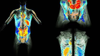
MIBG is labeled with iodine-123, which gives off gamma rays that a gamma camera detects. The camera builds a picture of where MIBG has gathered, and a combined SPECT/CT scan shows the exact anatomical location.
Iodine-131 labeled MIBG is used for treatment rather than imaging, at much higher doses. The diagnostic scan uses iodine-123, which gives clearer pictures with less radiation.
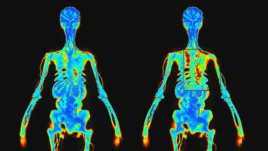
The scan shows function rather than shape. It finds tumor cells in bone marrow and bone that CT and MRI can miss.
Main uses of the MIBG scan
Condition
Question answered
Who it is for
Neuroblastoma
Extent of disease, response to treatment, relapse
Children
Pheochromocytoma and paraganglioma
Location, spread, suitability for MIBG therapy
Adults and children
Heart failure
Sympathetic nerve damage in the heart, risk
Adults
Parkinson disease and Lewy body dementia
Loss of cardiac sympathetic nerves supports the diagnosis
Adults
Some neuroendocrine tumors, such as carcinoid, also take up MIBG, though other scans are usually preferred for them.
Neuroblastoma
Neuroblastoma is the commonest solid tumor outside the brain in young children. It spreads to bone and bone marrow in about half of cases at diagnosis. Around nine in ten neuroblastomas take up MIBG, which makes the scan the reference test for staging.
Curie score after initial chemotherapy, 280 children with stage 4 neuroblastoma
Curie score
Three-year event-free survival
Meaning
2 or below
44.9 percent
Good early response
Above 2
15.4 percent
Poor response, other treatment considered
0 after transplant
49.3 percent
No remaining MIBG-avid disease
The Curie score divides the body into ten regions and grades each from 0 to 3, giving a total that can be compared scan to scan. Europe also uses the SIOPEN score. Both turn the images into numbers the oncology team can act on.
Metaiodobenzylguanidine scans are the most sensitive and specific method of staging and response evaluation in neuroblastoma, particularly when used with a semi-quantitative scoring method (Matthay and colleagues, British Journal of Cancer, 2010).
1
When scans are done
At diagnosis, after induction chemotherapy, before and after stem cell transplant, at the end of treatment and during follow-up. Tumors that do not take up MIBG are followed with FDG PET/CT instead.
Pheochromocytoma and paraganglioma
Pheochromocytoma grows in the adrenal gland and paraganglioma along nerve chains in the neck, chest and abdomen. Both can release adrenaline and noradrenaline, causing high blood pressure, headaches, sweating and palpitations in sudden attacks that patients describe as frightening. Blood or urine tests for metanephrines come first.
Imaging then locates the tumor and shows whether there are others. Inherited forms, linked to genes such as SDHB, VHL and RET, often cause several tumors.
In recent years PET scans have taken over much of this role, since they are more sensitive for small and spread tumors. The MIBG scan remains important in one situation especially, deciding whether a patient can receive MIBG therapy.
Imaging options for pheochromocytoma and paraganglioma
Scan
Strength
When used
Ga-68 DOTATATE PET/CT
Highest sensitivity for most paragangliomas, especially SDHB-related
Staging and searching for spread
F-18 FDOPA PET/CT
Excellent for adrenal pheochromocytoma and head and neck tumors
Where available
I-123 MIBG scan
Confirms MIBG uptake
Before MIBG therapy, where PET is unavailable
The heart and nervous system
Heart failure
The two-year event rate was 15 percent for patients with a heart-to-mediastinum ratio of 1.60 or higher and 37 percent for those below, and MIBG imaging provided independent prognostic value in heart failure (Jacobson and colleagues, Journal of the American College of Cardiology, 2010).
Cardiac MIBG in 961 patients with heart failure
Heart-to-mediastinum ratio
Two-year cardiac events
Meaning
1.60 or higher
15 percent
Sympathetic nerves relatively preserved
Below 1.60
37 percent
Higher risk of worsening, arrhythmia and cardiac death
Cardiac MIBG also helps neurologists. In Parkinson disease and dementia with Lewy bodies, the heart's sympathetic nerves are lost early, so uptake is low, while in conditions that mimic them, such as multiple system atrophy and progressive supranuclear palsy, it stays normal. For cardiac and neurological scans, images are taken at 15 minutes and 4 hours on the same day.
Preparation and medicines
Which medicines must be stopped before the scan?
Many common medicines block MIBG uptake and can make a tumor invisible. They include labetalol, some calcium channel blockers, tricyclic antidepressants, some antipsychotics, cocaine and decongestants such as pseudoephedrine found in cold remedies. Most are stopped for one to two weeks before the injection, and never without the prescribing doctor agreeing. The nuclear medicine team reviews the full medicine list at booking.
1
Protecting the thyroid
Free radioactive iodine can collect in the thyroid. Potassium iodide or a similar drug, taken by mouth from the day before the injection for one to two days, fills the thyroid with ordinary iodine so it takes up none of the radioactive kind.
Blood pressure medicines may need replacing temporarily in patients with pheochromocytoma, and this is planned with the cardiologist or endocrinologist so that blood pressure stays safe. No fasting is needed.
Common medicines that interfere with MIBG uptake
Medicine group
Examples
Usual stop time
Combined alpha and beta blockers
Labetalol
About 72 hours to one week
Tricyclic antidepressants
Amitriptyline, imipramine
One to three weeks
Sympathomimetics and decongestants
Pseudoephedrine, phenylephrine
About 48 hours
Some calcium channel blockers
Nifedipine, amlodipine
About 48 hours
Iodine allergy
An allergy to iodine-containing X-ray contrast or shellfish does not prevent the scan, since the amount of iodine is tiny and chemically different. No special steps follow.
The two scan days
1
Day one
Thyroid blocking tablets are started. A cannula is placed and MIBG is injected slowly over a few minutes. You can then go home or to your hotel.
A typical schedule
Time
Step
Duration
Day before injection
Start thyroid blocking tablets
Once or twice daily
Day 1
Injection
About 30 minutes including preparation
Day 2, about 24 hours later
Whole-body images and SPECT/CT
One to two hours
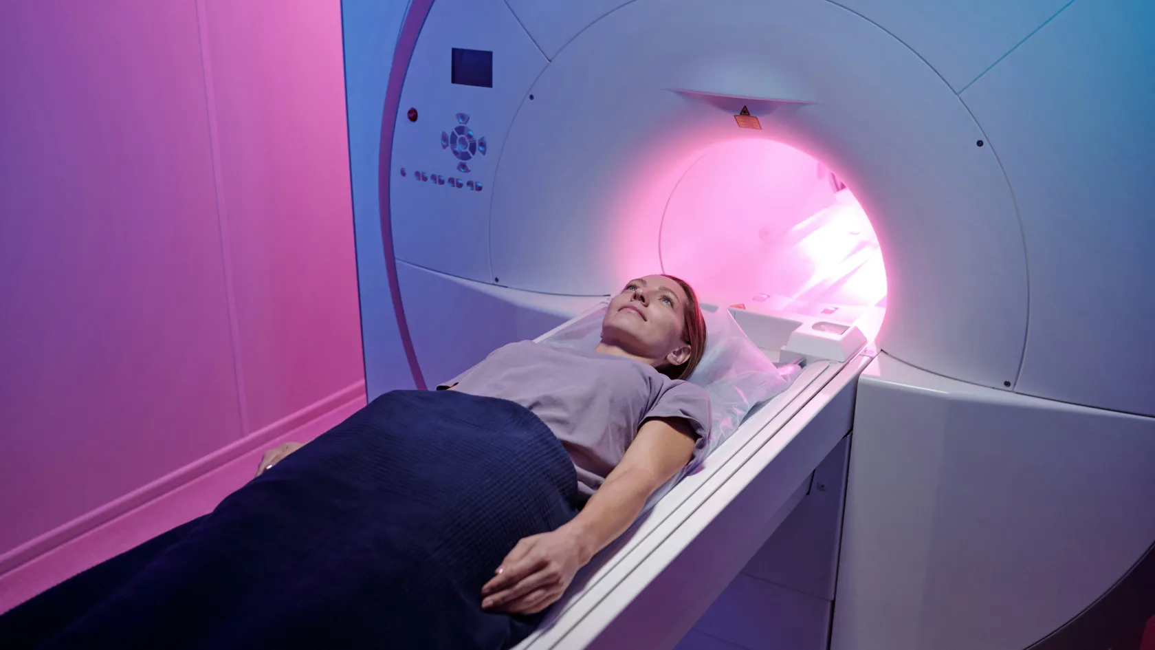
The camera does not surround you tightly and makes no loud noise. Lying still is the only requirement.
Images taken on day two
Image
What it covers
Time
Whole-body planar
Head to feet, front and back
About 30 to 45 minutes
SPECT/CT
3D images of selected areas, matched with CT
About 20 to 30 minutes
Why the scan takes two days
Time sharpens the picture.
Patience pays. Right after the injection, MIBG circulates in the blood and lights up many normal tissues. Over the next day the background clears through the kidneys while tumor cells hold on to the tracer, so images taken at 24 hours show the tumor against a much darker background. Uncertain findings get a second look at 48 hours. Cardiac and neurological scans follow a shorter schedule, with images at 15 minutes and 4 hours on a single day, because the nerve endings in the heart handle MIBG differently from tumors.
Scans in children
Young children find it hard to lie still for an hour. The nuclear medicine team uses blankets, films and a parent at the bedside, and many children over five manage without sedation. The dose of MIBG is adjusted to body weight.
A parent can stay with the child throughout the scan, except pregnant mothers.
1
Sedation
Babies and young children usually need light sedation or brief anesthesia for the images, given by a pediatric anesthetist. Fasting instructions for the sedation day are given in advance.
Before MIBG therapy
Iodine-131 MIBG therapy delivers radiation straight to tumors that take up MIBG, and it treats relapsed or resistant neuroblastoma and spread pheochromocytoma and paraganglioma. A diagnostic scan comes first. It confirms that enough of the disease takes up MIBG.
1
Dosimetry
In some programs the diagnostic scan also helps calculate the therapy dose, so that the tumor receives as much radiation as possible while the bone marrow stays within safe limits.
Reading the result
Normal organs take up MIBG too. Readers expect this.
The salivary glands, heart, liver, bowel and bladder all appear on a normal scan, and the normal adrenal glands sometimes show faintly. The nuclear medicine physician compares the pattern with what is expected, reads the SPECT/CT to pin down each spot, and reports areas of abnormal uptake, their location and, for neuroblastoma, the Curie or SIOPEN score. The written report follows within one to two working days. Images are compared with earlier scans whenever available, since the change over time decides treatment.
A negative scan needs context. One in ten neuroblastomas never take up MIBG, and for them a different scan is used from the start.
Safety
The radiation dose from an iodine-123 MIBG scan is modest, similar to a CT scan of the abdomen, and far below the therapy dose.
What to expect
A brief warm feeling or rarely a rise in blood pressure during injection, which is why it is given slowly.
No effect on how you feel afterwards in almost every case.
Tracer leaves the body through the urine over the following days.
Drink plenty of fluids and empty the bladder often after the injection to reduce the radiation dose to the bladder. Breastfeeding mothers stop for a set period, and the team gives exact instructions.
Pregnancy has to be excluded in women of childbearing age before the injection.
For patients from abroad
One coordinator from the international patients office handles arrangements from the first message through discharge and answers the same WhatsApp number once you are back home. The office works in English, Arabic, French, Russian, Serbian, Romanian and Spanish and books interpreters for other languages, and it arranges the visa invitation letter, airport transfers, daily transport and accommodation near the hospital. Halal, vegetarian and diabetic meals, a prayer room on site, and a female physician on request wherever the rota allows.
Send the list of current medicines, recent blood or urine metanephrine results, and earlier scans before booking. Medicines that interfere with the scan must be stopped before travel, under your doctor's guidance, so the list is reviewed early. MIBG is ordered for a specific date, and the coordinator plans arrival around it.
1
Length of stay and fitness to fly
Three to four days cover the thyroid tablets, injection, images and report. Patients are fit to fly after the day two images. Carry the report and images for your doctors at home.
Cost
What a quote for an MIBG scan covers
Item
Included
Priced separately
Tracer and imaging
Iodine-123 MIBG, whole-body images, SPECT/CT
Additional PET scans
Preparation
Medicine review, thyroid blocking tablets
Sedation or anesthesia for children
Report
Nuclear medicine report, images on disc
Consultation with the treating specialist
The price depends on whether SPECT/CT is added and whether a child needs sedation. The tracer is produced for each patient and ordered in advance, so the date is fixed once booked.
Ask for the quote once the medicine review is done, since changes to the date may affect the price.
References
Yanik GA, Parisi MT, Shulkin BL, et al. Semiquantitative mIBG scoring as a prognostic indicator in patients with stage 4 neuroblastoma. A report from the Children's Oncology Group. J Nucl Med. 2013;54(4):541-548.
Matthay KK, Shulkin B, Ladenstein R, et al. Criteria for evaluation of disease extent by 123I-metaiodobenzylguanidine scans in neuroblastoma. A report for the International Neuroblastoma Risk Group (INRG) Task Force. Br J Cancer. 2010;102(9):1319-1326.
Jacobson AF, Senior R, Cerqueira MD, et al. Myocardial iodine-123 meta-iodobenzylguanidine imaging and cardiac events in heart failure. Results of the prospective ADMIRE-HF study. J Am Coll Cardiol. 2010;55(20):2212-2221.
Bombardieri E, Giammarile F, Aktolun C, et al. 131I/123I-metaiodobenzylguanidine (mIBG) scintigraphy. Procedure guidelines for tumour imaging. Eur J Nucl Med Mol Imaging. 2010;37(12):2436-2446.
Editor's note
Written by the Biruni Hospital medical editorial team. Reviewed by Specialist Dr. Nesrin Aslan İlkmen, Nuclear Medicine.