
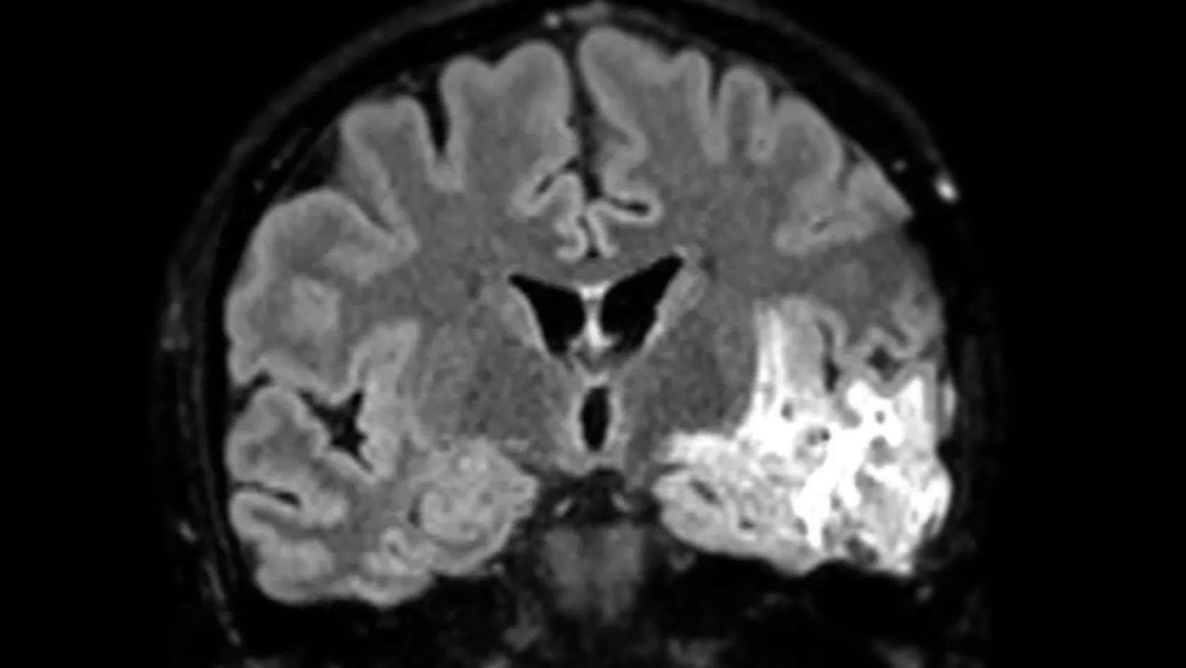
Meningioma Microsurgery
Many meningiomas are found by accident and never cause trouble, so the first decision is whether to operate at all. This page sets out who needs surgery, what a complete removal has to include, and how the position of the tumor changes everything that follows.
About This Department
Most meningiomas are benign. Whether yours needs an operation is a separate question.
Removing the tumor together with its attachment to the lining of the skull and any bone it has grown into is often curative, and few brain tumors allow that sentence to be written at all.
You were probably told two things in the same conversation. That a scan has found a tumor pressing on your brain, and that it is very likely benign. Those two sentences pull in opposite directions, and everything difficult in this diagnosis sits in the space between them.
Benign here has a precise meaning.
The tumor grows from the lining that wraps the brain, sits outside the brain itself, and pushes instead of invading, so removing it completely can end the problem for good, and the same pushing can take away your vision or your hearing while behaving perfectly benignly all the while.
So the questions this page answers run in an unusual order. Whether an operation is needed at all comes first, because a large share of these tumors are found by accident and never cause trouble. Then how much of it has to come out, which for a meningioma means a specific and old classification of what the surgeon did to the attachment instead of to the tumor, and then what location does to the risk, because a tumor on the surface of the brain and the same tumor at the base of the skull are different operations with different consequences. Then the treatment that sometimes follows, the recovery, and the two things a patient flying in from abroad has to plan around.
What a meningioma is, and what benign means here
Meningiomas grow from cells in the arachnoid membrane, one of the three layers that wrap the brain and spinal cord, and they are the most common primary tumor found inside the skull in adults, they occur more often in women than in men, and the great majority behave slowly enough that the word benign is honest. Each one attaches itself to the tough outer lining, draws its blood supply from that lining, and grows outward into whatever space it can find.
Why pushing matters more than invading
A glioma grows through brain tissue, mixing with it, so no operation ever removes all of it. A meningioma does the opposite. It sits outside the brain and displaces it, and a thin layer of arachnoid membrane stays between the two like a wrapper, so a surgeon working carefully can develop that plane and lift the tumor away from brain that has been squashed but never entered. That single anatomical fact is what makes a complete removal curative, and it explains why surgeons pay such attention to a boundary the scan barely shows.
Grade matters too.
Under the World Health Organization classification most meningiomas are grade 1, a minority are grade 2, called atypical, and a small number are grade 3, while grade 2 and grade 3 tumors come back more often and are managed differently, and the grade is read from the tissue after surgery instead of guessed from the scan before it.
Symptoms come from geography.
One over the motor area weakens a hand, one under the frontal lobes takes away the sense of smell and slowly changes personality, one on the optic nerves narrows the visual field so gradually that patients notice only when they fail an eye test, and one at the back of the skull affects balance, hearing and swallowing. Not one item on that list depends on the tumor being malignant.
Whether to operate at all
Scanners find meningiomas in people who came in for something else entirely, a headache, a car accident, a hearing test, and the European Association of Neuro-Oncology states plainly that a large share of these, particularly in patients who have no symptoms or who are elderly, are managed by watching and scanning. So the honest first conversation asks whether to leave it alone.
Researchers followed 441 patients with 459 incidentally discovered meningiomas for a median of 55 months, built a risk model from four imaging features, and produced three groups.
In all three groups the risk of progression flattened out after five years, which is a useful thing to know when a scan has been stable for a long time.
Against that, the case for operating is straightforward when it applies. A tumor causing symptoms should come out, one that is clearly growing should come out before it reaches the structures it is heading toward, and a large tumor with swelling around it will not improve on its own, and the common threshold quoted across specialist centers is a tumor above 3 cm that is producing symptoms, and a surgeon who tells you to wait when you meet that description has earned a second opinion.
Simpson grades, and why the dural base decides everything
Every meningioma operation gets a number afterward, on a scale a London neurosurgeon published in 1957 and which nobody has managed to replace.
What makes it strange, and important to understand before you consent to anything, is that it grades what happened to the tumor's attachment instead of what happened to the tumor.
- Grade I. The tumor is gone, the piece of lining it grew from has been cut out with it, and any bone it had invaded has been drilled away or removed. Nothing is left that can regrow.
- Grade II. The tumor is gone and the attachment has been burned rather than cut out, usually because cutting it out would have meant opening a vein that carries blood away from the brain.
- Grade III. The tumor is gone and the attachment has been left alone entirely, along with anything growing outside the skull lining.
- Grade IV. Part of the tumor has deliberately been left behind, most often because it is wrapped around a nerve or an artery that matters more than the last few percent of tumor does.
- Grade V. The operation took a sample and relieved pressure without removing the tumor.
Read grades I through III again and notice that in all three the tumor itself is completely out, and the scan afterward looks the same. What separates them is invisible on that scan, and it predicts whether you will be back. Which grade is achievable is decided by where the tumor sits and not by how hard the surgeon tries, since one sitting on the convexity of the skull, away from anything critical, allows a grade I removal almost every time, because the surgeon can take a margin of lining and a patch of bone and replace them with a graft. One lying against the wall of a major draining vein cannot, because that vein carries blood from half your brain and cutting it out to gain a Simpson grade would be a poor trade. So a good preoperative conversation names the grade the surgeon expects to reach and the reason for the ceiling.
What the numbers say about coming back
A series of 144 patients operated on over eleven years, followed for a median of four years, sorted them by two things, whether the removal reached Simpson grade I and whether the tumor looked dense on a diffusion sequence. Patients with neither risk factor had no progression or recurrence at all five years after treatment, and patients with both had a 45 percent cumulative incidence of it. The authors noted that this combination predicted who came back better than the tumor's grade under the microscope did, which is a striking thing to find and a good argument for asking which resection grade you were given, and not only what the pathology said.
Grade 2 changes the arithmetic
Fifty nine patients with WHO grade 2 meningiomas, managed at one institution over twenty years, recurred in 31 percent of cases after a Simpson grade I or II removal and in 73 percent after a grade III or IV removal, and almost all of those recurrences happened within five years, so an atypical meningioma incompletely removed is a tumor that comes back in three patients out of four, and the pathology result arriving a week after surgery can therefore change the whole plan.
None of these are numbers to memorize.
They exist to make one point clearly, which is that a meningioma's future depends on the operation to a degree that is unusual in neurosurgery, and that the operation therefore deserves both travel and hard questions.
Location decides the operation and the risk
The skull base is a different operation
Two patients hear the same words, a 3 cm benign meningioma, and face completely different days. On the convexity, the outer surface of the brain, the tumor is reached by lifting a window of bone directly above it, the dural base comes out with the tumor, and the main risks are the ordinary risks of any craniotomy, while at the base of the skull the tumor lies in a crowded floor where every cranial nerve and the carotid artery pass through small openings, the approach comes in through a corridor instead of from directly above, and the operation is measured by how much function is preserved as much as by how much tumor is removed.
This table scrolls sideways on a narrow screen. Swipe or drag to see every column.
| Position | What it usually causes | What limits the removal |
|---|---|---|
| Convexity | Seizures, or weakness if it sits over the motor strip | Usually nothing. A Simpson grade I removal is the expectation here |
| Parasagittal and falcine | Weakness in a leg, seizures, headache | The large vein running along the midline. Removing tumor from its wall risks a stroke from blocked drainage |
| Sphenoid wing | A bulging eye, double vision, loss of vision on one side | The carotid artery and the nerves that move the eye, which the tumor often surrounds |
| Olfactory groove | Lost sense of smell, personality change noticed by family before the patient | Sealing the floor afterward, since the tumor often erodes the bone separating brain from nose |
| Tuberculum sellae | Narrowing visual fields, found late because the loss creeps in from the edges | The optic nerves themselves, and the small vessels feeding them |
| Petroclival and the angle behind the ear | Facial numbness or weakness, hearing loss, unsteadiness, double vision | Seven or eight cranial nerves and the brainstem. Partial removal with radiation afterward is often the better plan |
| Foramen magnum | Neck pain, clumsy hands, weakness that spreads limb by limb | The vertebral artery and the junction where the brainstem becomes the spinal cord |
Find your own row before you read anything else on survival or recurrence, because a page written for convexity meningiomas answers almost none of the questions a petroclival tumor raises.
What microsurgery means here
Microsurgery is not a brand of operation and it is not a smaller one.
It means the surgeon works under an operating microscope at high magnification with instruments a few millimeters wide, and for a meningioma it describes a particular sequence that has barely changed in forty years because it works.
- The head is fixed and the navigation is registered to a scan taken shortly before, so the opening in the skull can be planned over the tumor's attachment instead of over its bulk.
- The dura is opened and the attachment is dealt with first. Cutting the tumor away from its blood supply at the base before touching the rest turns a bleeding operation into a dry one, and it is the single step that most separates an experienced meningioma surgeon from an occasional one.
- The middle of the tumor is hollowed out, often with an ultrasonic aspirator that breaks up tissue and suctions it away. Emptying the inside lets the shell collapse inward, away from the brain, instead of being pulled off it.
- The capsule is then dissected off the arachnoid layer millimeter by millimeter under the microscope, with continuous nerve monitoring where facial movement, hearing or the eye muscles are close, and an angled endoscope used where a corner needs to be seen.
- The attachment is removed or coagulated, which is the moment the Simpson grade is decided, and involved bone is drilled away and replaced.
- The lining is closed watertight, with a graft where a piece was removed, and the bone flap goes back with small plates.
Published series put the operation at two to six hours, with skull base cases running considerably longer. One night in intensive care and three to seven days on the ward is the usual admission for a straightforward case.
Blocking the blood supply first
Some meningiomas are fed by arteries reachable with a catheter. Where that is true, a radiologist can thread a catheter up from the groin a day or two before surgery and plug those feeding vessels with tiny particles, so the tumor arrives in the operating room softer, paler and considerably less bloody.
When it earns its place
Large tumors, tumors with a heavy blood supply visible on the scan, and tumors in positions where controlling bleeding early is hard. It carries a small risk of its own, since particles can travel where they were not intended, and it adds a procedure and a day or two to the stay, and centers that do a lot of this work use it selectively instead of routinely, and if it is proposed for you the reasonable question is which feature of your scan justifies the extra step.
Risks, named
Benign tumors in difficult places carry the risks of the place instead of the risks of the tumor, so this list changes shape depending on which row of the table above is yours.
Vision, when the tumor sits on the optic nerves
Vision deserves separating out from the list above, because it cuts both ways. Pressure released from an optic nerve often restores lost field, sometimes dramatically, and the chance of that recovery falls the longer the nerve has been compressed, so a tuberculum sellae meningioma with failing vision is one of the few meningiomas that is genuinely time sensitive. The same operation can also injure the nerve or its tiny blood supply and make sight worse. Ask for a formal visual field test before surgery and another afterward, because without the first one nobody can tell you which way it went.
The first two weeks
Fatigue dominates, and it surprises families more than pain does. You go home on a reducing course of steroids to settle the swelling around the cavity, on painkillers for a headache that eases over the first week, and with clips or stitches that come out at ten to fourteen days at a review that also confirms you are fit to travel. You also go home on an anti-seizure tablet if the tumor touched the surface of the brain. Desk work comes back at around four to six weeks and physical work later, the scar disappears under hair within a couple of months, and the flatness and the afternoon sleep that catch people out are normal and pass. Recovery after a skull base operation runs on a longer clock. A weak facial nerve or double vision improves over months instead of weeks, an eye that will not close needs drops and sometimes a temporary stitch to protect the cornea, and swallowing that has been affected is assessed before you are cleared to eat. None of that is unusual and all of it is planned for.
When radiation joins in
Surgery is the whole treatment for most grade 1 meningiomas removed completely, and nothing else is needed beyond scans.
Radiation enters the picture in three situations, namely a piece deliberately left behind, a grade 2 or grade 3 result on the pathology, or a tumor that has come back.
After an incomplete removal or a grade 2 result
NRG Oncology ran a phase 2 trial in an intermediate risk group, meaning newly diagnosed grade 2 tumors after a complete removal and recurrent grade 1 tumors after any extent of removal, with 54 Gy in 30 fractions, and three year progression free survival came out at 93.8 percent against a historical figure of 70 percent for surgery alone, local failure at 4.1 percent, and overall survival at 96 percent, with no severe side effects recorded. Radiotherapy now follows surgery for this group as a matter of course because of it.
Radiosurgery for a small residual
Small residual tumor sitting away from the optic nerves can take a single high dose session aimed precisely at it, an alternative to six weeks of daily treatment, and the European guideline supports that choice when the size and the position allow it. Where the residue is large or wrapped around a nerve, fractionated treatment spread over weeks is safer. That decision belongs to a radiation oncologist looking at the post-operative scan, and it is a reason to have both specialties in the same building.
If it comes back
Regrowth after a complete removal is uncommon and slow, and it is picked up on a routine scan long before it produces a symptom.
That is the entire purpose of the follow up schedule.
Three options, and the case for each
A second operation makes sense where the regrowth is reachable, where a better Simpson grade is achievable this time, and where you are well, while radiation makes sense where the tumor sits somewhere a surgeon would prefer not to go twice, and it controls small volumes well. Watching makes sense where the change is a few millimeters on one scan in an older patient, since a tumor that took nine years to become visible again is unlikely to demand anything in the next twelve months. A team that offers all three, and says which one it would choose for you and why, is giving you a clinical opinion instead of a service. If regrowth turns up on a scan at home, send that scan and the original operation note back to the team that operated, because the useful thing at that point is a direct comparison against the baseline study, which a fresh report written without it cannot provide.
What moves the cost
No figure appears here, and the table above explains why.
Take a 3 cm convexity tumor removed in three hours and discharged on day four, against a petroclival tumor approached through the temporal bone with a neurophysiology team in the room for eight hours, are separated by a factor nobody can guess without seeing your imaging.
Six things carry most of the difference. Its position, which sets everything else. The number of hours in the operating room. Whether embolization is planned beforehand, which adds an angiography suite, a radiologist and a day. Whether cranial nerve and motor monitoring are needed, which adds a neurophysiologist and equipment for the whole case. How many intensive care nights the plan assumes. And whether radiotherapy or radiosurgery follows, which is decided only after the pathology and the post-operative scan.
Your own situation moves it too. Age, other illnesses, blood thinning medication, diabetes, previous surgery or radiation at the same site, and how much rehabilitation a cranial nerve problem might need all change what gets planned.
Packages published by hospitals and agencies in this market run from the airport pickup to the final appointment before departure and include the pre-operative tests, the surgeon and anesthesia fees, a stated number of intensive care and ward nights, the scan afterward, an interpreter and hotel nights for a patient and one companion. Flights, insurance, extra nights and the treatment of a complication sit outside. On a meningioma quote the two lines to check hardest are the monitoring and the radiation, since both are decided by findings the quote was written before anyone had. Five questions make two quotations comparable. Which Simpson grade does this plan aim for, and what would stop it? Are cranial nerve and motor monitoring inside the figure, together with the neurophysiology staff that each of them needs? Is embolization included or added later if the angiogram says it is needed? Are intensive care nights counted, and how many? Is radiotherapy or radiosurgery quoted here, or priced separately once the pathology comes back?
Send the MRI as original files and any previous reports. A neurosurgeon reading them will tell you which operation your tumor allows and whether it needs one at all, and that review costs nothing and commits you to nothing.
Planning the trip
Pack the imaging as original files on a disc or a drive, because a surgeon needs to move through every sequence to plan an approach and a photograph of a screen shows none of that. Two studies taken a year apart answer the growth question that one study cannot, so bring any earlier scans too. If your vision or hearing is affected, bring the formal test results, and if you have none, ask for them at home before you travel.
Who comes with you
One person should travel with you. A second bed in the room means they stay through every night of the admission, accommodation for you both on the nights either side is arranged along with the airport transfers and every trip between hotel and hospital, and having a second person in the consent conversation matters here because that conversation is about which specific nerve is at risk and what the surgeon will do if the tumor is stuck to it. Being understood forms part of the clinical work here. The international patients team works in English, Arabic, French, Russian, Serbian, Romanian and Spanish, interpreting in other languages is arranged on request, and one coordinator carries your case from the first message through to discharge and stays reachable on the same WhatsApp number after you fly home. Meningiomas are diagnosed more often in women than in men, so say so in writing if it matters to you. A request for a woman surgeon or physician goes to the department and is met wherever the rota allows, and the time to ask is your first message.
An appointment confirmation and an invitation letter naming the hospital and your doctor, the paperwork most consulates ask for with a medical visa, are issued about ten days before you travel. The hospital kitchen covers halal, vegetarian and diabetic requirements, and there is a prayer room on site.
Radiotherapy and radiosurgery are delivered at Biruni Hospital on the same site as the surgery, so a patient whose pathology turns out to be grade 2 continues with the same team and the same scans, and a patient who would rather have that treatment at home leaves with the operation note, the imaging, the full pathology and a written recommendation addressed to their own oncologist.
Once you are home
Meningioma follow up lasts years, and it runs on scans compared against each other. The usual shape looks like this.
Local scans go back for review by the team that operated, and a photograph of a report or a question on a dose reaches your coordinator on the number you have used all along.
Some things need a doctor the same day, wherever you are.
Clear fluid dripping from a nostril or leaking from the wound. Fever with headache and a stiff neck. A first seizure, or a seizure that runs past five minutes without stopping. New weakness, new double vision, or sight that drops away without warning. A wound that opens, or turns red and painful. A swollen painful calf or breathlessness arriving suddenly. For any of them the nearest emergency department comes first, with your operation note in your hand, and a message to your coordinator afterward.
Meningioma microsurgery FAQ
Does a meningioma always need surgery?
Can meningioma microsurgery cure it completely?
How long is the operation and the hospital stay?
When can I fly home?
Will my husband or wife be able to stay with me?
Will my vision come back after the tumor is removed?
Written by the Biruni Hospital medical editorial team. Reviewed by Dr Yunus Emre Yavuz, Neurosurgery.
References
- Goldbrunner R, Stavrinou P, Jenkinson MD, Sahm F, Mawrin C, Weber DC, et al. EANO guideline on the diagnosis and management of meningiomas. Neuro-Oncology. 2021;23(11):1821-1834.
- Islim AI, Kolamunnage-Dona R, Mohan M, Moon RDC, Crofton A, Haylock BJ, et al. A prognostic model to personalize monitoring regimes for patients with incidental asymptomatic meningiomas. Neuro-Oncology. 2020;22(2):278-289.
- Hwang WL, Marciscano AE, Niemierko A, Kim DW, Stemmer-Rachamimov AO, Curry WT, et al. Imaging and extent of surgical resection predict risk of meningioma recurrence better than WHO histopathological grade. Neuro-Oncology. 2016;18(6):863-872.
- Nanda A, Bir SC, Konar S, Maiti T, Kalakoti P, Jacobsohn JA, et al. Outcome of resection of WHO grade II meningioma and correlation of pathological and radiological predictive factors for recurrence. Journal of Clinical Neuroscience. 2016;31:112-121.
- Rogers L, Zhang P, Vogelbaum MA, Perry A, Ashby LS, Modi JM, et al. Intermediate-risk meningioma. Initial outcomes from NRG Oncology RTOG 0539. Journal of Neurosurgery. 2018;129(1):35-47.
Editor's note
Written by the Biruni Hospital medical editorial team. Reviewed by Assistant Professor Özcan ÇIKLATEKERLİO, Neurosurgery.
Medically reviewed by
Assistant Professor Özcan ÇIKLATEKERLİO
Neurosurgery
Related Treatments
View All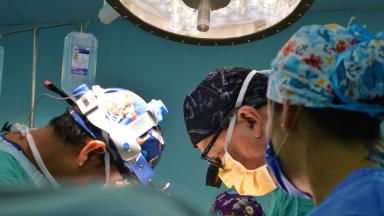
ALIF - Anterior Lumbar Interbody Fusion Surgery
Coming in from the front restores the curve of the lower back better than any posterior route and leaves the back muscles untouched. It also carries one risk most pages will not name, and this page names it.

Brain AVM Surgery - Arteriovenous Malformation Surgery
An unruptured brain AVM bleeds at roughly 2.2 percent a year, and surgery spends its whole risk on a single morning. This page sets those two shapes of risk side by side, explains what the ARUBA trial does and does not say about your case, and shows how a Spetzler-Martin grade changes the answer.
Brain Tumor Surgery - Brain Tumor Removal
Brain surgery is one trade repeated all day, how much tumor comes out against what the brain around it still does. This page puts the published numbers on both sides of that trade.
Cervical Microdiscectomy - Minimally Invasive Neck Disc Surgery
Neck disc surgery is done from the front, through the space between your windpipe and your carotid artery, and almost everything that goes wrong that is not the disc happens in that corridor. What has been measured in it.
Chiari Malformation Surgery
A Chiari malformation is measured in millimeters and treated according to symptoms, and those two things come apart more often than patients are told. Most people with the finding never need an operation. This page sets out who does, what posterior fossa decompression removes, why surgeons still argue over opening the dura, and what the pooled studies say about both sides of that argument.