
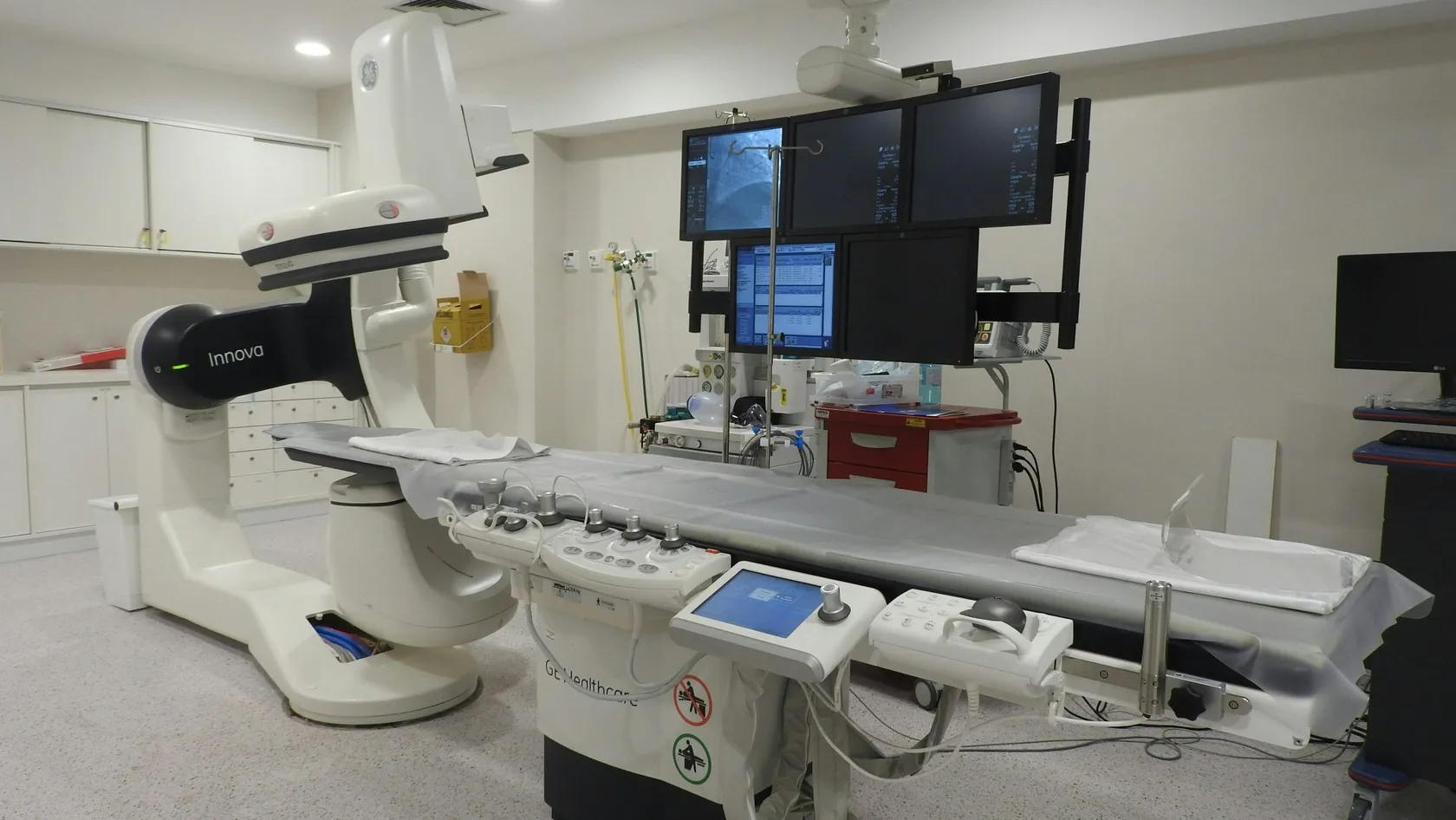
Intraoperative Radiation Therapy - IORT
Intraoperative radiation therapy gives a single dose of radiation to the tumor bed during breast-conserving surgery, so that selected women with early breast cancer finish radiation on the day of the operation. This page explains who qualifies, what the TARGIT-A and ELIOT trials found and why they differ, what happens in the operating room, recovery and side effects, the other cancers treated this way, and how patients from abroad arrange it in Istanbul.
About This Department
For selected women with early breast cancer, the whole course of radiation can be given in the operating room, in the twenty minutes after the tumor comes out. One dose. No return visits.
Intraoperative radiation therapy, or IORT, delivers a single dose of radiation to the bed of a tumor while the patient is still under anesthesia, through an applicator placed into the wound. The TARGIT-A trial randomized 2,298 women aged 45 and over with small breast cancers to a single intraoperative dose or to three to six weeks of external radiation, and found local recurrence at five years of 2.11 percent against 0.95 percent, inside the margin the trial had set for equivalence, with no difference in survival from breast cancer over a median of 8.6 years (Vaidya and colleagues, BMJ, 2020). Around one woman in five needed external radiation added when the pathology showed higher risk. This page explains who qualifies, what the two large trials found and where they disagree, what happens in the operating room, the side effects, the other cancers treated this way, and how patients from abroad arrange it at Biruni Hospital in Istanbul.
What IORT is
Radiation after breast-conserving surgery exists to kill the cancer cells that may remain in the tissue around the cavity. Nine out of ten recurrences in the breast happen within a few centimeters of where the tumor was. IORT takes that observation literally. Once the surgeon has removed the tumor and checked the margins, an applicator the size of the cavity is placed into it, the tissue is gathered around it, and a dose that would take three to five weeks to deliver from outside is given in a single exposure lasting twenty to thirty minutes. The staff leave the room. The anesthesia continues. Then the applicator comes out and the wound is closed as usual.
Two ways to deliver it
One system generates low-energy X-rays at the tip of a probe inside a spherical applicator. The dose peaks at the surface of the cavity and falls away within a centimeter, which spares the skin, the ribs, the lung and the heart almost entirely. The other uses a mobile linear accelerator wheeled into the operating room to deliver electrons through a cylinder placed against the tumor bed, reaching a little deeper and treating a wider area in a few minutes. The trials below tested one of each. Which system the hospital uses for a given patient depends on the size and depth of the tumor bed, and the case review will say.
Who qualifies
A minority of women with breast cancer. The right minority.
- Age. Forty-five and over in the TARGIT-A trial, 48 to 75 in ELIOT. Younger women have a higher rate of recurrence and are treated with whole breast radiation.
- Size. A single tumor of 3.5 centimeters or less on imaging, and in practice under 2.5 centimeters, of the ordinary ductal type.
- Lymph nodes. Clinically clear before surgery, confirmed by a sentinel node biopsy at the operation.
- Receptors. Estrogen receptor positive and HER2 negative disease carries the lowest risk of recurrence and gains the most.
- Margins. Clear on the frozen section at surgery, and confirmed on the final pathology.
What if the pathology report after surgery shows higher risk?
The risk-adapted approach
Can IORT replace radiation entirely?
| Feature | Suitable | Better served by whole breast radiation |
|---|---|---|
| Age | 50 and over | Under 45 |
| Tumor size | Up to 2.5 cm, single | Over 3 cm, or more than one |
| Type | Invasive ductal, grade 1 to 2 | Lobular, grade 3, extensive DCIS |
| Nodes | Negative | Positive |
| Receptors | ER positive, HER2 negative | Triple negative or HER2 positive |
| Genetics | No known mutation | BRCA carrier |
What the trials show
Two large randomized trials. They agree on survival and disagree on recurrence, and the reason for the disagreement is the lesson.
Is a single dose as effective as three weeks of radiation?
| Measure | TARGIT-A (X-rays, 2,298 women) | ELIOT (electrons, 1,305 women) |
|---|---|---|
| Local recurrence at 5 years | 2.11 percent IORT, 0.95 percent external | 4.2 percent IORT, 0.5 percent external |
| Local recurrence at long follow-up | No significant difference in local recurrence-free survival at 8.6 years | 12.6 percent IORT, 2.4 percent external at 15 years |
| Overall survival | No difference, non-breast deaths lower after IORT | No difference at 5, 10 and 15 years |
| External radiation added when pathology showed risk | Yes, about 20 percent | No |
| Who was enrolled | Age 45 and over, ductal cancer up to 3.5 cm, risk-adapted | Age 48 to 75, tumor up to 2.5 cm, all subtypes |
- ELIOT gave the single dose to every woman randomized to it, whatever the final pathology showed, and its recurrence rate was more than four times that of whole breast radiation, although survival was identical (Orecchia and colleagues, Lancet Oncology, 2021).
- TARGIT-A added external radiation when the pathology showed risk, and its recurrence rate stayed within a percentage point.
- Both trial groups reached the same conclusion in their own words, that ELIOT should be offered to selected patients at low risk of recurrence and that TARGIT-IORT should be discussed with eligible patients when breast-conserving surgery is planned.
Reading the numbers as a patient
A recurrence in the breast after IORT is treated, usually by a second operation and external radiation, and it did not shorten life in either trial. What a patient weighs, then, is a small extra risk of a second local treatment against the certainty of avoiding three to five weeks of daily visits, the skin reaction, the tiredness and the exposure of the heart and lung. For a woman of 70 with a small hormone-sensitive tumor, the trade is easy. For a woman of 46 with a grade 3 tumor, it is a trade the criteria exist to prevent.
The operation day
The same operation as any breast-conserving surgery, made longer by about forty minutes.
If the frozen section says no
What happens if the node or the margin is positive during surgery?
Recovery and side effects
Fewer than after external radiation, and different in timing.
Will I need any other treatment?
What the trials did not measure
Beyond the breast
The randomized evidence lives in breast cancer. The technique itself predates it by decades, and specialist centers use it wherever a tumor lies against a structure that cannot be given a full dose from outside, because the surgeon can lift the bowel, the ureter or the nerve out of the field with a retractor and the dose can go where no external beam could safely reach.
- Locally advanced and recurrent rectal cancer, where a single electron dose to the pelvic sidewall after removal of the tumor raises local control in patients whose margins are close.
- Pancreatic cancer, where the dose goes to the bed of the tumor after resection or, in unresectable disease, to the tumor itself for pain control.
- Soft tissue sarcoma of the limb or retroperitoneum, where IORT to the tumor bed reduces the external dose needed and protects the nerves and bowel nearby.
- Recurrent gynecologic and head and neck cancers in previously irradiated tissue, where no further external dose is possible.
What it does not replace
Chemotherapy, hormone therapy and surgery itself. IORT changes how radiation is given. Nothing on this page changes whether radiation is needed.
Deciding, and coming from abroad
IORT rewards planning. Imaging and biopsy settle the decision before surgery, the frozen section confirms it during, and a patient who arrives with the right studies can have the operation within days of the consultation. What the team needs is the mammogram and ultrasound as images, the biopsy report with receptor status and grade, an MRI if one was done, the surgical plan proposed at home, and the list of medicines. Two things shorten the process more than anything else. A biopsy report that states the receptor status, the HER2 result and the grade, since without those three the case cannot be assessed and the biopsy would have to be repeated, and imaging sent as files rather than as a written report, because the radiologist here will measure the tumor again and look for a second focus that would rule IORT out. A patient who has already been told at home that she needs a mastectomy, or that her tumor is too large for IORT, should still send the file, because the criteria vary between centers and the answer is sometimes different.
How long do I need to stay?
TARGIT-IORT should be discussed with eligible patients when breast conserving surgery is planned (Vaidya and colleagues, 2020).
Who decides whether I qualify?
One coordinator from the international patients office handles arrangements from the first message through discharge and answers the same WhatsApp number once you are back home. The office works in English, Arabic, French, Russian, Serbian, Romanian and Spanish and books interpreters for other languages. It arranges the visa invitation letter, airport transfers, daily transport and accommodation near the hospital. The kitchen prepares halal, vegetarian and diabetic meals, a prayer room is on site, and a request for a female surgeon is met wherever the rota allows. Patients are fit to fly after the wound check.
Cost
An estimate follows the file review and covers the surgery, anesthesia, the intraoperative dose with the physicist's time, the applicator, pathology and the hospital night. Hospitals in this market quote breast-conserving surgery with IORT as one package, with any external radiation that the pathology later requires priced as a separate course.
Two questions. What the figure includes, and what the added course would cost if the pathology calls for it.
References
- Vaidya JS, Bulsara M, Baum M, et al. Long term survival and local control outcomes from single dose targeted intraoperative radiotherapy during lumpectomy (TARGIT-IORT) for early breast cancer. TARGIT-A randomised clinical trial. BMJ. 2020;370:m2836.
- Orecchia R, Veronesi U, Maisonneuve P, et al. Intraoperative irradiation for early breast cancer (ELIOT). Long-term recurrence and survival outcomes from a single-centre, randomised, phase 3 equivalence trial. Lancet Oncol. 2021;22(5):597-608.
- Murray Brunt A, Haviland JS, Wheatley DA, et al. Hypofractionated breast radiotherapy for 1 week versus 3 weeks (FAST-Forward). 5-year efficacy and late normal tissue effects results from a multicentre, non-inferiority, randomised, phase 3 trial. Lancet. 2020;395(10237):1613-1626.
Editor's note
Written by the Biruni Hospital medical editorial team. Reviewed by Prof. Dr. Metin GÜDEN, Radiation Oncology.
Medically reviewed by
Prof. Dr. Metin GÜDEN
Radiation Oncology
Related Treatments
View All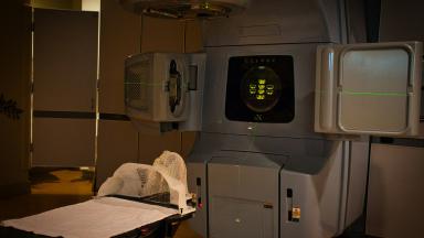
Brachytherapy - Internal Radiation Therapy
A beam from outside has to cross healthy tissue to reach a tumor. Brachytherapy avoids that by placing a rice-sized radioactive source inside the tumor for a few minutes, then taking it out again. It is an essential part of curing cervical cancer and a tested option for womb, prostate and early breast cancer.
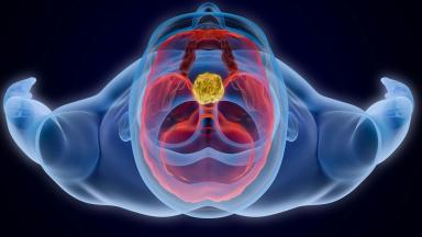
Brain Tumor Radiation Therapy
The plan for a brain tumor starts with what must be protected, the optic nerves, the brainstem, the hippocampus, and then shapes the dose around them. Radiosurgery in one to five sessions now treats most metastases, and six weeks of radiotherapy with temozolomide remains the standard after surgery for glioblastoma.

Chemoradiation Therapy - Concurrent Chemoradiotherapy
Blood test on Monday, a drip on Tuesday, radiotherapy every weekday, and the same again for six or seven weeks. Chemoradiation asks more of a patient than either treatment alone and, for several cancers, cures more. See the survival gains trial by trial, the side effects they came with, who is fit for it and how a long stay is organized.
Elekta Versa HD Radiation Therapy
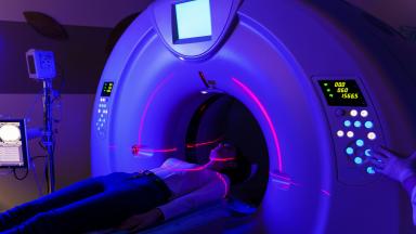
The Elekta Versa HD is the linear accelerator on which Biruni Hospital delivers external beam radiation therapy, with a 160-leaf collimator, high dose rate beams and cone beam CT imaging before every session. This page covers the techniques, from VMAT to radiosurgery, the cancers treated, what large trials show about shorter courses for breast, prostate and metastases, the path from first visit to last session, side effects, and how patients from abroad arrange treatment.
Glioblastoma Radiation Therapy
A glioblastoma has no edge, so surgery alone is never enough. Radiation covers the cavity and a margin of brain around it, together with a daily temozolomide capsule, over six weeks or three depending on age and fitness.