
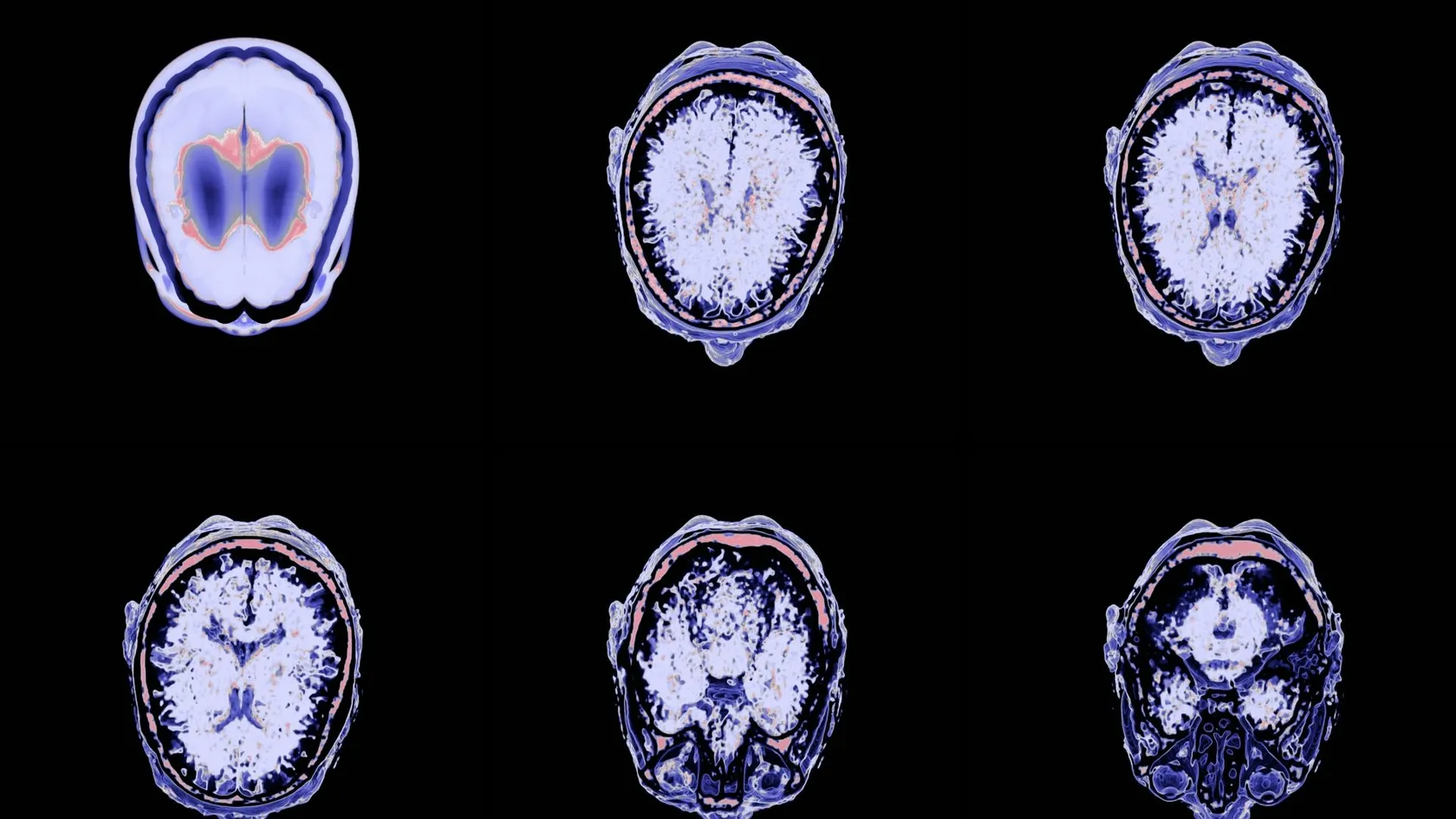
Glioblastoma Surgery
Surgery does not cure a glioblastoma, and no honest page says otherwise. What it changes is how much tumor is left for radiotherapy to work on, and the published survival gap between resection groups is measured in years rather than weeks.
About This Department
Somebody has already given you a number. The published range behind it is much wider.
Across 761 patients at one center, younger people whose surgeons cleared both the enhancing and the non enhancing tumor had a median survival of 37.3 months, against 16.5 months where the non enhancing part was left in place.
Someone has probably already said a number out loud to you, and it was probably somewhere between twelve and fifteen months. That figure is a median from a trial population, which means half the people in it lived longer, some of them a great deal longer, and it was measured across every age and every subtype together.
Medians describe groups. You are one patient.
Four things move where a person sits in that range, and only one of them is still open on the day you are reading this. Age is fixed. The methylation status of the MGMT promoter in your tumor is fixed. How well you are functioning right now is mostly fixed. How much tumor comes out at the operation is the one that has not happened yet, and the published spread attached to it is wide enough to be worth traveling for. This page sets out what the operation can and cannot do, what the numbers behind extent of removal say, what happens in the three to six weeks afterward, and the two decisions that catch out every international patient. The first decides whether the radiotherapy course that follows runs three weeks or six. The second decides whether you sit it out here or fly home for it.
What actually moves the outlook
Any neuro oncologist weighs the same four things before saying anything at all. Three of them arrived with you.
That last one is the reason people get on airplanes.
What the operation is for
Surgery does three separate jobs on the same afternoon, and it helps to keep them apart. It takes pressure off the brain, which is what lifts a headache, clears a fog and often restores a weak arm within days, and it supplies the tissue that the pathology and the molecular panel are read from, which is what decides every drug and every dose afterward. And it reduces the number of tumor cells that radiotherapy and chemotherapy then have to deal with, which is where the survival difference between resection groups comes from.
What it cannot do
No operation cures a glioblastoma, and any page that implies otherwise is selling something. The tumor sends cells out along nerve fibers well past the bright rim on the scan, into brain that looks and works properly, and no instrument reaches those without taking the brain with them, so the tumor returns in essentially every patient, and the honest description of what surgery buys is more time and better function during that time, with a cure out of reach. Buying that is not nothing. It falls short of what people hope they are buying, and being told the difference clearly at the start beats optimism.
How much comes out, and why age changes the target
On an MRI a glioblastoma shows two territories.
One shows as a bright rim that takes up contrast, the dense part of the tumor with its leaky new blood vessels. The other spreads out as a wider gray zone around it on the FLAIR sequence, holding swelling and tumor cells mixed together. Surgeons have chased the bright rim for decades. The argument of the last few years concerns the gray zone.
What 761 patients showed, split by ageA study of 761 patients treated at one university center, checked afterward against 107 patients at a second and 99 at a third, sorted patients by how much of each territory was removed. Younger patients whose surgeons cleared the enhancing tumor and went on into the non enhancing tumor had a median survival of 37.3 months, which is close to what patients with the far gentler IDH mutant tumors achieve. Younger patients whose enhancing tumor was cleared but whose non enhancing tumor stayed behind had a median survival of 16.5 months. Older patients gained from clearing the enhancing tumor, at a median of 12.4 months, and did not gain measurably from going further. The pattern held whether or not the MGMT promoter was methylated.
Read the difference between 37.3 and 16.5 months carefully, because it is the same tumor and the same enhancing rim removed in both groups, and what separated them is how far past the rim the surgeon was able to go. Going past the rim is only possible where the brain past the rim can be spared, and that is decided by anatomy rather than by ambition. A tumor in the front of the right frontal lobe allows a wide margin. The same tumor sitting on the speech area allows almost none. This is why two surgeons looking at two different scans give two different answers on how aggressive they intend to be, and why an opinion given without your imaging in front of the person is not an opinion at all.
What makes a bigger removal safe
Four things in the room do the work, and none of them replaces the surgeon's judgment on where to stop.
Ask which of these your operation will use, and ask what the plan is if the answer to any of them is no.
When a biopsy is the right operation
Some glioblastomas cannot be resected safely, and hearing that early from a surgeon who has read your scan is more useful than hearing an optimistic answer from someone who has not.
Tumors deep in the thalamus or the brainstem sit behind structures that cannot be crossed, and tumors that have grown across the corpus callosum into both hemispheres cannot be taken out on either side without a heavy cost. Disease that has appeared in several places at once is already spread beyond what any resection reaches. And a patient who is bedbound before surgery loses more to a long operation than the removal gives back.
What a biopsy still gives you
Everything the treatment plan depends on. A navigated needle through a small opening takes enough tissue for the diagnosis, the grade and the full molecular panel, the whole procedure takes around an hour, and you leave the next day, and radiotherapy and chemotherapy are then chosen from the same markers as they would be after a large resection, so a biopsy does not put you on a lesser pathway. What it does not do is take pressure off, so steroids carry more of the load afterward.
The operation and the days around it
Five stages, from the scan to the ward
The molecular results follow separately, seven to fourteen working days later. Book your return around that report rather than around the discharge date.
Risks, and how each is handled
Everything below appears on the consent form, paired with its management in the same sentence.
- A new weakness or speech difficulty. The complication that matters most. Most are temporary, driven by swelling around the cavity instead of by injury, and they lift over days to weeks with steroids and therapy. A minority do not, and mapping exists to keep that minority small.
- Bleeding into the cavity. Uncommon and usually announced within hours, which is what the first night in intensive care is for. A repeat scan settles it, and a small number of patients return to the operating room.
- Seizures. Common before surgery and possible afterward. Anti-seizure medication is given around the operation and continued for months, and a single seizure in the first days is treated on the ward rather than as a disaster.
- Wound problems. Steroids slow healing and radiotherapy slows it more, so the scar is checked before radiotherapy is allowed to begin. An infection here delays treatment, and delay is the real damage.
- Blood clots. Patients with a glioblastoma clot more readily than almost any other surgical group. Stockings, early walking and blood thinning injections once the bleeding risk passes are the standard answer, and a swollen calf or sudden breathlessness is an emergency anywhere in the world.
- Fluid collecting under the scar. A soft swelling appears in some patients as the cavity settles. Most are watched and shrink on their own.
Steroids deserve their own paragraph
Dexamethasone is the drug that makes a patient with a large tumor look transformed within two days, and it is also the drug families ask most about once the transformation wears thin, since it brings hunger, a round face, broken sleep, a short temper, muscle weakness in the thighs and higher blood sugar, none of which signals that anything is going wrong. The dose comes down on a written schedule instead of on how you feel, because stopping abruptly after weeks on it makes people unwell, so take the schedule seriously, keep it in writing, and tell any doctor you see at home that you are on it.
What starts three to six weeks later
Radiotherapy is the treatment that follows every glioblastoma operation, it begins once the wound has healed, and its length is decided mainly by your age. That fact carries more logistical weight than anything else on this page for a patient who has flown in.
If you are under 65
The standard course delivers 60 Gy spread over six weeks, with a low dose of temozolomide taken every single day alongside it, followed by monthly cycles of the same tablet, and six weeks means thirty visits, Monday to Friday, and the tablet continues through the weekends.
What those six weeks feel like
Waiting rooms take longer. Each appointment takes a few minutes on the table after a first session that is longer, because a mask is made to hold your head in exactly the same position every day. Many patients work or potter through the first two or three weeks. Fatigue builds from week four, hair falls out in the path of the beams, and appetite drops. None of that is a sign the treatment is failing, and all of it recovers over the months afterward.
If you are 65 or older
Researchers randomized 562 patients aged 65 and above, median age 73, between a short course of 40 Gy in 15 fractions given alone and the same short course with temozolomide added. Median survival ran 9.3 months with the drug against 7.6 months without it, quality of life came out similar in both groups, and among the 165 patients whose MGMT promoter was methylated the gap was much wider, at 13.5 months against 7.7. So an older patient is offered three weeks of radiotherapy rather than six, which halves the time you have to stay wherever you decide to have it, and the MGMT result is what decides whether the tablet joins in.
Three weeks or six. Ask which one applies to you before you book anything.
This table scrolls sideways on a narrow screen. Swipe or drag to see every column.
| Schedule | Who it is offered to, and what it involves | Time in one place |
|---|---|---|
| Standard course | Adults under 65, and fitter patients above that age. 60 Gy over six weeks with a low daily dose of temozolomide, then monthly cycles of the tablet | Six weeks of daily visits, Monday to Friday |
| Short course | Patients of 65 and above, median age 73 in the trial. 40 Gy in 15 fractions, with temozolomide added, and the widest gain where MGMT is methylated | Three weeks of daily visits, Monday to Friday |
| Both | Start three to six weeks after surgery, once the wound has healed, and need a mask fitted at a planning session first | Add a week for planning and the first scan afterward |
The scan that looks worse and is not
Nothing on this page will save you more distress than the next two paragraphs, and almost no page written for patients contains them.
Pseudoprogression, in numbersRadiotherapy inflames the tissue it treats, and that inflammation takes up contrast on an MRI exactly the way tumor does. In a cohort of 284 glioblastoma patients, 19.4 percent of the scans done three months after radiotherapy showed what looked like progression and turned out to be treatment effect, falling to 7.0 percent at six months. It was more common in patients whose MGMT promoter was methylated, which is to say in the patients whose treatment was working best, and more common in those who had no new neurological symptoms. The authors recommended continuing the temozolomide cycles when the imaging is unclear rather than abandoning a treatment that may be succeeding.
Roughly one scan in five at that point is telling you something other than what it appears to say. If you are back in your own country when it happens, and a radiologist who has never seen your case writes the word progression in a report, the correct next step is a conversation with the team that treated you and a repeat scan six to eight weeks later before anything in the plan changes.
Send the images. That is what the coordinator and the WhatsApp number are for.
Tumor treating fields, and what to ask about them
A wearable device delivers low intensity alternating electric fields through four adhesive arrays stuck to a shaved scalp, and it interferes with tumor cells as they divide, it is worn at least 18 hours a day, and it is added to the monthly temozolomide and replaces nothing. Compliance decides whether it works. The trial behind it randomized 695 patients who had finished chemoradiotherapy. Median survival came out at 20.9 months with the device against 16.0 months without, and progression free survival at 6.7 months against 4.0. Skin reactions under the arrays affected 52 percent of the patients using it, and the practical cost is a shaved head, a battery pack to carry and a device that has to be worn most of the day to work. Availability, licensing and funding for it differ sharply between countries, so the useful question is whether it is obtainable and paid for where you live, and that is one for your own oncologist rather than for any hospital's website.
Treatment here, or the same treatment at home
Surgery costs you a week.
What follows costs three weeks or six, and it has to begin within three to six weeks of the operation, so the whole trip has to be planned backwards from that.
What each route needs from you
Staying works because radiotherapy and chemotherapy are delivered at Biruni Hospital on the same site as the surgery, so the radiation oncologist plans from the scans already in the system, the pathology never moves, and nobody has to be persuaded to accept another hospital's report. Nothing has to be re-argued. Hotel rooms for you and one companion and the transport to every appointment are arranged by the international patients office, and the coordinator who answered your first message is the same person managing week five. For a patient over 65 on the short course this means three weeks in one city, which many families find easier to organize than they expect. Going home works too, and for someone with children, a job or an oncologist they already trust it is often better. It works on one condition. The appointment at home is arranged before you leave rather than after you land, because the window is measured in weeks and a referral started from a hotel room is the thing that goes wrong. You travel with the operation note, the post-operative MRI on a disc, the complete pathology including the MGMT result, and a written treatment recommendation addressed to your own oncologist, so nothing needs repeating and no time is lost re-testing tissue.
Your surgeon signs the fitness to fly opinion at the wound check, usually between day 10 and day 14 and once the scar has dried, and puts a date on it for the airline and the insurer.
When it comes back
A second operation is possible for some patients and pointless for others, and the evidence is unusually specific on which is which, since an eight center study followed 681 patients through a first recurrence, 310 of whom had a second operation. Re-resection was associated with longer survival even after adjusting for molecular and clinical differences, and the benefit tracked one measurement, namely how much enhancing tumor was left afterward. Patients left with one cubic centimeter or less did better than patients managed without surgery at all. Going further and chasing the non enhancing tumor a second time was not associated with longer survival and was frequently followed by a new neurological deficit. That is a narrow, useful conclusion. Operate again where the enhancing recurrence can be cleared and the patient is well enough to go on to more treatment afterward, and do not operate again simply because an operation is possible. The other options at recurrence are a different chemotherapy, a second and smaller course of radiotherapy where the first one finished long enough ago, a clinical trial, and treatment aimed purely at symptoms. Any team that presents all of those honestly, including the last one, is a team worth listening to.
What moves the cost
Publishing a price here would mislead almost everyone who read it, because a glioblastoma quote covers wildly different amounts of work depending on what the scan turns out to allow, and an hour long needle biopsy and a seven hour awake resection followed by six weeks of daily radiotherapy are not the same purchase, and nobody knows which one applies to you until a neurosurgeon has looked at your imaging. Six things carry most of the variation. Which operation you are having and how long it runs. Whether fluorescence, intraoperative ultrasound and nerve monitoring are used, and whether the case needs awake mapping with a neurophysiologist present. How many intensive care nights the plan assumes. How wide the molecular panel is, since a methylation array costs considerably more than a basic set of stains and it is the line a quotation skips most reliably. Whether the radiotherapy course is three weeks or six. Quotations rarely spell these out. And whether that radiotherapy and the tablets alongside it sit inside the same quotation or arrive as a second one after the pathology.
Your own situation moves it as well. Age, how well you are functioning, diabetes made worse by steroids, blood thinning medication, heart or lung disease, and how much rehabilitation the case is likely to need all change what is planned and therefore what it costs.
Published packages in this market run from the airport pickup to the last appointment before departure, taking in the pre-operative workup, the operating room and anesthesia fees, an agreed number of intensive care and ward nights, the scan afterward, an interpreter and hotel nights for you and one companion, while flights, insurance, any night the plan did not anticipate and the treatment of a complication sit outside it. Read both lists. Five questions make two quotations comparable.
- Does this figure assume a biopsy, a debulking or a maximal resection, and how many hours in the operating room?
- Are fluorescence, monitoring and any awake mapping included, with the staff each of those needs?
- Which molecular tests are inside the figure, and is MGMT methylation one of them?
- How many intensive care nights are counted, and what happens financially if a seventh ward night is needed?
- Is radiotherapy quoted here, and if so for three weeks or six?
Send the MRI, any pathology you already have and your list of medicines. A neurosurgeon reading them will tell you which operation your tumor allows, and that review costs nothing and commits you to nothing.
Planning the trip
Bring the imaging as original files on a disc or a drive, since a surgeon has to scroll through every sequence to plan an approach and cannot do it from photographs of a screen, and bring any tissue slides or blocks from an earlier operation, because the molecular tests can often be run on them. Write out your medicines with their doses, and mark the steroid clearly.
Who travels with you, and why it matters here
One person should come, and not only to carry the bags. A glioblastoma can blunt memory and concentration before anyone notices, steroids do the same for different reasons, and the conversations you will have in the first two weeks carry decisions on radiotherapy schedules and travel dates that are hard to hold onto alone. Patient rooms have a companion bed, so that person stays every night of the admission, with accommodation for you both on the nights either side and every transfer between airport, hotel and hospital arranged for you. Bring a notebook. Have your companion take notes in every consultation, and have the team write the plan down. Being understood matters more here than on most pages, because the decisions come quickly and they are permanent. Interpreting in other languages is arranged on request, the team itself works in English, Arabic, French, Russian, Serbian, Romanian and Spanish, and one coordinator carries your case from the first message through to discharge and stays on the same WhatsApp number once you are home. If you would rather see a woman surgeon or physician, say so in that first message, and the department accommodates it wherever the rota allows.
An appointment confirmation and an invitation letter naming the hospital and your doctor, the document most consulates want with a medical visa application, go out about ten days before you travel, and the hospital kitchen covers halal, vegetarian and diabetic requirements, which matters while steroids are pushing blood sugar up, and there is a prayer room on site.
Once you are home
Follow up runs on scans, with an MRI a month after radiotherapy finishes and then every two to three months, each one compared against the scan taken in the first 48 hours after surgery, so keep that early study safe, on a disc, in more than one place. Scans are the whole conversation. Local scans can be sent back to the team that operated for a second opinion, and a photograph of a report or a question on a dose reaches your coordinator on the number you have been using all along.
Leave with a file your own oncologist can act on without telephoning anybody. The operation note stating what was removed, the post-operative MRI, the complete pathology with the MGMT result and its date, the steroid reducing schedule, the anti-seizure medication with its dose, the radiotherapy plan and the date of the next scan.
Six things need a doctor the same day, wherever in the world you are.
A seizure that runs past five minutes, or one seizure rolling into the next without you waking in between. New weakness, new confusion, or words that will not come. Fever with headache and a stiff neck, which after brain surgery is treated as meningitis until proven otherwise. A wound that opens or leaks. A headache that wakes you and brings vomiting with it. A swollen painful calf or breathlessness arriving out of nowhere. For any of them the nearest emergency department comes first, with your operation note in your hand, and a message to your coordinator afterward.
Glioblastoma surgery FAQ
How long does the operation take and how long will I be in hospital?
Do I have to stay for the radiotherapy?
When can I fly home after glioblastoma surgery?
My scan after radiotherapy looks worse. Is the tumor growing?
Can my wife or husband stay in the room with me?
Is a second operation worth it if the tumor returns?
Written by the Biruni Hospital medical editorial team. Reviewed by Dr Yunus Emre Yavuz, Neurosurgery.
References
- Molinaro AM, Hervey-Jumper S, Morshed RA, Young J, Han SJ, Chunduru P, et al. Association of maximal extent of resection of contrast-enhanced and non-contrast-enhanced tumor with survival within molecular subgroups of patients with newly diagnosed glioblastoma. JAMA Oncology. 2020;6(4):495-503.
- Perry JR, Laperriere N, O'Callaghan CJ, Brandes AA, Menten J, Phillips C, et al. Short-course radiation plus temozolomide in elderly patients with glioblastoma. New England Journal of Medicine. 2017;376(11):1027-1037.
- Stupp R, Taillibert S, Kanner A, Read W, Steinberg D, Lhermitte B, et al. Effect of tumor-treating fields plus maintenance temozolomide vs maintenance temozolomide alone on survival in patients with glioblastoma. A randomized clinical trial. JAMA. 2017;318(23):2306-2316.
- Blakstad H, Mendoza Mireles EE, Heggebø LC, Magelssen H, Sprauten M, Johannesen TB, et al. Incidence and outcome of pseudoprogression after radiation therapy in glioblastoma patients. A cohort study. Neuro-Oncology Practice. 2023;11(1):36-45.
- Karschnia P, Dono A, Young JS, Juenger ST, Teske N, Häni L, et al. Prognostic evaluation of re-resection for recurrent glioblastoma using the novel RANO classification for extent of resection. A report of the RANO resect group. Neuro-Oncology. 2023;25(9):1672-1685.
Editor's note
Written by the Biruni Hospital medical editorial team. Reviewed by Assistant Professor Fikret BAŞKAN, Neurosurgery.
Medically reviewed by
Assistant Professor Fikret BAŞKAN
Neurosurgery
Related Treatments
View All
ALIF - Anterior Lumbar Interbody Fusion Surgery
Coming in from the front restores the curve of the lower back better than any posterior route and leaves the back muscles untouched. It also carries one risk most pages will not name, and this page names it.

Brain AVM Surgery - Arteriovenous Malformation Surgery
An unruptured brain AVM bleeds at roughly 2.2 percent a year, and surgery spends its whole risk on a single morning. This page sets those two shapes of risk side by side, explains what the ARUBA trial does and does not say about your case, and shows how a Spetzler-Martin grade changes the answer.
Brain Tumor Surgery - Brain Tumor Removal
Brain surgery is one trade repeated all day, how much tumor comes out against what the brain around it still does. This page puts the published numbers on both sides of that trade.
Cervical Microdiscectomy - Minimally Invasive Neck Disc Surgery
Neck disc surgery is done from the front, through the space between your windpipe and your carotid artery, and almost everything that goes wrong that is not the disc happens in that corridor. What has been measured in it.
Chiari Malformation Surgery
A Chiari malformation is measured in millimeters and treated according to symptoms, and those two things come apart more often than patients are told. Most people with the finding never need an operation. This page sets out who does, what posterior fossa decompression removes, why surgeons still argue over opening the dura, and what the pooled studies say about both sides of that argument.