
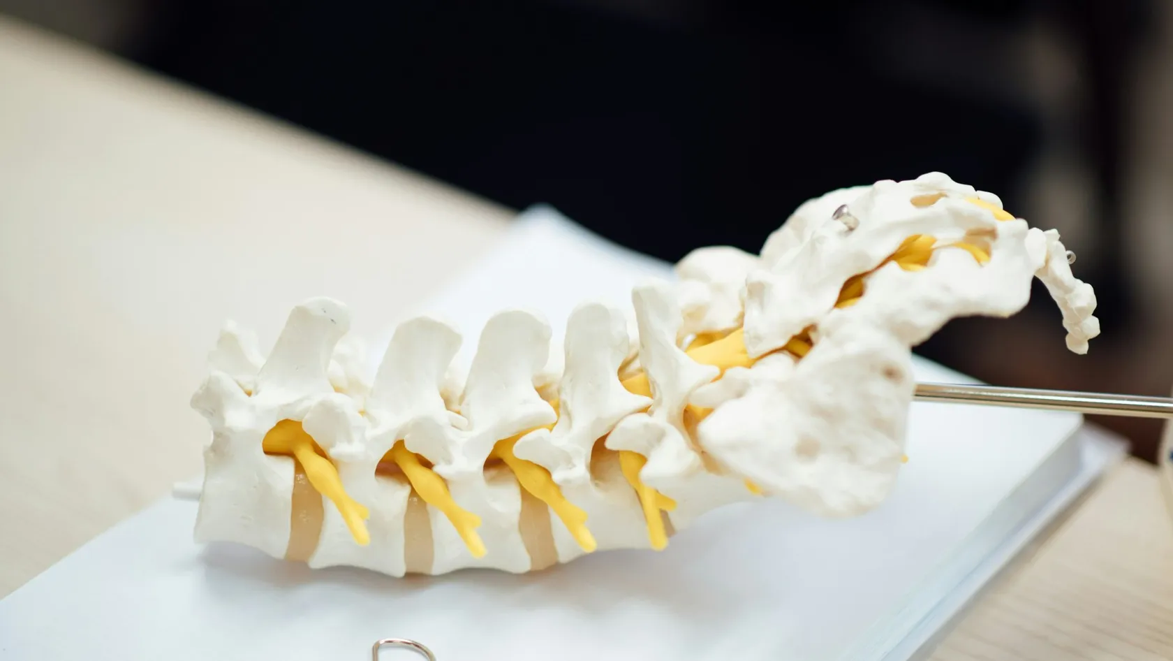
Herniated Disc Surgery - Bulged Disc Surgery
Bulge, protrusion, extrusion and sequestration are four different things on your radiology report, and only some of them are ever operations. What each word means for the back and the neck, and which findings dissolve on their own.
About This Department
In a study that scanned 1,211 people with no neck symptoms whatsoever, 87.6 percent had a bulging disc, including 73.3 percent of the men and 78.0 percent of the women in their twenties. A bulge is not a herniation, three professional societies have agreed a naming standard that keeps them in separate categories, and radiology reports mix the words anyway. This page is about the words on your report, which of them describe something an operation can fix, and which of them describe what a normal spine looks like on a scan.
Free consultation
Send the report and the images, and have someone read them against your symptoms
Send the radiology report in full together with the image files themselves, a description of where the pain, numbness or weakness actually goes in the arm or the leg, how long it has been there and whether it is changing, and a note of anything already tried. A spine surgeon and a radiologist read the report against your symptoms and tell you which finding on it could plausibly be responsible, which findings are incidental, and whether an operation is even the question. No fee, no obligation, and a coordinator replies in your own language, usually within the same working day.
Six words, six things
Almost everyone who searches for disc surgery does so with a piece of paper in front of them, and almost everyone has been left to interpret that paper alone. The document typically contains several findings at several levels, described in words that sound interchangeable and are not. Bulge, protrusion, extrusion, sequestration, degeneration and annular fissure name six different states of a disc, with different implications, different natural histories and different relationships to whether anything hurts.
Combined task forces of three professional bodies agreed the naming convention that separates them in 2014, those bodies being the North American Spine Society, the American Society of Spine Radiology and the American Society of Neuroradiology, updating a version first published in 2001. It is a naming convention and not a piece of evidence, which is worth saying plainly, and it exists because the same appearance on a scan was being reported in incompatible ways by different departments. Its central structural point is that a bulge sits outside the herniation family altogether.
That distinction is not a technicality. It changes what happens to the disc over the next year, it changes how strongly the finding associates with symptoms at all, and it changes whether anyone should be discussing an operation.
This page walks through the vocabulary, then applies the same treatment to the neck, which follows different rules and has an entirely separate set of operations.
How a disc fails, in order
What each word means
Read your own report with this beside it. The findings are listed roughly in order of how much they matter, which is not the order they usually appear in.
Wide table. Swipe or drag it sideways on a narrow screen, because it scrolls instead of shrinking.
| Word on the report | What it describes | What it means for you |
|---|---|---|
| Sequestration | A piece of disc has broken off completely and lies free in the spinal canal, no longer connected to the disc it came from. | Sounds the worst and behaves the best. In pooled data, 87.77 percent of sequestrations shrank away without surgery. |
| Extrusion | Disc material has pushed out through the outer ring and the escaped part is wider than the neck connecting it. | The finding most strongly associated with symptoms, with odds of 4.38 and a confidence interval of 1.98 to 9.68. |
| Protrusion | A focal outpouching where the base is wider than the part sticking out. Still a herniation, still contained. | Associated with symptoms at odds of 2.65, and shrinks less often than an extrusion at 37.53 percent. |
| Bulge | The whole disc extends slightly beyond the edge of the bone all the way round. Not focal, and not classed as a herniation. | Present in 87.6 percent of people with no neck symptoms at all. Almost never a reason to operate. |
| Annular fissure | A tear in the fibrous outer ring, sometimes reported as a high-intensity zone or an annular tear. | In a pooled comparison of people with and without back pain, the association was not statistically significant. |
| Degeneration or desiccation | The disc has dried out and lost height. Often reported as a black disc or given a grade from one to five. | In a population study of 975 people, degeneration on its own was not associated with back pain. |
| Modic or endplate signal change | A signal change in the bone immediately above or below a disc, graded in three types. | Median prevalence 43 percent in people with back pain and 6 percent in people without. Not an operation. |
| Facet arthropathy | Wear in the small paired joints at the back of each spinal segment. | Found in about 60 percent of men and 67 percent of women in a community sample, with no association with back pain. |
What happens over a year
The bulge, specifically
Which findings track with pain
What the comparison found
A meta-analysis pooled fourteen studies covering 3,097 adults aged fifty and under, half of them with self-reported back pain and half without, and compared what their scans showed. Extrusion was the finding most clearly associated with symptoms, at odds of 4.38 with a confidence interval of 1.98 to 9.68. Protrusion followed at odds of 2.65 with an interval of 1.52 to 4.62. Disc degeneration came in at 2.24 with an interval of 1.21 to 4.15. Those three are the precise ones, and they line up exactly with the sequence described earlier, since the further disc material has traveled from where it belongs, the more strongly it associates with hurting. None of those three intervals is narrow enough to fix a number for any individual, so the sensible way to read the ordering is as a ranking of which finding is most likely to be the explanation rather than as a measure of how much each one hurts. What survives the caveats is the ordering itself, and it is the reason the exact word on your report is worth extracting from whoever wrote it rather than accepting a summary sentence that lumps six different states of a disc under one reassuring or one frightening heading.
What did not associate at all
Four findings showed no statistically significant association with back pain in that analysis. Any Modic change, taken as a group, gave odds of 1.62 with an interval from 0.48 to 5.41. High-intensity zones gave 2.10 with an interval from 0.73 to 6.02. Annular fissures gave 1.79 with an interval from 0.97 to 3.31, which just includes no effect. Spondylolisthesis gave 1.59 with an interval from 0.78 to 3.24. Every one of those four appears routinely in reports and is routinely read by patients as an explanation for their pain.
Two things that analysis cannot tell you
The first is direction. Every study in it was cross-sectional, meaning the scan and the symptom were recorded at the same moment, so nothing establishes which came first. The second is that the population was restricted to people aged fifty and under, and the exposure was self-reported back pain rather than a verified diagnosis. Association at a population level also says nothing about an individual, and the whole clinical skill lies in deciding whether the finding on your scan explains your particular pattern of symptoms.
Findings that are not operations
Five entries appear on almost every spine report and none of them is a surgical diagnosis on its own. Each is worth understanding, and none is worth losing sleep over.
- Disc degeneration or desiccation. A population study of 975 people with a mean age of 66 found degeneration alone in 30.4 percent, and its authors concluded that degeneration on its own was not associated with low back pain. What did associate was the combination of degeneration with an endplate signal change, present in 26.6 percent, which carried odds of 2.17 overall in the lumbar spine with an interval of 1.2 to 3.9.
- Modic or endplate signal change. A systematic review of 82 samples found a median prevalence of 43 percent in people with back pain or sciatica and 6 percent in people from non-clinical populations, and noted that reported prevalence was lower in the better-quality studies. A later meta-analysis of 31 studies, only one of which was at low risk of bias, found 15 of them reported a significant positive association and its only pooled figure applied to a narrow subgroup assessed by an invasive and contested test.
- Facet arthropathy. In 188 people from a community cohort assessed by computed tomography, facet joint osteoarthritis was present in 59.6 percent of men and 66.7 percent of women, rising to 89.2 percent of those in their sixties. The authors reported that individuals with facet arthritis at any spinal level showed no association with low back pain, and stated in their conclusion that they had failed to find one.
- Schmorl's nodes. Small indentations where disc material has pushed vertically into the bone of the vertebra above or below. In the same population study of 975 people they appeared alone in only 1.5 percent, and combined with other findings far more often. They are a structural curiosity rather than a target.
- High-intensity zone. A bright spot in the outer ring of the disc, sometimes reported as evidence of a painful tear. A systematic review found six usable studies and reported prevalence ranging from 3 to 61 percent in people with back pain against 2 to 3 percent in people without, with only three of the six finding a significant association. A twenty-fold spread across six studies is not a finding on which to base an operation.
Which ones dissolve
If one number on this page justifies learning the vocabulary, it is this one. A meta-analysis pooled 31 studies of 2,233 people whose herniations were treated without surgery and rescanned, and sorted the results by exactly the categories described above.
Overall, 70.39 percent of herniations shrank spontaneously. Broken down by type, the figures were 87.77 percent for sequestrations, 66.91 percent for extrusions, 37.53 percent for protrusions and 13.33 percent for bulges. Resorption occurred mainly within the first six months of conservative treatment. The authors concluded that ruptured herniations resorb more often than contained ones.
That gradient runs precisely opposite to how alarming the words sound. The finding that reads as catastrophic on a report, a free fragment sitting loose in the canal, is the one most likely to be gone within a year, because the immune system recognizes displaced disc material as something to clear away and the more thoroughly displaced it is, the better it can get at it. The finding that sounds mildest, a bulge, is barely displaced at all and therefore has nothing to be cleared.
Two cautions belong with those numbers. The studies pooled were observational, with no confidence intervals published for any of the proportions and no measure of how much they varied between studies, and the reported rates differed by country across a range the authors themselves flagged as unexplained. There is also a selection problem built into the design, since only people who agree to a second scan appear in the denominator, and whether that inflates or deflates the figures depends on who tends to come back. Treat the gradient as robust and the individual percentages as approximate. Confidence in the direction of the gradient rests on a mechanism that makes sense independently of the data, which is why it deserves more weight than any single percentage inside it, and mechanisms that explain their own numbers are rare enough in this field to be worth pointing out when one turns up. Nobody should plan a year around 66.91 rather than 60 percent.
How reliably words are applied
Having a standard vocabulary is not the same as applying it consistently, and several groups have tested how well readers agree. Kappa is the statistic used, where values above 0.8 count as almost perfect, 0.61 to 0.80 as substantial, 0.41 to 0.60 as moderate and 0.21 to 0.40 as fair.
Another wide one. Swipe or drag it sideways on a narrow screen, because it scrolls instead of shrinking.
| What was measured | Result | Who and how many |
|---|---|---|
| Whether a displacement is there at all | 85 percent agreement, kappa 0.68 | Three readers, 122 scans, two imaging centers. Readers were chiropractic radiologists. |
| Which category it belongs to | 76 percent agreement, kappa 0.60 | The same study. Naming the finding is measurably harder than spotting it. |
| The same reader, reading twice | Kappa 0.38 to 0.46 for classification | Forty-four scans reread. The authors call this lower than expected, and it is. |
| Disc contour under two naming systems | Fair to moderate between readers | Five general radiologists from three hospitals, 53 patients, four readings each. |
| Grading nerve root compromise | Kappa 0.62 to 0.67 between readers | Three observers, 500 nerve roots in 250 patients, with 94 checked against surgery. |
| Grading degeneration one to five | Kappa 0.69 to 0.81 between readers | Three observers, 300 discs in 60 patients. The most reproducible measure of the set. |
What to ask about your report
Five things turn a page of vocabulary into a conversation. Each of these has a specific answer that a competent clinician can give in a sentence.
- Which single finding on this report explains my symptoms. Not which findings are present, since several always are, but which one is the culprit and why. If the answer is a list instead of a single item with a reason, the report has been read to you instead of interpreted for you.
- Does the side and the level match where my pain goes. A left-sided finding does not explain right-sided symptoms and a finding at one level does not explain a nerve from another. Have the pattern of your symptoms traced and then pointed at on the image.
- Is it a bulge, a protrusion, an extrusion or a sequestration. Get the exact word. It changes the chance the problem resolves on its own from roughly one in eight to roughly seven in eight, which is the single largest piece of information available about your likely course.
- Which of these findings would you expect in someone my age without symptoms. A clinician who can answer this is reading your scan in context. Bulging discs were present in 87.6 percent of asymptomatic volunteers in one large study of the neck, so their presence is information about age rather than about disease.
- What would change if I did nothing for three months. The honest answer for most people is that the fragment would likely shrink and the pain would likely settle. If a specific reason exists to expect otherwise in your case, hearing it stated is far more useful than any adjective on the report.
Pressure and chemistry
Why size and symptoms come apart
The mechanical story, that a lump presses on a nerve and the nerve complains, is intuitive and incomplete. It cannot explain why enormous herniations are sometimes silent, why small ones are sometimes agonizing, or why pain frequently resolves months before the fragment does. The missing element is chemistry, because escaped disc material is not just an object in the wrong place, it is biologically active tissue that the body responds to.
What has been measured
A study of 37 patients classified their discs at the time of surgery into bulging, contained and non-contained groups and measured inflammatory mediators in tissue taken from each. Leukotriene B4 and thromboxane B2 differed significantly between contained and non-contained herniations, with the highest concentrations in the non-contained group, so the categories on your report correspond to measurably different biochemistry, and that same study found no tumor necrosis factor alpha and no interleukin-6 at all, which is a useful corrective to the impression that the inflammatory story is settled. A later systematic review of 16 studies and 1,212 patients with sciatica catalogued a long list of inflammatory markers including tumor necrosis factor alpha and phospholipase A2, found a moderate correlation between tumor necrosis factor alpha in biopsy tissue and pain, and concluded that there was insufficient evidence to draw firm conclusions about the relationship between inflammation and symptoms. Taken together the two studies say that something measurable is happening in the tissue and that nobody has yet shown which molecule matters, which is a more honest position than the confident inflammatory narrative circulating in patient material and in advertising for injections.
What happened when someone tried to treat it
If inflammation drives the pain, blocking it should help, and that has been tested. A systematic review pooled six studies of drugs targeting tumor necrosis factor alpha in sciatica, five of them randomized and one not. The medium-term global effect gave odds of 2.7 with a confidence interval of 1.0 to 7.1, short-term leg pain improved by a weighted mean difference of 13.6 points with an interval from 0.4 to 26.8, and the improvements were no longer statistically significant when the analysis was restricted to the randomized trials alone. Their conclusion was that there was insufficient evidence to recommend these agents and sufficient evidence to justify larger trials, which remains the position. Read in sequence, the biochemical measurements and the drug trials describe a mechanism real enough to detect in tissue and not yet understood well enough to interfere with usefully, which is roughly where a great deal of spine medicine currently sits. That gap is uncomfortable and it is where the field is.
The neck, in numbers
Herniated discs in the neck produce pain, numbness or weakness down an arm rather than a leg, and almost everything about them differs from the lumbar version. A records-linkage study of an entire American county over fifteen years produced the reference figures.
Wide again. Drag the table sideways on a narrow screen, because it scrolls instead of shrinking.
| Question | Answer | Worth knowing |
|---|---|---|
| How common is it | 83.2 per 100,000 per year | 107.3 in men and 63.5 in women, peaking at 202.9 per 100,000 between fifty and fifty-four. |
| Did an injury cause it | In 14.8 percent of cases | Physical exertion or trauma preceded onset in fewer than one in six. Most people cannot identify a cause because there usually is not one. |
| Is a disc always responsible | A confirmed disc protrusion in 21.9 percent | Spondylosis, disc, or both accounted for 68.4 percent. This cohort predates routine scanning, so read it as a caution rather than a modern proportion. |
| Which nerve is usually involved | C7, then C6 | Consistent with the levels where compression is most often seen on scans of people with no symptoms, at C5-C6 and C6-C7. |
| How does it usually end | 90 percent asymptomatic or mildly affected | At a median follow-up of 4.9 years. Note that 26 percent of that cohort had surgery, so it is not a pure non-operative figure. |
| Does it come back | Recurrence in 31.7 percent | And 41 percent had previously had a lumbar radiculopathy, which says something about who develops these problems. |
Questions to take with you
The first four apply wherever the problem is. The last six are specific to the neck, where the choice of operation is more consequential than in the lower back.
- Name the exact category of my herniation, and tell me what that implies about it resolving on its own.
- Show me on the image which structure is touching which nerve, and confirm the side matches my symptoms.
- List which findings on my report you would expect to see in a person my age with no pain.
- Tell me what you expect to happen over the next three months without any intervention.
- For a neck problem, explain whether you are proposing to go in from the front or the back, and why that one.
- Where a fusion is proposed, say what the evidence is on the level next door needing surgery later.
- Should a disc replacement be proposed, name the device and say who funded the trials behind it.
- Confirm whether I have any signs of spinal cord involvement rather than nerve root involvement.
- Tell me which symptoms should make me seek help urgently rather than wait for a follow-up appointment.
- Give me the operative note and the images to take home, whatever is decided.
When the neck is urgent
Choosing a neck operation
The level next door
The number everyone argues about
Fusing one segment of the neck is thought to load the segments above and below, and whether that causes trouble later is the central argument in cervical spine surgery. The reference study followed 374 patients through 409 anterior cervical fusions for up to twenty-one years. New radiculopathy or myelopathy at an adjacent level occurred at 2.9 percent per year over the decade after surgery, and survival analysis predicted that 25.6 percent of patients, with a confidence interval of 20 to 32 percent, would develop it within ten years. More than two thirds of those needed a further operation. Those numbers describe a genuine risk and they do not settle what causes it, because a fused segment and its neighbors share the same owner, the same genetics and the same decades of load, so any figure of this kind bundles the consequence of the operation together with whatever was going to happen anyway. The study that produced it also produced the awkward result below.
The finding that complicates the story
One result in that cohort ran against what its own authors expected, and they published it anyway. The risk of adjacent segment disease was significantly lower after a multilevel fusion than after a single-level one, which is the opposite of what the mechanical theory predicts, since fusing more segments should load the neighbors more. The most likely reading is that some of what gets called adjacent segment disease is simply the natural progression of a degenerative process that was going to affect those levels regardless, and that the patients who need a single-level fusion are the ones with more disc left to degenerate. If that is right, replacing the fusion with a moving implant would not prevent as much as the argument for it assumes. None of that removes the risk of needing surgery at another level later, and it does change what the risk is evidence of, which matters a great deal when the argument is being used to steer you toward a moving implant instead of a fusion.
What the neck trials show
Every randomized comparison of disc replacement against fusion in the neck is a manufacturer's regulatory trial. The results are consistent and the sponsorship is universal, so both belong in the same sentence.
This table is wide too. Swipe it sideways on a narrow screen, because it scrolls instead of shrinking.
| Study and comparison | Result | What has to be said with it |
|---|---|---|
| Disc replacement against fusion, seven years | At two levels, overall success 60.8 percent against 34.2 percent. At one level, 55.2 percent against 50 percent, which was not significant. | Manufacturer trial across 24 US sites. The success measure includes absence of further surgery, so it and the reoperation result are not independent. |
| Surgery at the level next door | At one level, 3.7 percent after replacement against 13.6 percent after fusion. At two levels, 4.4 percent against 11.3 percent. | The most clinically meaningful advantage reported for the moving implant, and it is the outcome the device is designed to affect. |
| Disc replacement at ten years | Further surgery at the operated level rose from 6.6 percent at seven years to 10.3 percent at ten. Adjacent-level surgery reached 13.8 percent. | A single-arm follow-up of the implant group. Bone formation around the implant grade four rose from 1.2 to 4.6 to 9.0 percent at two, seven and ten years. |
| Pooled across twenty randomized trials | 4,004 patients, 2,212 with a disc replacement and 1,792 with a fusion. No difference in blood loss or length of hospital stay. | Eight different prostheses across the included studies, so this pools devices that are not interchangeable. |
| Foraminotomy from behind against fusion from the front | Success by the standard criteria was 88 percent after the posterior operation and 76 percent after the anterior one, meeting the trial's threshold for being no worse. | 265 patients in nine Dutch hospitals, investigator-blinded. One of the two arm-pain endpoints crossed the non-inferiority margin, so the result is not uniform. |
| Who this evidence covers | Single-level, one-sided nerve root compression in the foraminotomy trial. Patients with pure neck pain and no arm symptoms were excluded. | None of this evidence speaks to operating on someone whose only complaint is a sore neck. |
The trial about knowing
Given that this entire page is about understanding a radiology report, one study deserves prominence precisely because it complicates the premise. Somebody randomized whether patients were told what their scan showed.
Two hundred and forty-six patients with acute back pain or radiculopathy all had a scan. Half were given the results within 48 hours, and for the other half both the patient and the treating doctor were kept blind to them. Everyone then had six weeks of conservative management. Clinical outcome at six weeks was similar in both groups, and general health on a standard questionnaire improved more in the group who had not been told, with a p value of 0.008.
Read that carefully before drawing a conclusion from it. The general health finding is one subscale among many comparisons with no correction described, so it is suggestive and not proven, and the patients had pain of less than three weeks' duration, which is not the situation of someone who has been struggling for a year. The abstract provides no group sizes and no scores, only the direction and that single p value.
What it does establish is that knowing your result did not make you better, and may have made you feel slightly worse about your health overall, in a trial where both groups had exactly the same findings inside them. That is not an argument against understanding your report. It is an argument for understanding it accurately rather than alarmingly, which is the difference between reading that you have a bulging disc and reading that most people your age have one too.
What a report changes
The largest test of report wording ever run
Investigators inserted a line into lumbar spine imaging reports at 98 primary care clinics across four American health systems, stating how common each finding is in people of the same age without back pain. Then they counted what happened to 238,886 patients over the following year. The main measure of subsequent spine care showed a difference of 0.7 percent with a confidence interval running from minus 2.9 to plus 1.5 percent, which is nothing. A follow-up analysis of the same trial found no effect on injections and no effect on operations, with odds of 1.01 and 0.99 respectively. Nothing larger has ever been attempted, and the primary result was flatly null, which is worth stating plainly on a page built around the idea that the words on a report change how people think about their own spines, and the null held across four health systems and a full year of follow-up. The effect on care was indistinguishable from zero.
The one signal, and how weak it was
Opioid prescribing within a year fell slightly, with odds of 0.95 and a confidence interval of 0.91 to 1.00, at a p value of 0.04. The upper bound of that interval sits exactly on no effect and it was a secondary outcome in a trial whose primary outcome was null, which is about as fragile as a positive result gets. The investigators randomized clinics instead of individual patients, so the quarter of a million people describe exposure rather than assignment.
Why that is worth telling you
Any page whose premise is that understanding the words matters should say honestly that when somebody put those words into a quarter of a million reports, the health system carried on doing exactly what it was doing. The trial measured what got done to patients. Whether patients felt better informed or less frightened by the added line went unmeasured, by that trial and by everyone since. Understanding your own report is worth doing for your own sake, which is a smaller claim than the one usually made and is the one the evidence supports.
Coming to Istanbul
Send the images before you book anything
Nothing is more useful before you travel than having someone read your images against your symptoms and tell you whether an operation is even the question. Send the image files rather than the report alone, because the report is one radiologist's wording while the images are the evidence, and a second reading frequently reclassifies a finding, and given how much the category changes the expected course, that reclassification can change the whole plan without anyone touching you.
Length of stay if an operation is right
A single-level neck operation typically means one or two nights in hospital and five to seven days in Istanbul in total, allowing a day beforehand for assessment and several days afterward for a wound check before flying. Swallowing is uncomfortable for the first week after an operation through the front of the neck, and that is expected and not a complication. Where the cord is involved rather than a single nerve root, expect a longer stay and a longer conversation, since those decisions are less reversible. Plan the return flight with a few days of slack rather than to the earliest date that looks possible, because the commonest reason people end up rebooking is not a complication but a wound check that gets moved by a day, and rebooking a long-haul ticket takes more time and effort than the extra nights ever would. Nobody regrets the spare afternoon.
Follow up after you return home
You leave with the operative note naming the exact level and what was implanted, the device card if a disc replacement was used, your imaging, and a named contact here. Give all of it to your own doctor instead of filing it, because the level next door needs surgery in about a quarter of fused patients within ten years and the person managing that in eight years' time will need to know precisely what was done. Contact somebody the same day for new weakness, for clumsiness in the hands, for any change in walking or bladder function, or for spreading redness at the wound. Whoever looks after you afterward will not have been in the room, so the paperwork is the only durable record of what was done and at which level, and a folder sitting in a drawer at home is no use at all to a doctor eight years and two countries away. Hand it over at the first appointment.
Herniated and bulging disc FAQ
Is a bulging disc the same as a herniated disc?
Is there an operation for a bulging disc?
Which type of herniation gets better on its own?
My report lists degeneration and facet arthritis. Do those need treating?
Do radiologists agree on these terms?
How is a neck disc different from a back disc?
If I have a neck fusion, will the next level fail?
What symptoms mean I should not wait?
References
- Fardon DF, Williams AL, Dohring EJ, Murtagh FR, Rothman SLG, Sze GK. Lumbar disc nomenclature, version 2.0, recommendations of the combined task forces of the North American Spine Society, the American Society of Spine Radiology and the American Society of Neuroradiology. The Spine Journal. 2014;14(11):2525-2545.
- Nakashima H, Yukawa Y, Suda K, Yamagata M, Ueta T, Kato F. Abnormal findings on magnetic resonance images of the cervical spines in 1211 asymptomatic subjects. Spine. 2015;40(6):392-398.
- Brinjikji W, Diehn FE, Jarvik JG, Carr CM, Kallmes DF, Murad MH, Luetmer PH. MRI findings of disc degeneration are more prevalent in adults with low back pain than in asymptomatic controls, a systematic review and meta-analysis. American Journal of Neuroradiology. 2015;36(12):2394-2399.
- Zou T, Liu XY, Wang PC, Chen H, Wu PG, Feng XM, Sun HH. Incidence of spontaneous resorption of lumbar disc herniation, a meta-analysis. Clinical Spine Surgery. 2024;37(6):256-269.
- Zeng Z, Qin J, Guo L, Hirai T, Gui Z, Liu T, Su C, Yu D, Yan M. Prediction and mechanisms of spontaneous resorption in lumbar disc herniation, a narrative review. Spine Surgery and Related Research. 2024;8(3):235-242.
- Orief T, Orz Y, Attia W, Almusrea K. Spontaneous resorption of sequestrated intervertebral disc herniation. World Neurosurgery. 2012;77(1):146-152.
- Arana E, Kovacs FM, Royuela A, Estremera A, Sarasibar H, Amengual G, Galarraga I, Martinez C, Muriel A, Abraira V, Zamora J, Campillo C. Influence of nomenclature in the interpretation of lumbar disk contour on magnetic resonance imaging, a comparison of the agreement using the combined task force and the Nordic nomenclatures. American Journal of Neuroradiology. 2011;32(6):1143-1148.
- Cooley JR, Danielson CD, Schultz GD, Hall TA. Posterior disk displacement, morphologic assessment and measurement reliability, lumbar spine. Journal of Manipulative and Physiological Therapeutics. 2001;24(5):317-326.
- Pfirrmann CWA, Dora C, Schmid MR, Zanetti M, Hodler J, Boos N. MR image-based grading of lumbar nerve root compromise due to disk herniation, reliability study with surgical correlation. Radiology. 2004;230(2):583-588.
- Pfirrmann CW, Metzdorf A, Zanetti M, Hodler J, Boos N. Magnetic resonance classification of lumbar intervertebral disc degeneration. Spine. 2001;26(17):1873-1878.
- Jensen TS, Karppinen J, Sorensen JS, Niinimaki J, Leboeuf-Yde C. Vertebral endplate signal changes (Modic change), a systematic literature review of prevalence and association with non-specific low back pain. European Spine Journal. 2008;17(11):1407-1422.
- Herlin C, Kjaer P, Espeland A, Skouen JS, Leboeuf-Yde C, Karppinen J, Niinimaki J, Sorensen JS, Storheim K, Jensen TS. Modic changes, their associations with low back pain and activity limitation, a systematic literature review and meta-analysis. PLoS One. 2018;13(8):e0200677.
- Teraguchi M, Yoshimura N, Hashizume H, Muraki S, Yamada H, Oka H, Minamide A, Nakagawa H, Ishimoto Y, Nagata K, Kagotani R, Tanaka S, Kawaguchi H, Nakamura K, Akune T, Yoshida M. The association of combination of disc degeneration, end plate signal change, and Schmorl node with low back pain in a large population study, the Wakayama Spine Study. The Spine Journal. 2015;15(4):622-628.
- Teraguchi M, Yim R, Cheung JPY, Samartzis D. The association of high-intensity zones on MRI and low back pain, a systematic review. Scoliosis and Spinal Disorders. 2018;13:22.
- Kalichman L, Li L, Kim DH, Guermazi A, Berkin V, O'Donnell CJ, Hoffmann U, Cole R, Hunter DJ. Facet joint osteoarthritis and low back pain in the community-based population. Spine. 2008;33(23):2560-2565.
- Nygaard OP, Mellgren SI, Osterud B. The inflammatory properties of contained and noncontained lumbar disc herniation. Spine. 1997;22(21):2484-2488.
- Jungen MJ, ter Meulen BC, van Osch T, Weinstein HC, Ostelo RWJG. Inflammatory biomarkers in patients with sciatica, a systematic review. BMC Musculoskeletal Disorders. 2019;20(1):156.
- Williams NH, Lewis R, Din NU, Matar HE, Fitzsimmons D, Phillips CJ, Sutton A, Burton K, Hendry M, Nafees S, Wilkinson C. A systematic review and meta-analysis of biological treatments targeting tumour necrosis factor alpha for sciatica. European Spine Journal. 2013;22(9):1921-1935.
- Radhakrishnan K, Litchy WJ, O'Fallon WM, Kurland LT. Epidemiology of cervical radiculopathy, a population-based study from Rochester, Minnesota, 1976 through 1990. Brain. 1994;117(Pt 2):325-335.
- Radcliff K, Davis RJ, Hisey MS, Nunley PD, Hoffman GA, Jackson RJ, Bae HW, Albert T, Coric D. Long-term evaluation of cervical disc arthroplasty with the Mobi-C cervical disc, a randomized, prospective, multicenter clinical trial with seven-year follow-up. International Journal of Spine Surgery. 2017;11(4):31.
- Gornet MF, Burkus JK, Shaffrey ME, Schranck FW, Copay AG. Cervical disc arthroplasty, ten-year outcomes of the Prestige LP cervical disc at a single level. Journal of Neurosurgery: Spine. 2019;31(3):317-325.
- Xie L, Liu M, Ding F, Li P, Ma D. Cervical disc arthroplasty versus anterior cervical discectomy and fusion for treatment of symptomatic cervical disc disease, a meta-analysis of randomized controlled trials. SpringerPlus. 2016;5(1):1188.
- Broekema AEH, Simoes de Souza NF, Soer R, Koopmans J, van Santbrink H, Arts MP, Burhani B, Bartels RHMA, van der Gaag NA, Verhagen MHP, Tamasi K, van Dijk JMC, Reneman MF, Groen RJM, Kuijlen JMA. Noninferiority of posterior cervical foraminotomy versus anterior cervical discectomy with fusion for procedural success and reduction in arm pain among patients with cervical radiculopathy at 1 year, the FACET randomized clinical trial. JAMA Neurology. 2023;80(1):40-48.
- Hilibrand AS, Carlson GD, Palumbo MA, Jones PK, Bohlman HH. Radiculopathy and myelopathy at segments adjacent to the site of a previous anterior cervical arthrodesis. The Journal of Bone and Joint Surgery, American Volume. 1999;81(4):519-528.
- Fehlings MG, Wilson JR, Kopjar B, Yoon ST, Arnold PM, Massicotte EM, Vaccaro AR, Brodke DS, Shaffrey CI, Smith JS, Woodard EJ, Banco RJ, Chapman JR, Janssen ME, Bono CM, Sasso RC, Dekutoski MB, Gokaslan ZL. Efficacy and safety of surgical decompression in patients with cervical spondylotic myelopathy, results of the AOSpine North America prospective multi-center study. The Journal of Bone and Joint Surgery, American Volume. 2013;95(18):1651-1658.
- Martin AR, Kalsi-Ryan S, Akbar MA, Rienmueller AC, Badhiwala JH, Wilson JR, Tetreault LA, Nouri A, Massicotte EM, Fehlings MG. Clinical outcomes of nonoperatively managed degenerative cervical myelopathy, an ambispective longitudinal cohort study in 117 patients. Journal of Neurosurgery: Spine. 2021;34(6):821-829.
- Ash LM, Modic MT, Obuchowski NA, Ross JS, Brant-Zawadzki MN, Grooff PN. Effects of diagnostic information, per se, on patient outcomes in acute radiculopathy and low back pain. American Journal of Neuroradiology. 2008;29(6):1098-1103.
- Jarvik JG, Meier EN, James KT, Gold LS, Tan KW, Kessler LG, Suri P, Kallmes DF, Cherkin DC, Deyo RA, Sherman KJ, Halabi SS, Comstock BA, Luetmer PH, Avins AL, Rundell SD, Griffith B, Friedly JL, Lavallee DC, Stephens KA, Turner JA, Bresnahan BW, Heagerty PJ. The effect of including benchmark prevalence data of common imaging findings in spine image reports on health care utilization among adults undergoing spine imaging, a stepped-wedge randomized clinical trial. JAMA Network Open. 2020;3(9):e2015713.
- Suri P, Meier EN, Gold LS, Marcum ZA, Johnston SK, James KT, Bresnahan BW, O'Reilly M, Turner JA, Kallmes DF, Sherman KJ, Deyo RA, Luetmer PH, Avins AL, Griffith B, Heagerty PJ, Rundell SD, Jarvik JG, Friedly JL. Lumbar spine imaging reports and subsequent spine procedures, secondary outcomes of the Lumbar Imaging with Reporting of Epidemiology randomized trial. Pain Medicine. 2021;22(6):1272-1280.
Editor's note
Written by the Biruni Hospital medical editorial team. Reviewed by Assistant Professor Özcan ÇIKLATEKERLİO, Neurosurgery.
Medically reviewed by

Assistant Professor Özcan ÇIKLATEKERLİO
Neurosurgery
Related Treatments
View All
ALIF - Anterior Lumbar Interbody Fusion Surgery
Coming in from the front restores the curve of the lower back better than any posterior route and leaves the back muscles untouched. It also carries one risk most pages will not name, and this page names it.

Brain AVM Surgery - Arteriovenous Malformation Surgery
An unruptured brain AVM bleeds at roughly 2.2 percent a year, and surgery spends its whole risk on a single morning. This page sets those two shapes of risk side by side, explains what the ARUBA trial does and does not say about your case, and shows how a Spetzler-Martin grade changes the answer.
Brain Tumor Surgery - Brain Tumor Removal
Brain surgery is one trade repeated all day, how much tumor comes out against what the brain around it still does. This page puts the published numbers on both sides of that trade.
Cervical Microdiscectomy - Minimally Invasive Neck Disc Surgery
Neck disc surgery is done from the front, through the space between your windpipe and your carotid artery, and almost everything that goes wrong that is not the disc happens in that corridor. What has been measured in it.
Chiari Malformation Surgery
A Chiari malformation is measured in millimeters and treated according to symptoms, and those two things come apart more often than patients are told. Most people with the finding never need an operation. This page sets out who does, what posterior fossa decompression removes, why surgeons still argue over opening the dura, and what the pooled studies say about both sides of that argument.