
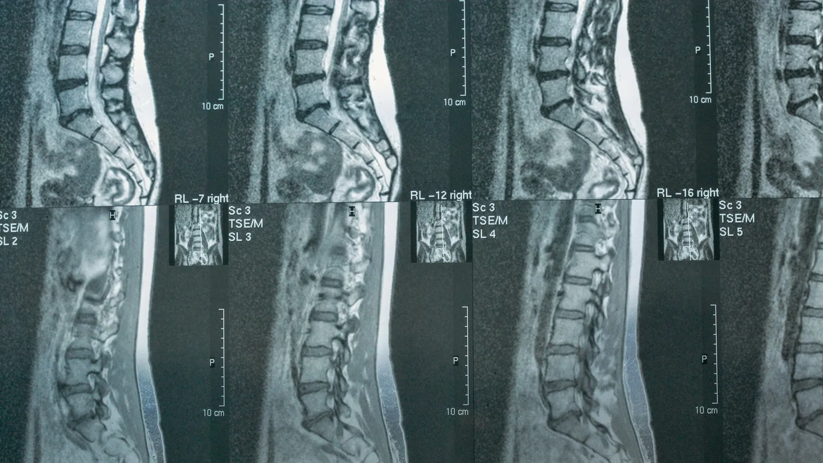
Herniated Disc Removal - Disc Herniation Surgery
Across 52705 patients, two different neck operations gave matching pain scores. They parted on one line only, which was the rate of a second operation.
About This Department
Three regions of the spine can push a disc into a nerve, and each one is a different operation carrying a different risk. A pooled analysis of 52705 patients compared the two standard ways of doing it in the neck. Pain scores matched. Disability scores matched. The difference showed up somewhere else entirely. In the mid back, where herniation is rarest, a pooled series of 258 operated patients recorded neurological worsening in 3.5 percent, which is a number no honest page should bury. The low back is the common one and usually the least dramatic. So herniated disc removal is one phrase covering at least six separate operations, and the question that matters is rarely whether to operate. It is which operation, at which level, and how long you can safely wait.
Free consultation
Find out which operation your own scan actually calls for
There is no charge and no obligation. Send the MRI images themselves and not only the radiology report, since the report describes the disc while the images show what it is touching. Say which arm or leg the pain runs down, whether anything has gone numb or weak, how long this has been going on and what you have already tried. A neurosurgeon reads the films and answers in plain language.
The distinction this page keeps returning to
Taking the fragment out and rebuilding the disc space are separate requests
Some operations lift out the piece pressing on the nerve and leave the rest of the disc where it is. Others cannot reach the fragment without going through the whole disc, so the disc has to be replaced with a cage or an implant afterward. Anatomy settles which of the two you are having, usually before anyone has asked your preference.
What a herniated disc actually is
A spinal disc is a thick fibrous ring with a soft center, and the two parts have names worth knowing, since your report will use them. The ring is the annulus. The soft core is the nucleus. When the annulus tears or weakens anywhere around its circumference, nucleus material migrates outward through the gap under the load the spine carries all day. If it stops there, nothing much happens and most people never learn it occurred. If it reaches a nerve root or the spinal cord, the picture changes within days.
Two separate things then cause symptoms. The first is mechanical, since a lump of displaced material is physically compressing a nerve. The second is chemical, because nucleus material was never meant to be in contact with nerve tissue and provokes an inflammatory reaction when it is. That second mechanism explains why some people whose scans look alarming improve steadily on anti-inflammatory treatment while others with small herniations are in genuine trouble. Surgery deals with the first mechanism directly and the second indirectly.
The words on your radiology report, in ascending order
Reports use four terms and patients often read all four as the same emergency. They are not.
- Bulge. The whole ring pushes out evenly, all the way around. This is common with age, usually causes nothing, and is not a herniation in the surgical sense.
- Protrusion. A focal outpouching, wider at its base than at its tip. The ring is stretched but intact.
- Extrusion. Material has come through the ring. The tip is now wider than the neck it came through. This is what most people mean by a slipped disc.
- Sequestration. A fragment has broken off completely and sits free in the canal. It can migrate up or down and it can also shrink on its own, sometimes dramatically.
None of those four words tells anyone whether to operate. The scan describes the disc. The examination describes the nerve. A large extrusion in a comfortable patient gets left alone, and a modest protrusion sitting exactly on a root in someone whose foot is dropping does not.
Why the level decides the operation
Ask what a surgeon has to get past to reach the disc and the whole subject reorganizes itself. In the low back, the spinal cord has already ended and the canal contains loose nerve roots that tolerate being gently moved aside. In the neck, the cord is right there. In the mid back, the cord is right there and the ribcage is in the way as well.
That single anatomical fact drives everything downstream, including how long the operation takes, whether an implant is needed, what the complication rate looks like and how long you stay.
- Cervical, meaning the neck. Roughly one in five surgically treated herniations. Symptoms run down an arm. The cord sits directly behind the disc, so most surgeons approach from the front through the neck, which means removing the disc entirely and filling the space. A smaller subset can be reached from the back without touching the disc space.
- Thoracic, meaning the mid back. Well under one operated case in a hundred. The cord is present, the ribs restrict access, and the disc is often calcified into something closer to bone. Every approach involves a detour, and the numbers reflect it.
- Lumbar, meaning the low back. The large majority. Symptoms run down a leg. The fragment can usually be lifted out through a small window in the bone with the rest of the disc left in place.
This is why a friend's account of their disc surgery may bear no resemblance to yours. They may have had a forty minute procedure through a two centimeter incision and gone home the next morning. You may be looking at an implant, a plate and a soft collar. Same diagnosis. Different level. Different world.
When removal is the right answer
Most disc herniations do not need an operation. Bodies reabsorb displaced disc material, inflammation settles, and a nerve that was furious in week two is often calm by week eight without anyone having done anything surgical. The honest framing is that surgery for a herniated disc is usually a decision about time. It is less often a decision about outcome. It buys relief sooner. Over a long enough horizon the gap between operated and non-operated patients narrows considerably in the ordinary cases.
That framing collapses in three situations, and the collapse is the reason this page exists.
The findings that take the decision out of your hands
The first is loss of bladder or bowel control together with numbness in the area that would touch a saddle, which points to compression of the whole bundle of lumbar nerve roots. That is an emergency measured in hours. The right response is the nearest hospital, and the email to a clinic abroad can wait until afterward.
The second is spinal cord compression in the neck or mid back, which announces itself quietly. Handwriting deteriorates. Buttons become difficult. Walking feels unsteady in the dark or on uneven ground. People often attribute all of this to age for a year before anyone scans them. Cord compression damages tissue that recovers poorly, so here the operation protects tissue as well as relieving symptoms, and waiting costs something that cannot be bought back.
The third is progressive motor weakness. Not pain. Weakness. A foot that catches on stairs, a grip that drops a cup, a calf that will not push off. Pain is miserable and pain waits. Weakness that is deepening week on week does not.
Outside those three, the case for operating is built on how long this has gone on, how much of your life it has taken, and what has already been tried properly. Six weeks of genuine physiotherapy counts. Two weeks of rest and painkillers does not.
Two ways into a disc in the neck
Cervical disc herniation typically presents as pain running from the neck into one shoulder, down the arm and into two or three fingers, often with pins and needles in exactly the territory of one nerve root. The pattern is specific enough that an experienced examiner can usually name the level before opening the scan. Where the fragment sits then determines which of two very different operations is on the table.
Going in from the front
The anterior route reaches the spine through a small horizontal incision in a skin crease at the front of the neck. The windpipe and gullet are held gently to one side, the disc comes into view directly, and the surgeon removes it completely along with whatever has escaped behind it. Because the disc is gone, the space has to be filled. A cage packed with bone graft goes in, sometimes with a small plate across the front, and over the following months the two vertebrae grow together into one. That is anterior cervical discectomy and fusion, which most people encounter as its initials.
In selected patients an artificial disc is used instead of a cage, which preserves movement at that level instead of ending it. Candidacy is narrower than the marketing suggests, since the joints at the back of the level have to be reasonably healthy and the level has to still move. Whether you qualify is a scan question, and it gets answered early or not at all.
The front route has one commanding advantage. It sees the entire back wall of the disc, including the part sitting centrally against the cord, and it takes pressure off from the direction the pressure is coming from. The trade is that the disc itself is spent.
Going in from the back
The posterior route approaches through the muscles at the back of the neck and enlarges the bony tunnel the nerve root travels through, lifting out any fragment that has wandered into it. The disc space is never entered. Nothing is implanted. Nothing is fused. Movement at that level continues as before.
This works when the compression is out to the side, in the tunnel, pressing on the root alone. It does not work when the material sits centrally against the cord, because getting to a central fragment from behind would mean moving the cord, which no one does. So the approach is not a preference. It is a description of where your fragment happens to be.
What the pooled data showed
For the group where both operations are technically possible, meaning one level, one side, a root problem rather than a cord problem, the two approaches have been compared directly. A meta-analysis gathered three randomized trials and twelve retrospective studies, 52705 patients in total, and set the front route against the back route.
| Measure | What the pooling found |
|---|---|
| Neck disability index | No significant difference between the two |
| Pain scores | No significant difference |
| Patient satisfaction | No significant difference |
| Complication rate | Equivalent between the groups |
| Operating time | Shorter with the back route |
| Length of hospital stay | Shorter with the back route |
| Total hospital cost | Lower with the back route |
| Reoperation rate | Higher after the back route |
Read the table from top to bottom and the back route wins six lines and loses one. Read it from the bottom up and the single line it loses is the one you will still be living with in five years. Neither reading is dishonest. They are simply different questions.
The pooled analysis concluded that the posterior approach is a sufficient alternative with a shorter operation, a shorter stay and lower cost, while noting that its higher reoperation rate should also be taken into consideration. A surgeon offering you the back route and never mentioning that last clause has told you most of the truth.
There is a further wrinkle worth understanding. Fusing a level transfers load to the levels above and below it, and over years that transfer shows up as new trouble next door. So the front route trades a lower early reoperation rate for a later one, arising for a different reason. Anyone who tells you one of these operations is simply better has compressed a genuinely open question into a slogan.
The mid back, where numbers get harder
Thoracic disc herniation is uncommon enough that many spine surgeons operate on only a handful in a career. It also behaves unlike its neighbors. The symptoms are vague, frequently mistaken for a cardiac or abdominal problem, and often described as a band of tightness around the chest or belly instead of pain shooting down a limb. By the time the diagnosis is made, a fair number of patients already have subtle signs of cord involvement in the legs.
The technical problem is access. The spinal cord occupies the canal and has no tolerance for retraction. In front of the vertebrae sit the lungs, the great vessels and the diaphragm. Many thoracic herniations calcify over years and end up welded to the covering of the cord. So the surgeon has to reach around the cord, since moving it is out of the question, and every recognized approach is a variation on that theme, whether through the chest, along the side of the vertebra, or through the joint at the back with the pedicle preserved.
The last of those, which goes through the joint at the back while preserving the pedicle, has now been studied in pooled form across eleven published series covering 328 patients operated between 2009 and 2022. Pain improved with a standardized mean difference of 0.749. Myelopathy scores improved with a standardized mean difference of 0.775. Both favor the operation clearly.
Then the complication figures. Across the 258 patients in whom complications were reported, the pooled overall rate was 12.4 percent, and neurological worsening occurred in 3.5 percent. That second number deserves to be stated without softening. Roughly one operated patient in thirty came out of a thoracic disc operation neurologically worse than they went in, in a pooled series that the authors themselves describe as favorable compared with other approaches. Nobody publishes a figure like that with enthusiasm, and it is precisely the figure a patient weighing a mid back operation needs, because the alternative is not a comfortable status quo. Untreated cord compression at this level has its own trajectory, and the comparison that matters is between two risks rather than between risk and safety.
Practically, this means a thoracic herniation is worth a second opinion in a way that a routine lumbar one is not. Ask how many the surgeon has done. Ask which approach they intend and why that one. Ask what neurological monitoring runs during the case.
The low back, in short
Lumbar herniation is the version most people picture, and it is covered in depth on our lower back disc surgery page. The summary is that the cord has ended above this level, so the surgeon works among mobile nerve roots rather than beside a cord, and the standard operation removes only the escaped fragment through a window of a centimeter or two. The disc that remains is left in place. No implant, no fusion, and usually one night in hospital.
Where the endoscope fits
Endoscopic removal reaches the same fragment through a tube around seven millimeters wide, either from the side or between the laminae, with the surgeon working from a screen. Muscle damage is smaller and people are often up the same afternoon. Fragments that have migrated a long way, or that sit in awkward corners, remain easier to retrieve under the microscope, so the endoscope has a territory rather than a monopoly.
Inside the operating room
A single-level removal in the low back takes roughly forty five to ninety minutes. A one-level neck operation from the front runs a little longer once the implant is placed. A thoracic case can occupy most of a morning. What follows describes the common thread through all three.
Marking the level with a film in the room
A needle is placed and an image taken before the incision, so the level being opened is confirmed against your own anatomy instead of counted from memory. Wrong-level surgery is rare in spine practice and this is one of the reasons it stays rare.
Opening a window instead of a corridor
Muscle is separated along its fibers instead of cut across them, and a retractor holds the gap open. A small amount of bone at the edge of the lamina is thinned to expose the nerve. In the neck from the front there is no muscle to divide at all, since the approach runs between natural planes.
Finding the nerve before finding the disc
The compressed root is identified and protected first. Only then is the fragment approached, from a direction that moves it away from the nerve instead of through it. This ordering is the difference between an uneventful operation and a difficult one.
Deciding how much disc to take
Removing more of the remaining disc lowers the chance of another fragment escaping later and raises the chance of the space collapsing and the back aching. Removing less preserves height and accepts a slightly higher recurrence risk. In the neck this question does not arise, because the disc comes out entirely and something replaces it.
Closure is unremarkable. Most people wake with the limb pain already gone, which is the single most reported experience after this operation, and with a sore incision that was never the problem in the first place.
The risks, stated plainly
Every figure below shifts with the level, the patient and the surgeon. Treat them as orders of magnitude. The thoracic column in particular rests on a small pooled literature, and no one should read it as a settled number.
| Problem | Where it matters most | Rough frequency |
|---|---|---|
| Tear in the covering of the nerves | All levels, more with revision surgery and calcified discs | A few percent, repaired during the same operation |
| Neurological worsening | Thoracic above all, since the cord is in the way | 3.5 percent pooled in thoracic series, far lower in the low back |
| Hoarse voice or swallowing difficulty | Neck, front approach only | Common early, settling within weeks in the large majority |
| Wound infection | All levels, more with longer operations | Around one percent for a simple removal |
| Same disc herniating again | Lumbar, where the rest of the disc stays behind | 3.6 percent pooled after endoscopic removal |
| Numbness that never fully returns | Any level where compression was prolonged | Variable, and strongly related to how long the nerve was squashed |
| Clot in a leg vein | Anyone immobile, and anyone flying soon afterward | Uncommon, and the reason early walking is pushed hard |
One risk sits outside that table because it is not a complication at all. Surgery reliably relieves the pain that travels down the limb. It is far less reliable against pain in the neck or back itself. Patients whose dominant complaint is central back pain, with only a little radiating pain, are the group most often disappointed by a technically perfect operation. Being clear with yourself about which pain is worse is worth more than any second opinion.
Ask before you agree
What proportion of my pain is in the limb rather than the spine
Put a rough percentage on it out loud. If the answer is eighty percent leg or arm, the operation is aimed at your problem. If the answer is eighty percent back or neck, ask the surgeon directly what they expect the operation to do for that eighty percent, and listen carefully to the answer.
Why some discs herniate again
In the low back the operation removes the escaped fragment and leaves the rest of the disc in place, which means the hole in the ring is still there afterward. Whether more material comes through that hole is largely a question of how big the hole is.
Seven comparative studies covering 1653 patients were pooled to test exactly that. Thirty percent of them had a large defect in the ring and seventy percent a small one. Symptoms came back roughly two and a half times as often in the large-defect group, and reoperation was about two and a third times as likely, over a median follow-up close to three years. The finding held through every sensitivity analysis the authors ran.
Timing has been mapped as well. Pooled across studies of endoscopic removal, recurrence ran at 3.6 percent overall, and 61.7 percent of those recurrences happened within the first six months. Age above fifty and a body mass index above twenty five both raised the rate, roughly doubling it in the case of weight. Upper lumbar levels recurred more often than the two lowest ones, where most herniations sit.
Devices exist to close the defect. Pooled across four studies, 24 symptomatic recurrences occurred among 811 patients who received an annular closure or repair device against 51 among 645 who did not, an odds ratio of 0.34 with no heterogeneity between studies. The authors are careful to call these early results and to ask for longer follow-up before anyone treats the question as closed. Whether such a device is appropriate for you depends on the size of your defect, which is measured during the operation rather than predicted beforehand.
The window that matters
Six months, and most of it in the first three
Since three recurrences in five arrive within half a year, the restrictions given at discharge are not arbitrary caution. Heavy lifting, repeated bending and long unbroken hours of sitting all load a ring that has not yet scarred over. People ignore this in month two. That is precisely when they feel entirely well and the ring does not.
Recovery, stage by stage
Nerves recover on their own schedule and it is slower than the schedule of the wound. Pain that shot down the limb usually vanishes on the table. Numbness and weakness follow much later, sometimes over a full year, because a nerve rebuilds its insulation at roughly a millimeter a day and no operation accelerates that.
The first forty eight hours
You get up and walk the same day or the next morning, which counts as treatment and not as encouragement. After a neck operation from the front, swallowing feels odd and the voice may tire quickly. After a low back operation, sitting is the least comfortable position and standing the easiest, which surprises almost everyone.
Weeks one to three
Walking distance builds daily. The rule is short and often. Long and heroic comes later. Lifting is capped at a few kilograms, and the way you pick something up matters more than what it weighs. Desk work becomes possible toward the end of this window, in short blocks with frequent standing.
Weeks four to twelve
Structured rehabilitation begins in earnest, aimed at the deep muscles that stabilize the spine more than at general fitness. Swimming and stationary cycling come back first. Manual work and contact sport wait for clearance. After a fusion in the neck, this is the period when the graft is quietly turning into bone.
Three months to a year
Whatever numbness remains at three months may still improve, slowly. Strength usually comes back further than sensation does. A patch of altered feeling on the outside of the foot or in a fingertip is the commonest permanent souvenir, and most people stop noticing it.
Smoking deserves its own sentence. It impairs bone healing badly enough to change the odds on any operation involving a fusion, and it is the single modifiable factor with the largest effect on that outcome.
Planning the trip to Istanbul
Two questions decide the shape of the trip, and they are the level being operated on and whether an implant is going in. A one-level removal in the low back sits at the short end. A thoracic case, or a two-level neck fusion, sits at the long end and should be planned as such from the beginning, since extending a trip halfway through goes badly.
- Before you book anything. Send the MRI images as files, since photographs of a screen lose the detail that matters, and include any earlier scans of the same region. Two scans a year apart tell a surgeon something a single scan cannot. You get a written opinion naming the level, the proposed operation and the expected stay.
- Days one and two. Arrival, examination in person, blood tests, a cardiology or anesthetic review where age or medication calls for it, and fresh imaging if what you sent is more than a few months old.
- Surgery, then one to three nights. A simple lumbar removal often means a single night. A neck fusion usually means two. A thoracic case means longer, and the team will say so before you travel.
- The gap before flying. Wound review, stitch or clip removal where used, an x-ray if an implant went in, and a walking assessment. This part is unglamorous and it is what the extra days are for.
- Fitness to fly, decided last. Discharge from hospital and clearance to sit in a pressurized cabin for hours are two separate judgments made on two separate days. Book a changeable return ticket and let the second judgment fall where it falls.
For a straightforward single-level removal, ten to fourteen days in the country covers all of it comfortably. Add a week for a fusion, and plan individually for anything thoracic. Companions are welcome and genuinely useful in the first week, particularly for anyone who cannot yet turn their head to look over a shoulder.
Once you are back home, follow-up continues remotely. Photographs of the wound in the first fortnight, a written report and imaging for your own doctor, and a route back to the surgical team by WhatsApp if something changes. Arrange in advance who will remove any remaining stitches locally and who will supervise physiotherapy, because those two things are far easier to organize before you leave than after you land.
The cost questions worth asking
Prices are quoted individually because the same diagnosis produces very different operations, so anything published as a single figure is describing one version of the procedure and quietly excluding the others. What can be described here is which variables move the number, which is more useful anyway.
The level and the number of levels come first. Whether an implant is used comes second, and a cage, a plate or an artificial disc each carry their own cost. Operating time follows, since theater and anesthesia are billed by duration and a calcified thoracic disc takes several times as long as a soft lumbar fragment. Then the length of stay, then whether neurophysiological monitoring runs through the case, which it should for anything near the cord. Revision surgery on a previously operated level costs more than a first operation because scar tissue slows everything down.
When you compare quotes between hospitals, compare what is inside them. Ask whether the price covers the implant, the imaging in the operating room, the anesthetic team, the nights on the ward, the physiotherapy sessions and the follow-up review before your flight. Ask what happens financially if you need an extra two nights. A quote that answers those questions in writing is comparable with another quote. A number in an email is not.
Choosing who does it
Herniated disc removal is performed by both neurosurgeons and orthopedic spine surgeons, and the training route matters less than what the surgeon does week in and week out. At Biruni University Hospital these operations sit within the Neurosurgery department, working alongside neurology, physical therapy and rehabilitation, radiology and anesthesia.
Three questions separate a considered plan from a generic one. Ask which specific approach is proposed for your fragment and what makes that approach right for where it sits. Ask what would make the plan change once the surgeon is looking at the level directly. Ask what the realistic expectation is for the numbness or weakness you currently have, as opposed to the pain, since those recover differently and a surgeon willing to say so plainly is telling you something useful about the rest of their advice.
The consultation costs nothing and commits you to nothing. Send the images, describe what has changed over the last months, and ask whatever you want answered. If the honest answer is that your disc is likely to settle without surgery, that is the answer you will get.
Frequently asked questions
Will my neck be stiff forever after a fusion at one level?
My scan shows a herniation but my pain is improving. Should I still operate?
Why is a mid back herniation treated so much more cautiously?
If the fragment is removed, why can it come back?
Is the back-of-the-neck operation better because nothing gets fused?
How soon can I sit on a plane after disc surgery?
References
- Fang W, Huang L, Feng F, et al. Anterior cervical discectomy and fusion versus posterior cervical foraminotomy for the treatment of single-level unilateral cervical radiculopathy. A meta-analysis. J Orthop Surg Res. 2020;15(1):202.
- Hamid S, Moradi F, Bagheri SR, et al. Evaluation of clinical outcomes, complication rate, feasibility, and applicability of transfacet pedicle-sparing approach in thoracic disc herniation. A systematic review and meta-analysis. J Orthop Surg Res. 2023;18(1):516.
- Miller LE, McGirt MJ, Garfin SR, Bono CM. Association of annular defect width after lumbar discectomy with risk of symptom recurrence and reoperation. Systematic review and meta-analysis of comparative studies. Spine (Phila Pa 1976). 2018;43(5):E308-E315.
- Yin S, Du H, Yang W, Duan C, Feng C, Tao H. Prevalence of recurrent herniation following percutaneous endoscopic lumbar discectomy. A meta-analysis. Pain Physician. 2018;21(4):337-350.
- Choy WJ, Phan K, Diwan AD, Ong CS, Mobbs RJ. Annular closure device for disc herniation. Meta-analysis of clinical outcome and complications. BMC Musculoskelet Disord. 2018;19(1):290.
Editor's note
Written by the Biruni Hospital medical editorial team. Reviewed by Assistant Professor Özcan ÇIKLATEKERLİO, Neurosurgery.
Medically reviewed by

Assistant Professor Özcan ÇIKLATEKERLİO
Neurosurgery
Related Treatments
View All
ALIF - Anterior Lumbar Interbody Fusion Surgery
Coming in from the front restores the curve of the lower back better than any posterior route and leaves the back muscles untouched. It also carries one risk most pages will not name, and this page names it.

Brain AVM Surgery - Arteriovenous Malformation Surgery
An unruptured brain AVM bleeds at roughly 2.2 percent a year, and surgery spends its whole risk on a single morning. This page sets those two shapes of risk side by side, explains what the ARUBA trial does and does not say about your case, and shows how a Spetzler-Martin grade changes the answer.
Brain Tumor Surgery - Brain Tumor Removal
Brain surgery is one trade repeated all day, how much tumor comes out against what the brain around it still does. This page puts the published numbers on both sides of that trade.
Cervical Microdiscectomy - Minimally Invasive Neck Disc Surgery
Neck disc surgery is done from the front, through the space between your windpipe and your carotid artery, and almost everything that goes wrong that is not the disc happens in that corridor. What has been measured in it.

Chiari Malformation Surgery
A Chiari malformation is measured in millimeters and treated according to symptoms, and those two things come apart more often than patients are told. Most people with the finding never need an operation. This page sets out who does, what posterior fossa decompression removes, why surgeons still argue over opening the dura, and what the pooled studies say about both sides of that argument.