
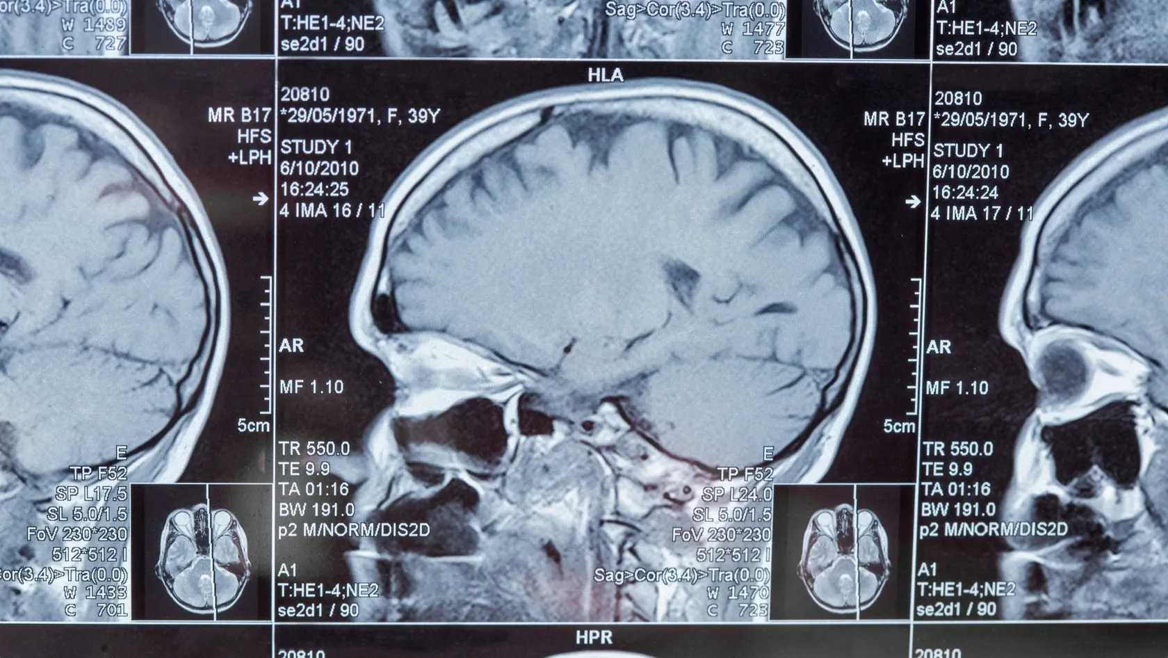
Glioma Surgery
Early surgery for a grade 2 glioma was followed by a median survival of 14.4 years in one Norwegian region and 5.8 years in the region that watched and waited. This page explains what the operation decides and what the pathology report decides after it.
About This Department
Two tumors carrying the same name can behave nothing alike. The laboratory report tells them apart.
Where two Norwegian regions treated the same grade 2 gliomas differently for eleven years, patients whose tumors were removed early lived a median of 14.4 years against 5.8 years for those who were watched.
Two regions of Norway treated the same tumor differently for eleven years, and the numbers that fell out of it are the strongest argument anyone in this field has, since adults with grade 2 gliomas lived a median of 14.4 years in the region where surgeons removed the tumor early and 5.8 years in the region where the habit was to watch and wait.
Nobody designed that experiment. Geography ran it.
Glioma covers tumors a person lives with for two decades and tumors measured in months, and what separates them turns up in a laboratory result read from tissue the operation takes out. Glioma surgery does two jobs at once. It removes tumor, and it produces the answer that decides every treatment after it. This page follows that order, starting with the three molecular results and what each one changes, then how much tumor surgeons aim to remove and the volumes the published classifications use, then what the operation costs you in risk, and finally the part that decides an international patient's whole trip. Six weeks of daily radiotherapy begins three to six weeks after the operation. Whether you sit those weeks out here or fly home for them is a real decision with real consequences, and almost nobody writing for patients abroad warns you it is coming.
One word, three different diseases
The 2021 World Health Organization classification stopped naming gliomas by appearance alone and started naming them by their molecular makeup, which changed what a diagnosis means, so a tumor that used to be called a grade 2 astrocytoma may now be an IDH mutant astrocytoma with a twenty year outlook, or an IDH wildtype tumor that behaves like a glioblastoma and gets treated like one.
Three results do most of that work.
The three lines on your report that matter
Each result answers a different question, and read together they give the tumor its name.
This table scrolls sideways on a narrow screen. Swipe or drag to see every column.
| Result | What the laboratory is looking for | What it changes for you |
|---|---|---|
| IDH mutation | A change in one enzyme, present in most grade 2 and grade 3 gliomas of younger adults and absent in glioblastoma | Sets which disease you have. A mutant tumor grows more slowly, carries a far longer outlook, and now has drugs aimed at the mutation itself |
| 1p/19q codeletion | Loss of matching pieces of two chromosomes, found only in tumors that are already IDH mutant | Names the tumor an oligodendroglioma, the glioma that responds best to chemotherapy and the one with the longest published survival |
| MGMT promoter methylation | A chemical switch that silences a repair gene inside the tumor cells | Predicts how well temozolomide works. A methylated promoter changes the plan, and for an older patient it can decide whether chemotherapy is given at all |
| Grade 2, 3 or 4 | Now assigned from molecular features as well as from how the cells look under the microscope | Sets how fast treatment starts and how often you are scanned afterward |
Timing matters here in a way that catches travelers out. Ordinary staining comes back within two or three days, so the surgeon can tell you the broad category before you leave the ward. The molecular panel takes longer, seven to fourteen working days, and a methylation array longer still. Let that gap set your return date, because the second conversation is the one that changes your life.
How much of it has to come out
A glioma grows into brain rather than pushing it aside, so no clean edge exists to lift the thing out along, and tumor cells sit among working brain cells for centimeters beyond anything a scan shows, which leaves a tumor described as completely removed still able to return and makes the operation a judgment rather than an extraction. What surgeons can measure is how much visible tumor is left on the scan afterward, and that measurement turns out to predict a great deal.
What the resection categories are worthAn international group collected 1,008 adults with newly diagnosed IDH wildtype glioblastoma and sorted them by the tumor volume left behind. Clearing all of the contrast enhancing tumor beat clearing some of it, which beat a biopsy alone. Going further and reducing the non enhancing tumor to five cubic centimeters or less was worth more again, and that category is what the field now calls a supramaximal resection. A separate study built a survival model on 622 patients and confirmed it in 536 more, reporting a median survival of 57.3 months for younger patients who had a methylated MGMT promoter and a supramaximal resection, against 14.3 months for patients with poor performance status, an unmethylated promoter and an incomplete removal. One diagnosis, a fourfold difference, decided partly by biology and partly by what happened in the operating room.
Only one of those three factors was still open when you arrived.
The limit on the third one is function, and it is a hard limit. A surgeon who takes another ten percent of tumor and leaves you unable to find words has made your life worse and your treatment harder, because a patient with a fresh neurological deficit often starts radiotherapy late and sometimes never starts it at all. Extent of removal and neurological function get traded against each other in every glioma operation ever performed, and every piece of equipment in the room, the navigation, the ultrasound, the fluorescent dye, the nerve monitoring and the awake testing, exists to shift that trade in your favor. None of it abolishes the trade. Ask a surgeon where they would stop on your scan. A straight answer to that question tells you more about the team than any figure on a website.
A slow growing glioma is still an argument for operating
Grade 2 gliomas usually announce themselves with a seizure in an adult in their thirties or forties who was well the day before, and the scan then shows something that has clearly been growing quietly for years while the patient feels normal, so the pull toward watching it is strong on both sides of the desk. Two decades of practice went that way in many centers.
What two Norwegian regions showed by accident
Two Norwegian university hospitals covering neighboring populations held opposite policies between 1998 and 2009. One favored watching, the other favored early removal, and which one you got depended on your home address rather than on your tumor. Researchers followed 153 adults to 2016. In the watching region 29 percent had early surgery, against 86 percent in the operating region, and median overall survival came out at 5.8 years against 14.4 years, a gap that survived adjustment for the molecular markers and therefore rules out the obvious objection that the operated region simply had gentler tumors. Waiting does not keep a grade 2 glioma still. These tumors grow continuously, at a few millimeters a year, and most of them eventually turn into something more aggressive. Time is not on your side here. Watching a tumor that is going to transform means operating later on a larger tumor in a patient whose brain has had less chance to shift function away from it, which is a worse operation done in worse circumstances. Where the tumor sits somewhere reachable and the patient is well, early removal is the stronger choice, and the burden of argument sits with the person proposing to wait.
Seizures respond to the same logic. Removing the tumor, and a margin of the irritable brain around it, controls seizures better than medication alone in a large share of these patients, which matters more than it sounds. Seizure freedom is what returns a driving license, a job with machinery and a night's uninterrupted sleep.
When a biopsy is the right operation
Some gliomas should not be resected, and a team that says so early is doing its job rather than avoiding difficulty.
A tumor sitting in the brainstem or deep in the thalamus has no safe corridor into it. A glioma that has crossed the corpus callosum into both hemispheres cannot be removed on either side without taking function with it. Disease that appears in several separate places at once behaves as a whole rather than as a lump, and a patient who is already bedbound from the tumor rarely gains anything from a long operation, because the recovery costs more than the removal returns. In all of those situations a needle biopsy through a small opening, guided by navigation, gets the tissue that produces the molecular report, and that report is what starts treatment, so the operation runs about an hour, you go home the next day, and the treatment that follows is chosen by exactly the same markers as it would be after a full resection. Nothing about the drugs or the radiotherapy depends on how the tissue was obtained.
Recommending a biopsy is not the same as giving up. A surgeon who says it has declined to trade your speech for a better looking picture, and that judgment is what you traveled to get.
Inside the operating room
Published series put a glioma resection at roughly three to eight hours depending on the site and on whether mapping is used, with a straightforward biopsy far shorter than that. The sequence below is the common one.
- A planning MRI is done shortly before surgery and loaded into the navigation system. Where the tumor lies near speech or movement, a functional MRI and a tractography study map those pathways first.
- Your head is fixed in a frame and the opening in the skull is cut to the size the tumor needs, no larger.
- Finding the boundary is the hard part. Navigation gives the plan, ultrasound gives a live picture that stays accurate as the brain shifts, and for high grade tumors a fluorescent dye taken by mouth beforehand makes tumor tissue glow under blue light while normal brain does not.
- Removal runs against a map of function. Under general anesthesia that means continuous electrical monitoring of the motor pathways. Where speech is at risk it usually means waking you for the middle part of the operation and testing you while the surgeon works.
- An MRI is repeated within 24 to 48 hours, before healing changes start to light up on the scan and hide any tumor left behind. That scan decides which resection category you fall into, and the surgeon's impression does not.
Awake testing sounds worse than patients report it to be. You are asleep for the opening and the closing, awake and comfortable in between, naming pictures or counting or moving a hand while the surgeon stimulates the brain around the tumor and watches for the moment the task fails. That moment marks a boundary, and the boundary is respected. Language testing only works in a language you speak fluently, so ask early which languages the team can test in and whether an interpreter can stand at the head of the table.
One night in intensive care, then three to six days on the ward.
Risks, and what is done about each one
Every item below appears on the consent form a neurosurgeon goes through with you, each one paired with its management, since a risk named without its remedy frightens people for no useful reason.
Seizures deserve their own paragraph
Seizure control shapes daily life more than any scan result does, and driving rules vary by country and are strict everywhere, with a common threshold of six to twelve months free of seizures before a license comes back, which affects work, school runs and independence long after the wound has healed. Tell your team what you drive and what you do for a living before the operation, because they build the plan around it.
The first two weeks
Tiredness is the dominant experience, and it surprises families more than pain does. Headache after a craniotomy is real and answers ordinary painkillers, and it settles over the first week. Sleep when you need to. What lingers is a flatness and a need to sleep in the afternoon that can last a month, which is normal and is not a sign that something was missed.
Three things you go home onA steroid, usually dexamethasone, to control swelling around the cavity, on a written reducing schedule that you follow to the day, even once you feel better. An anti-seizure tablet, taken at fixed times, which interacts with several ordinary medicines and should be checked against anything you already take. And a wound with clips or stitches that come out at 10 to 14 days, at a review that also confirms you are fit to travel. Steroids bring appetite, disturbed sleep, a shortened temper and higher blood sugar, all of which fade as the dose comes down, and all of which are worth warning your family about before they see it.
Walking on the first day is expected. Strenuous exercise and heavy lifting wait four to six weeks, desk work comes back sooner than physical work, and hair grows over the scar within a couple of months.
What the report starts
If the report says glioblastoma
A trial published in 2005 randomized 573 patients between radiotherapy alone and radiotherapy given together with a daily low dose of temozolomide, followed by six monthly cycles of the same drug. Radiotherapy ran at 2 Gy a day, five days a week, for six weeks. Median survival came out at 14.6 months against 12.1 months, and survival at two years at 26.5 percent against 10.4 percent, results that have made this schedule the backbone of glioblastoma treatment ever since and that put it on this page instead of an oncology page, because of the calendar it imposes. Treatment begins three to six weeks after surgery and then requires you in one place, five days a week, for six consecutive weeks.
This table scrolls sideways on a narrow screen. Swipe or drag to see every column.
| The report says | What normally follows surgery | How long it keeps you in one place |
|---|---|---|
| Glioblastoma, IDH wildtype, grade 4 | Six weeks of daily radiotherapy with temozolomide taken alongside it, then monthly cycles of the tablet | Six weeks on site, then roughly six months of tablets that can be taken anywhere |
| IDH mutant astrocytoma, grade 2 | Scans alone where the tumor was fully removed and you are young, or radiotherapy and chemotherapy where tumor was left behind | A scan every three to six months, or the same six week block if treatment starts |
| Oligodendroglioma, 1p/19q codeleted | Radiotherapy followed by combination chemotherapy in most cases, since this tumor answers chemotherapy better than any other glioma | Six weeks, then several months of cycles |
| IDH mutant astrocytoma, grade 3 or 4 | Radiotherapy with temozolomide, on a schedule close to the glioblastoma one | Six weeks, then maintenance cycles |
| Biopsy only, no tumor removed | Whatever the markers call for, exactly as above | The same timetable, since the treatment follows the report rather than the operation |
If the report says IDH mutant
Something changed for this group in 2023. A phase 3 trial gave 331 patients with residual or recurrent grade 2 IDH mutant glioma, all of whom had been treated with surgery and nothing else, either an oral drug aimed at the mutated enzyme or a placebo. Progression free survival ran 27.7 months against 11.1 months, the time until the next treatment was needed stretched out with it, and serious side effects were more common on the drug than on placebo, at 22.8 percent against 13.5 percent, with raised liver enzymes the main one at 9.6 percent. For a thirty five year old with a partly removed grade 2 tumor, that result means the choice is no longer only between radiotherapy now and scans until something changes. Whether the drug is licensed and funded where you live is a question for your own oncologist, and it belongs in the conversation before you decide where to have your surgery, because the decision downstream depends on what the tissue shows.
Six weeks of treatment, here or at home
Here is the question that decides an international patient's whole trip, and the one almost no page raises. Surgery takes a week of your life. Plan for six weeks. The treatment that follows it takes six, and it has to start within three to six weeks of the operation to be given as the evidence describes it.
Two answers are available and both are legitimate.
What staying looks like, and what going home looks like
Radiotherapy and chemotherapy are delivered at Biruni Hospital on the same site as the surgery, so a patient who stays continues with the team that operated, using the same scans and the same pathology, without a referral and without a transfer of records. In practice that means a planning session with a mask made to fit your face, then a short daily appointment Monday to Friday for six weeks, with the tablet taken at home every day including weekends. Most people manage the first three weeks with little more than mild tiredness and then feel the fatigue build through the last three. Hotel accommodation and the transport to and from the hospital for every one of those appointments are arranged by the international patients office, and the coordinator assigned to you at your first message stays with you through all of it. Both routes end in the same treatment. Going home is the other real option, and it is the right one for plenty of patients who have family, work and an oncologist waiting. What makes it work is the handover. You leave with the operation note, the post-operative MRI, the full pathology including every molecular result, and a written treatment recommendation addressed to your own oncologist, so the radiotherapy can be planned at home without repeating anything. Arrange that appointment before you travel, because the three to six week window closes quickly and finding a slot from a hotel room in another country is the part that goes wrong.
Fitness to fly is confirmed at the wound review around day 10 to 14, once the scar is dry and no seizure has occurred. Your surgeon writes that opinion with a date on it, which airlines and insurers ask for.
When it comes back
Gliomas recur, and honest pages say so. Because tumor cells spread beyond anything the scan shows, the disease returns in most patients, usually at the edge of the old cavity, and the interval before it does depends far more on the molecular subtype than on anything else. A second operation is one of several options at that point, and it is offered when five things line up.
- The recurrence sits somewhere a surgeon can reach without taking function on the way in.
- You are still walking, working or otherwise functioning well, since performance status predicts the benefit better than the scan does.
- Enough time has passed since the first operation for the tissue to have settled.
- The imaging distinguishes tumor from the swelling radiotherapy leaves behind, which can look identical and sometimes needs a repeat scan or a biopsy to separate them.
- Something useful follows the operation, whether that is a different chemotherapy, a second course of radiotherapy or a trial.
The volume rule applies the second time as well. Where a repeat operation clears the enhancing tumor, survival runs longer than where tumor is left or where no operation is done, and where it leaves a deficit behind the benefit disappears, because a patient who cannot start the next treatment gains nothing from the surgery that preceded it.
What moves the cost
No figure appears on this page, and for glioma that omission matters more than it does anywhere else on this site, because a needle biopsy and an eight hour awake resection followed by six weeks of radiotherapy are different pieces of work by an order of magnitude, and nobody knows which one you need until a neurosurgeon has read your scan.
Six things carry most of the difference. Whether the operation is a biopsy, a debulking or a maximal resection, and how many hours it runs. Whether awake mapping and a neurophysiology team are part of it, since that adds two more people to the room and an hour or more to the operation. Which tools guide the removal, since intraoperative MRI, ultrasound and fluorescence are not equally available and are not equally priced. How many intensive care nights the plan assumes. Which molecular tests are ordered, because a full panel with a methylation array costs considerably more than a basic IDH stain and is the line most often left out of a quote. Quotes rarely say which. And whether radiotherapy and the chemotherapy that runs beside it belong to the same episode of care or get quoted separately later. Your own situation moves it too. Age, how well you are functioning, diabetes made worse by steroids, blood thinning medication, heart or lung disease and earlier surgery at the same site all change the plan and therefore the number. So does the amount of inpatient rehabilitation the case is likely to need.
Packages published by hospitals and medical travel agencies in this market cover airport transfers, pre-operative tests, the surgeon and anesthesia fees, a stated number of nights including intensive care, the post-operative scan, an interpreter, a set number of hotel nights and the appointments before departure. They leave out flights, travel insurance, nights beyond the plan, unplanned intensive care and the treatment of a complication, so on a glioma quote read the pathology line and the radiotherapy line hardest, because those two are assumed most often and written down least.
Five questions make two quotes comparable. Does this figure assume a biopsy, a debulking or a maximal resection, and for how many hours in the operating room? Are awake mapping and the neurophysiology staff inside it? Which molecular tests are included, and is a methylation array one of them? How many intensive care nights are counted? Are radiotherapy and the chemotherapy given with it quoted here, or separately once the pathology is known?
Send the MRI, any previous pathology and the list of medicines you take. A neurosurgeon reading them will tell you which operation your tumor allows, and that opinion costs nothing and commits you to nothing.
Planning the trip
Bring your scans as original files on a disc or a drive, never as photographs of a screen, because a surgeon needs to scroll through the sequences to plan an approach, and if you have had a previous operation anywhere, ask that hospital for the pathology blocks or slides as well, since the molecular tests can often be run on old tissue and that saves time. Label everything. Write down every medicine with its dose, and mark the steroid and the anti-seizure tablet clearly.
Traveling with someone who has seizures
Do not come alone. A patient who has had a seizure should not be traveling unaccompanied in any case, and after a craniotomy there are weeks with no lifting, no driving and an afternoon that has to be slept through. Patient rooms carry a companion bed, so one person stays with you every night of the admission, and accommodation for you both on the nights either side is arranged along with the airport transfers and every trip between hotel and hospital. The international patients team works in English, Arabic, French, Russian, Serbian, Romanian and Spanish, with interpreting in other languages arranged on request, and one coordinator holds your case from the first message through to discharge and stays reachable on the same WhatsApp number afterward. That language question is clinical here rather than administrative, because awake testing has to be done in a language you are fluent in.
Say in your first message if you would prefer a woman surgeon or physician, since the request goes to the department and is met wherever the rota allows.
An appointment confirmation and an invitation letter naming the hospital and your doctor, which is the document most consulates ask for with a medical visa application, are issued about ten days before you travel, and meals from the hospital kitchen cover halal, vegetarian and diabetic requirements, which matters more than usual on steroids, with a prayer room on site.
Once you are home
Glioma follow up runs on scans. An MRI is repeated at the end of radiotherapy, then every two to three months during chemotherapy and every three to six months afterward, and comparing each one against the post-operative scan is the entire point of keeping that first study safe. Local scans can be sent back for review by the team that operated, and a photograph of a report or a question on a dose gets an answer from your coordinator on the same WhatsApp number you used at the beginning.
Leave with a file your own oncologist can work from without telephoning anyone. The operation note naming the extent of the resection, the post-operative MRI on a disc, the complete pathology with every molecular result and its date, the steroid reducing schedule, the anti-seizure medication with its dose, the radiotherapy plan or the written recommendation for one, and the date of the next scan. Keep a copy yourself.
Some things need attention the same day, wherever you are.
For any of those the nearest emergency department comes first, with your operation note and your medication list in your hand, and a message to your coordinator afterward.
Glioma surgery FAQ
Can a glioma be removed completely?
How long will I be in hospital?
Do I have to stay for the radiotherapy?
When can I fly home after glioma surgery?
Will anyone speak my language during the operation?
What if the pathology is worse than we expected?
Written by the Biruni Hospital medical editorial team. Reviewed by Dr Yunus Emre Yavuz, Neurosurgery.
References
- Karschnia P, Young JS, Dono A, Häni L, Sciortino T, Bruno F, et al. Prognostic validation of a new classification system for extent of resection in glioblastoma. A report of the RANO resect group. Neuro-Oncology. 2023;25(5):940-954.
- Park YW, Choi KS, Foltyn-Dumitru M, Brugnara G, Banan R, Kim S, et al. Incorporating supramaximal resection into survival stratification of IDH-wildtype glioblastoma. A refined multi-institutional recursive partitioning analysis. Clinical Cancer Research. 2024;30(21):4866-4875.
- Jakola AS, Skjulsvik AJ, Myrmel KS, Sjåvik K, Unsgård G, Torp SH, et al. Surgical resection versus watchful waiting in low-grade gliomas. Annals of Oncology. 2017;28(8):1942-1948.
- Stupp R, Mason WP, van den Bent MJ, Weller M, Fisher B, Taphoorn MJB, et al. Radiotherapy plus concomitant and adjuvant temozolomide for glioblastoma. New England Journal of Medicine. 2005;352(10):987-996.
- Mellinghoff IK, van den Bent MJ, Blumenthal DT, Touat M, Peters KB, Clarke J, et al. Vorasidenib in IDH1- or IDH2-mutant low-grade glioma. New England Journal of Medicine. 2023;389(7):589-601.
Editor's note
Written by the Biruni Hospital medical editorial team. Reviewed by Assistant Professor Fikret BAŞKAN, Neurosurgery.
Medically reviewed by
Assistant Professor Fikret BAŞKAN
Neurosurgery
Related Treatments
View All
ALIF - Anterior Lumbar Interbody Fusion Surgery
Coming in from the front restores the curve of the lower back better than any posterior route and leaves the back muscles untouched. It also carries one risk most pages will not name, and this page names it.

Brain AVM Surgery - Arteriovenous Malformation Surgery
An unruptured brain AVM bleeds at roughly 2.2 percent a year, and surgery spends its whole risk on a single morning. This page sets those two shapes of risk side by side, explains what the ARUBA trial does and does not say about your case, and shows how a Spetzler-Martin grade changes the answer.
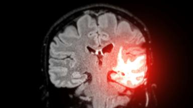
Brain Tumor Surgery - Brain Tumor Removal
Brain surgery is one trade repeated all day, how much tumor comes out against what the brain around it still does. This page puts the published numbers on both sides of that trade.
Cervical Microdiscectomy - Minimally Invasive Neck Disc Surgery
Neck disc surgery is done from the front, through the space between your windpipe and your carotid artery, and almost everything that goes wrong that is not the disc happens in that corridor. What has been measured in it.
Chiari Malformation Surgery
A Chiari malformation is measured in millimeters and treated according to symptoms, and those two things come apart more often than patients are told. Most people with the finding never need an operation. This page sets out who does, what posterior fossa decompression removes, why surgeons still argue over opening the dura, and what the pooled studies say about both sides of that argument.